A Case Report of Tsukamurella pulmonis Infection Misidentified as Atypical Mycobacteria
Ah Ra Cho1, Hye Ryoun Kim1, Mi-Kyung Lee1, Seong Ho Choi2, Sin Weon Yun3 Departments of 1Laboratory Medicine, 2Internal Medicine and 3Pediatrics,
Chung-Ang University College of Medicine, Seoul, Korea
We report a case of catheter-related bacteremia due to Tsukamurella pulmonis. T. pulmonis is a rare cause of opportunistic infection in immunosuppressed pa- tients and in cases of indwelling foreign materials.
This infection was nearly impossible to identify using conventional phenotyping methods because of its similarities to the related genera Nocardia, Rhodo- coccus, Gordonia, Streptomyces, Corynebacterium, and Mycobacterium. This organism was initially mis-
identified as Mycobacterium aubagnense through PCR-RFLP analysis. We correctly identified this or- ganism using 16S rRNA sequencing combined with phenotyping tests. (Korean J Clin Microbiol 2010;13:
93-97)
Key Words: Tsukamurella pulmonis, Catheter, Bacte- remia, Korea, Misidentification, Mycobacteria
Received 3 November, 2009, Revised 25 February, 2010 Accepted 25 March, 2010
Correspondence: Mi-Kyung Lee, Department of Laboratory Medicine, Chung-Ang University College of Medicine, 65-207, Hangangno 3-ga, Yongsan-gu, Seoul 140-757, Korea. (Tel) 82-2-748-9837, (Fax) 82-2-748-9929, (E-mail) [email protected]
93
Fig. 1. Tsukamurella pulmonis colonies on blood agar isolated from the blood culture The colony was yellow, dry, irregular and rough.
INTRODUCTION
Tsukamurella species are aerobic gram-positive organisms from the order Actinomycetes and environmental organisms found in soil, water and sludge. Tsukamurella is clinically considered to be a rare opportunistic pathogen because most of the reported cases were related to intravascular prosthetic devices and im- munosupression[1]. In the Korea, there have been only two re- ported cases of Tsukamurella infection[2,3]. These cases have in- cluded Tsukamurella inchonensis bacteremia in a patient who in- gested hydrochloric acid and catheter-related bacteremia of Tsukamurella pulmonis.
Tsukamurella species share many features with Nocardia, Rhodococcus, Gordonia, Streptomyces, Corynebacterium and rap- idly growing mycobacteria. Due to their similar phenotypic prop- erties, differentiation and speciation within these genera are diffi- cult using standard phenotyping tests.
T. pulmonis was first isolated from the sputum sample of a 92-year-old woman with pulmonary tuberculosis[4]. We now present the case of catheter-related bacteremia by T. pulmonis that was identified via 16S rRNA sequencing and phenotyping tests.
CASE REPORT
A 48-year-old man was presented with fever for the past several
days. He had undergone craniotomy due to intracerebral hemor- rhage 8 months earlier and was hospitalized for conservative therapy. Methicillin resistant Staphylococcus aureus was identi- fied at the suction tip culture and Acinetobacter baumannii was identified at urine culture. The blood culture grew gram-positive bacilli resembling a diphtheroid. The site of central catheter was noted to have minimal erythema and the catheter was changed.
The findings of the remainder of his physical examination were normal. Semiquantitative culture of the catheter tip showed 15∼
50 colonies and the isolates from his blood and catheter tip were thought as the same gram positive bacilli. Ceftazidime and vanco-
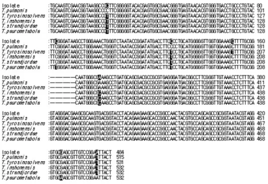
Fig. 2. Comparison of the DNA se- quences of the 16S rRNA genes from the isolate and Tsukamurella species. The accession numbers of the sequences are as follows: T.
pulmonis, AY741505; T. tyrosino- solvens, AY238514; T. inshonensis, AF283181; T. strandjordae, AF28- 3283; and T. paurometabola, AF2- 83280. The nucleotide sequences were deposited in GenBank
mycin were administered as empirical therapy, but intermittent low grade pyrexia was noted in the patient and subsequent five follow up blood cultures also showed the same organisms. It was decided to remove the catheter because it was believed to be the focus of the infection. After removing the catheter, the fever dis- appeared and the blood culture no longer showed this organism.
Blood cultures became positive after 54∼67 hours incubation and the isolate aerobically grew on blood agar at 36oC. This or- ganism initially appeared flat and spreading after about 48 hours and grew to irregular, rough, and dry yellowish colonies when in- cubation was extended (Fig. 1). The specimen was negative for acid-fast stain, but showed positivity to a modified acid-fast stain.
It was catalase-positive and alkaline-slant/alkaline-deep on triple sugar iron agar.
Phenotypic identification of these gram positive organisms was performed using VITEK 2 identification card (bioMérieux, Durham, NC, USA) and API Corynebacterium system (bioMérieux, Marcy l’Etoile, France). The isolates could not be identified with VITEK 2 identification test and gave the profile 4550004 in the API Corynebacterium system, which gave an identification of Brevibacterium sp. (58.4%), followed by Arthrobacter sp.
(17.6%), Rhodococcus sp. (12.2%). DNA was extracted and PCR-restriction fragment length polymorphism (RFLP) analysis was performed using Myco-ID (Molecules & Diagnostics, Wonju, Korea) to rule out Nocardia because of the colonies’ morpho- logical similarities and positivity of modified acid fast stain.
Digestion of the rpoB amplicon by MspI (Boehringer Mannheim Biochemicals, Manheim, Germany) revealed restriction fragments compatible with Mycobacterium aubagnense sp. nov pattern which was proposed for a novel, rapidly-growing Mycobacterium in 2006. It was unreasonable to be assured of Mycobacterium when considering the patient’s clinical manifestation and the or-
ganism’s characteristics.
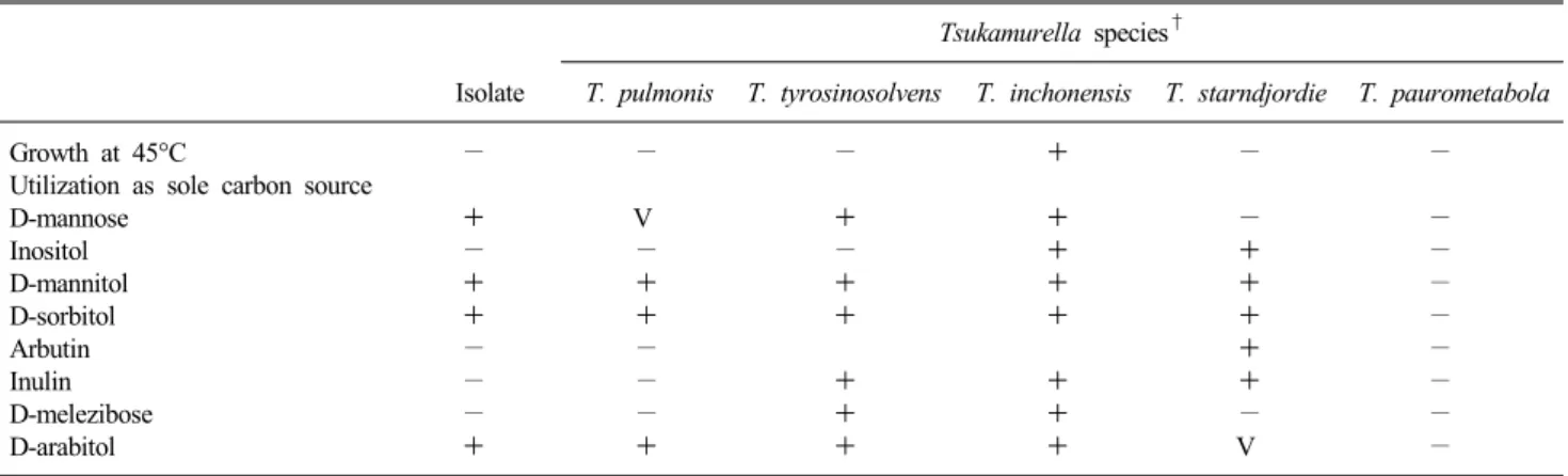
In order to make exact identification, the isolate was sub- sequently forwarded for molecular identification through PCR am- plification with the primers 16SF (5'-GCR KTC YTA ATA CAT GCA AGT CGA-3'), 16SR (5'-TTT CAC GAA CAA CGC GAC AA-3')[5] and direct sequencing of the partial region of the 16S rRNA gene on the ABI Prism 3100 DNA sequencer (Applied Biosystems, Foster City, CA, USA). On BLAST analysis, the se- quence analysis allowed reliable identification of T. pulmonis (AY741505, AY714240 and AY254698, 99.8% identity, 483/484 bases) followed by T. tyroinsolvens (AY238514, 99.2% identity, 480/484 bases), T. inchonensis (AF28318, 99.2% identity, 480/484 bases), T. strandjordi (AF283282, 99.2% identity, 480/484 bases) and T. paurometabola (AF283280, 99% identity, 479/484 bases) (Fig. 2). As seen above, the differences among the 16S rRNA gene sequences of various Tsukamurella species are small and therefore specific phenotypic tests were used to confirm T. pulmonis. Sugar assimilation results were obtained using API 50 CH systems (bioMérieux, Marcy-l’Etoile, France) and the tol- erance at various temperatures were tested. The organism grew at 28oC and 35oC but not at 45oC. Testing for sugar utilization with the API 50 CH system showed excellent correlation with the pub- lished data of tests using conventional biochemical kits[6-8]. This test was performed twice and evaluated by two observers. Test was generally reproducible on repeated testing and inconsistent results were considered to be negative. Results of sugar assim- ilation and temperature tolerance test are as seen in Table 1. It can be distinguished from T. tyroinsolvens, T. paurometabola and T. strandjordie by assimilation results of D-mannose, inositol, D-mannitol, D-sorbitol, arbutine, inulin, D-melezibose and D-ara- bitol, and also can be distinguished from T. inchonensis through the absence of growth at 45oC and lack of assimilation of inositol
Table 1. Phenotype characteristics of the isolate compared to those of other Tsukamurella species*
Tsukamurella species†
Isolate T. pulmonis T. tyrosinosolvens T. inchonensis T. starndjordie T. paurometabola
Growth at 45°C − − − + − −
Utilization as sole carbon source
D-mannose + V + + − −
Inositol − − − + + −
D-mannitol + + + + + −
D-sorbitol + + + + + −
Arbutin − − + −
Inulin − − + + + −
D-melezibose − − + + − −
D-arabitol + + + + V −
*+, presents; −, absent; V, variable; †For details, see references 6∼8.
and D-melezibose[6-8].
DISCUSSION
Tsukamurella species have emerged over the last decade as rare but significant causes of serious infections in immunocompro- mised individuals. The most common Tsukamurella infections in humans are indwelling device-related infections especially cathe- ter-related bacteremia[1,7,9]. Others are cutaneous infection, men- ingitis, lung infection, peritonitis, knee prosthesis infection and conjunctivitis[7-10]. Optimal management is uncertain due to the paucity of cases, but a combination of beta-lactam and amino- glycoside, along with the removal of medical devices appears to be the preferential treatment[1,9].
Their species are suspected initially with isolation of a gram-positive bacillus that has microbiological characteristics similar to Tsukamurella species, but differentiation and speciation are difficult in most clinical microbiology laboratories. The genus is phylogenetically related to genera Nocardia, Gordonia, Streptomyces, Rhodococcus, Corynebacterium and Mycobacterium.
At the genus level, the differentiation of Tsukamurella from sim- ilar species relies on gas-liquid chromatography or HPLC and dif- ferences in mycolic acid sizes and menaquinone compositions are invaluable[6,8,11]. In addition to this methodology, molecular ap- proaches are being developed. These include molecular analysis of the 16S rRNA, 16S-23S intergenic spacer region, groEL and heat shock protein gene[12,13]. But 16S rRNA gene sequencing is not discriminative enough for speciation within this genus be- cause the species share high 16S rRNA gene sequence sim- ilarities[7,13]. T. pulmonis could be distinguished on the basis of its sugar utilization profile and temperature tolerance in our case.
Tsukamurella are gram-positive, aerobic and partially acid-fast as a result of the presence of mycolic acid in the cell envelope.
However, it may rarely exhibit substantial acid-fast staining sim- ilar to that of the Mycobacterium species. The misidentification of Tsukamurella as Mycobacterium has been previously reported in several studies[11,13,14]. At the initial evaluation we performed
RFLP analysis of rpoB gene to exclude Norcardia. Phylogenetic trees were inferred with the PHYLIP 3.5 software package and made identification of M. aubagnense (98% similarity) possible.
The sequences of M. aubagnense were not registered on the BLAST database making comparative analyses impossible and the rpoB sequence results showed only 83% similarity between M.
gondii on the NCBI databases and our isolate. When considering positivity to modified acid-fast stain, colonial morphology and pa- tient’s symptoms, we could not absolutely define it as mycobacteria. Finally, by additionally executing 16s rRNA se- quencing and phenotyping testing, we identified it as T. pulmonis.
If this organism is isolated in pulmonary infections, there is high possibility of misidentification. T. pulmonis is rarely misidentified as mycobacteria in pulmonary infection cases[4,10,13,14]. The presentation of pulmonary infections with Tsukamurella bears a striking similarity to the clinical syndrome seen with mycobacte- rial infections and also the intensity of acid-fast staining is varia- ble, often confused with mycobacteria. We can usually suspect misidentification of species due to ineffective therapy with anti- mycobacterial drugs and failure to identify this organism on my- cobacterial molecular assays[4,10,14]. But there have been no cas- es of Tsukamurella being misidentified as mycobacteria in com- mercial mycobacterial molecular identification assays and to our knowledge, this is the first case.
The molecular analysis of the 16S rRNA gene was used as the first-line method for the identification of unusual mycobacterial isolates[15], but only the 16S rRNA gene assay was shown to be insufficient for identifying mycobacteria and so the rpoB gene as- say was developed as a suitable tool for accurate identification of nontuberculous mycobacteria (NTM)[16-18]. However, as evident in our case, the misidentification of genetically closely related or- ganism such as Tsukamurella was found to be possible even in the rpoB gene assay. Care must be taken to reliably identify all acid-fast bacilli from specimens which are being investigated for mycobacteria to exclude other acid-fast organisms from phyloge- netically related genera such as Tsukamurella. The importance of making an accurate microbiological diagnosis is underscored by
the fact that many of the antibiotics used to treat tuberculosis or nontuberculous mycobacteria are ineffective for other species.
In our case, it was initially considered as a contaminant or a saprophytic carriage rather than true pathogen and the organism was not identified as Tsukamurella until the patient’s condition improved. Due to the persistence of fever and subsequent blood culture results, this organism was considered to be a real pathogen. It is therefore important that Tsukamurella species are suspected to be true infections in the immunosuppressed patient especially with an indwelling catheter and gram-positive bacilli in blood. Good clinical outcomes are achievable by combining ap- propriate antibiotics with removal of a catheter as the case with our patient.
REFERENCES
1. Schwartz MA, Tabet SR, Collier AC, Wallis CK, Carlson LC, Nguyen TT, et al. Central venous catheter-related bacteremia due to Tsukamurella species in the immunocompromised host: a case series and review of the literature. Clin Infect Dis 2002;35:72-7.
2. Yassin AF, Rainey FA, Brzezinka H, Burghardt J, Lee HJ, Schaal KP. Tsukamurella inchonensis sp. nov. Int J Syst Bacteriol 1995;
45:522-7.
3. Shim HE, Sung HS, Baek SM, Namgung S, Kim MN, Kim YG, et al. A case of catheter-related bacteremia of Tsukamurella pulmonis. Korean J Lab Med 2009;29:41-7.
4. Yassin AF, Rainey FA, Brzezinka H, Burghardt J, Rifai M, Seifert P, et al. Tsukamurella pulmonis sp. nov. Int J Syst Bacteriol 1996;46:429-36.
5. Ninet B, Monod M, Emler S, Pawlowski J, Metral C, Rohner P, et al. Two different 16S rRNA genes in a mycobacterial strain. J Clin Microbiol 1996;34:2531-6.
6. Kattar MM, Cookson BT, Carlson LC, Stiglich SK, Schwartz MA, Nguyen TT, et al. Tsukamurella strandjordae sp. nov., a proposed new species causing sepsis. J Clin Microbiolol 2001;39:1467-76.
7. Woo PC, Ngan AH, Lau SK, Yuen KY. Tsukamurella conjunc-
tivitis: a novel clinical syndrome. J Clin Microbiol 2003;41:
3368-71.
8. Olson JB, Harmody DK, Bej AK, McCarthy PJ. Tsukamurella spongiae sp. nov. a novel actinomycete isolated from a deep-water marine sponge. Int J Syst Evol Microbiol 2007;57:1478-81.
9. Alcaide ML, Espinoza L, Abbo L. Cavitary pneumonia secondary to Tsukamurella in an AIDS patient. First case and a review of the literature. J Infect 2004;49:17-9.
10. Rey D, Fraisse P, Riegel P, Piemont Y, Lang JM. Tsukamurella infections. Review of the literature apropos of a case. Pathol Biol (Paris) 1997;45:60-5.
11. McNabb A, Shuttleworth R, Behme R, Colby WD. Fatty acid characterization of rapidly growing pathogenic aerobic actinomy- cetes as a means of identification. J Clin Microbiol 1997;35:1361-8.
12. Millar BC, Xu J, Moore JE. Analysis of 16S-23S intergenic spacer regions of the rRNA operons in Tsukamurella pulmonis. Br J Biomed Sci 2006;63:25-6.
13. Perez VA, Swigris J, Ruoss SJ. Coexistence of primary adenocar- cinoma of the lung and Tsukamurella pneumonia: a case report and review of the literature. J Med Case Reports 2008;2:207.
14. Stanley T, Crothers L, McCalmont M, Xu J, Millar BC, Goldsmith CE, et al. The potential misidentification of Tsukamurella pulmonis as an atypical Mycobacterium species: a cautionary tale. J Med Microbiol 2006;55:475-8.
15. Pauls RJ, Turenne CY, Wolfe JN, Kabani A. A high proportion of novel mycobacteria species identified by 16S rDNA analysis among slowly growing accuprobe-negative strains in a clinical setting. Am J Clin Pathol 2003;120:560-6.
16. Adékambi T, Colson P, Drancourt M. rpoB-based identification of non pigmented and late-pigmenting rapidly growing mycobacteria.
J Clin Microbiol 2003;41:5699-708.
17. Adékambi T and Drancourt M. Dissection of phylogenetic relation- ships among 19 rapidly growing Mycobacterium species by 16S rRNA, hsp65, sodA, recA, and rpoB gene sequencing. Int J Syst Evol Microbiol 2004;54:2095-105.
18. Turenne CY, Tschetter L, Wolfe J, Kabani A. Necessity of quality-controlled 16S rRNA gene sequence databases: identifying nontuberculous Mycobacterium species. J Clin Microbiol 2001;39:
3637-48.
=국문초록=
비정형성 마이코박테리아로 잘못 동정된
Tsukamurella pulmonis
균혈증 1예중앙대학교 의과대학 1진단검사의학교실, 2내과학교실, 3소아과학교실
조아라1, 김혜련1, 이미경1, 최성호2, 윤신원3
본 저자들은 카테터와 연관된 Tsukamurella pulmonis 균혈증을 경험하여 보고하고자 한다. Tsukamurella는 드물게 면역억 제자들에게 기회감염균으로 작용하며 특히 카테터 등 생체내 인공장치는 독립적인 위험인자로 작용한다. Tsukamurella는 계통발생학적으로 Nocardia, Rhodococcus, Gordonia, Streptomyces, Corenybacterium, Mycobacterium 등과 유사하여 일반적 인 표현형 검사로는 동정이 어렵다. 본원의 48세 남자환자는 처음 rpoB 유전자를 이용한 PCR-RFLP에서는 Mycobacterium aubagnense로 잘못 동정되었다. 그러나 16S rRNA 염기서열 분석과 다른 Tsukamurella 종과 구별하기 위해 시행한 생화학 적 검사로 T. pulmonis를 동정할 수 있었다. [대한임상미생물학회지 2010;13:93-97]
교신저자 : 이미경, 140-757, 서울시 용산구 한강로 3가 65-207 중앙대학교 용산병원 진단검사의학과
Tel: 02-748-9837, Fax: 02-748-9929 E-mail: [email protected]