Department of Periodontology, Research Institute for Periodontal Regeneration, College of Dentistry, Yonsei University
Abstract
The sinus floor elevation procedure has been used to be one of the predictable treatments for rehabilitation of atrophic and pneumatized edentulous posterior maxilla. Sinus elevation with simultaneous implant placement could be an optimal proce- dure due to reduction in surgical procedures and to patient’s convenience. This study reports the successful results of sinus elevation with the sand blasted, large grit, acid-etched (SLA) surfaced implant(ITI dental imiplant system, StraumannⓇ, Basel, Switzerland) placement simultaneously.
In the first case, the height of residual ridge was 2~4mm. Autogeneous bone from ramus and alloplast(Macroporous biphasic calcium phosphate-MBCP, BiomatlanteⓇSarl, Nantes, France) were applied after sinus elevation through lateral window approach. SLA surfaced implants(ITI) were installed simultaneously. Result after 17 months was showed success- fully.
In the second case, the height of residual ridge was 3~5mm. Allograft(Freeze dried bone allograft-FDBA, Oragraft, Lifenet health, Virginia Beach, VA, USA) and alloplast(MBCP) were used for sinus graft. All procedures were followed as in the first case. Result after 9 months was showed successfully.
In conclusion, this study presented successful results, when sinus elevation through window approach with mixed graft material was performed with simultaneous SLA surfaced implant placement on severe atrophied maxillary alveolar ridge, minimizing patient discomfort.
Key words: Sinus floor elevation, Simultaneous implant placement, window ostectomy, SLA surfaced implant
Sinus Floor Grafting with Simultaneous Implant Placement on Severe Artrophied Maxillary Ridge
Jin-Hyuk Yang, Ji-Youn Hong, Ui-Won Jung, Chang-Sung Kim, Kyoo-Sung Cho, Seong-Ho Choi 양진혁, 홍지연, 정의원, 김창성, 조규성, 최성호
연세대학교 치과대학병원 치주과학교실, 치주조직재생연구소
상악 구치부는 골양과 골질에 있어 다른 부위에 비해 많은 해부학적 취약점을 가진다. 치아 상실 시 치조골 흡수가 심하 며 상악동 내 함기화로 인해 임상가는 기능적, 생리적 회복을 위한 시술을 하기에 많은 한계점에 부딪힌다1,2). 960년대에 Boyne에 의해 보철물을 위한 상악동 거상술(Maxillary sinus)이 최초로 소개된 뒤3), Boyne와 James 가 1980년 상 악동하 골이식으로 자가골의 사용 등의 구체적인 술식을 보 고하였다4). 그 후 상악동 거상술은1996년 Sinus Consensus Conference에서 효과적인 치료법으로 인정받으며5) 필요에 따라서 여러 가지 형태로 변형, 발전하며 심하게 흡수된 상악 골에서 임프란트 식립을 위한 유용한 술식으로 임상에서 시 행되고 있다. 상악동 거상술의 적응증은 pneumatization(상 악동 함기화)이 심한 경우와 골질이 좋지 않은 경우, 또한 감 소된 골질과 골양에서 임프란트의 지지력을 높이고자 할 때 이다.
상악 구치부 폭은 악골 전체에서 비교적 빨리 흡수되는 부 위로 Misch6)는 상, 하악골의 치조골 흡수에 따라 5가지 형태 로 분류하였다. 이렇게 다양한 골양과 골질에 따른 분류에 따 라 치료방법에 달라져야 하는데, 2003년과 2008년 Chiapasco7,8)는 상악 구치부의 상태를 9개로 나누어 그에 따 른 치료법을 제시하였다. 여러 치료법 중에서 Sinus graft, onlay graft, Le Fort Ⅰ osteotomy with interpositional bone graft 등의 수술법이 있으며4, 9, 10), 이 중에서 상악동저 내에 골이식을 시행하여 부족한 상악골을 보강하는 sinus graft술식이 널리 쓰이고 있다.
상악동 골이식은 크게 2가지 방법이 있는데, 측방 접근을 통한 방법과 치조정을 통한 접근 방법이다4,11). 이러한 술식은 골 고경을 증가시키고 연약한 골을 압축시켜 골밀도가 증가 하여 임프란트를 식립할 수 있게 한다. 치조정을 통한 접근법
은 Summers11)에 의해 제안되었는데, 이 방법을 통해 임프란 트를 식립한 결과 높은 성공률을 보인다고 하였다12). Kent13) 와 Block12)이 Caldwell-Luc 술식을 변형한 측방 접근법을 통 한 상악동 거상술을 제안하였는데, 이 술식과 임프란트를 식 립한 경우 높은 성공률을 보인다고 보고되었기에13,14)최근 상 악동 거상술에 널리 사용된다. 측방 접근법은 판막을 거상하 여 외측창을 형성하여 시야가 확보된 상태에서 Schneiderian 막을 거상하고 골이식재를 함입시킬 수 있다. 이 술식은 높은 예지성을 가진 술식으로 인정받았으며 골절편(block bone) 보다는 골이식재를 사용하고 표면처리된 임프란트를 사용하 였을 때 높은 성공률을 보인다고 보고되었다15). 또한 동시 식 립을 한 경우나 단계적 식립을 한 경우의 성공률의 차이가 없 다고 보고 되었다15, 16).
본 연구는 측방 접근법 상악동 거상을 통한 골이식술과 SLA 표면처리를 한 임프란트를 동시 식립을 하였을 때 성공 적인 결과를 얻은 증례를 보고하고자 함이다.
증례 1
2007년 8월 56세 남자 환자가 임프란트 시술을 위해 보철 과로부터 의뢰되어 본과에 내원하였다. 환자는 상악 좌측 구 치부의 기능적 회복을 원하였으며, 약물 복용으로 잘 조절되 는 고혈압 이외에 특이한 전신적 질환을 가지고 있지 않았다.
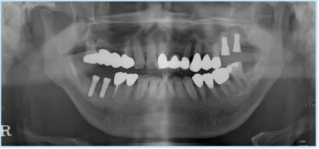
2년 전에 스케일링을 받았으며 상악 좌측 구치부는 무치악 상 태로 수년이 경과되었으며 부분의치도 착용하고 있지 않은 상태였다. 임상적 평가 결과, 상악 좌측 구치부 치조골의 수직 적, 수평적 골소실은 심한 상태는 아니었으나, 방사선적 평가 로는 상악동 함기화(Maxillary sinus pneumatization)가 심하여 잔존골의 두께는 2~4mm로 매우 얇은 상태임을 알
Ⅱ
I
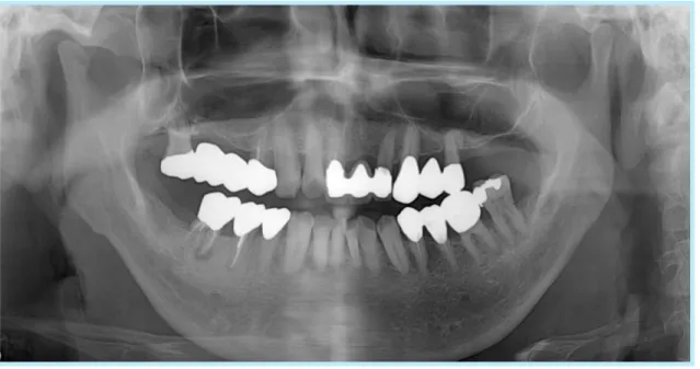
Fig. 2. 방사선 사진(파노라마, 술 전)
Jin-Hyuk Yang et al: Sinus Floor Grafting with Simultaneous Implant Placement on Severe Artrophied Maxillary Ridge. Implantology 2009
Fig. 1. 임상사진(술 전, AA:: 협측면, BB:: 교합면)
Jin-Hyuk Yang et al: Sinus Floor Grafting with Simultaneous Implant Placement on Severe Artrophied Maxillary Ridge. Implantology 2009
A B
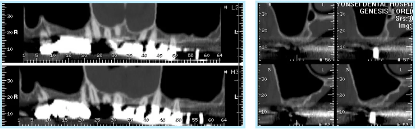
수 있었다(Fig. 1, 2). 정확한 잔존골의 측정을 위해 전산화 단 층 촬영(Computed tomography)를 시행하였는데, 잔존골이 심하게 부족한 #27번 부위에도 1mm 정도의 피질골(cortical bone)이 존재함을 확인할 수 있다(Fig. 3).
얇은 잔존골로 인해 좌측 상악동 거상술과 골이식을 한 후
#26, 27 부위에 임프란트 식립을 계획하였다. 일반적으로 잔 존골이 4mm 이하인 경우 임프란트의 기계적고정(mechan- ical stability)을 얻기 어렵기 때문에 측방 접근법을 통한 상
악동 거상술과 단계적 식립이 추천된다17). 본 증례의 경우도 잔존골이 2~4mm로 단계적 식립이 추천되나 수술의 횟수를 줄여 환자의 불편감을 감소시킬 수 있으며 이식한 골 이식재 에서 임프란트의 초기 고정을 추가적으로 얻을 수 있어 측방 접근법을 통한 상악동 거상술과 임프란트 동시식립을 시행하 기로 하였다. 골이식 재료로는 자가골과 합성골을 혼합하여 사용하기로 하였다. 통상적 방법대로 1:100,000을 함유한 2% lidocaine으로 술부인 좌측 상악 구치부와 하악 좌측구치 Fig. 3. 방사선 사진(전산화 단층 사진, 술 전)
Jin-Hyuk Yang et al: Sinus Floor Grafting with Simultaneous Implant Placement on Severe Artrophied Maxillary Ridge. Implantology 2009
Fig. 4. AA:: 좌측 하악지 부위, BB:: 분쇄된 골절편, CC:: 골 분쇄기
Jin-Hyuk Yang et al: Sinus Floor Grafting with Simultaneous Implant Placement on Severe Artrophied Maxillary Ridge. Implantology 2009
B C
A
부에 국소마취(침윤마취, 전달마취)를 시행한 후 상악 결절로 부터 잔존 치조제 근심치아의 근협측선까지 수평절개와 인접 치아의 열구절개를 시행하고 협측 수직절개를 시행하여 전층 판막을 거상하였다. 상악동 기저부로부터 최소 2mm 상방, 치조정으로부터 최소 4mm 상방에 외측창 골절제술(win- dow ostectomy)을 충분한 주수 하(irrigation)에 Piezo-
surgery kit bur로 시행하였다. 외측창(window)은 가로 세 로 각각 12×3 mm 크기로 형성하였다. 골절편을 주위골로부 터 분리하고 거상기구로 상악동막(sinus membrane)을 거상 하였다. 자가골 채취를 위해 하악 좌측 최후구치(#37) 후방에 충분한 절개를 시행한 후 Trephin bur로 자가골을 채취한 후 골분쇄기(Bone crusher)로 분쇄시켜 분말형태로 결정화시켰 다(Fig. 4). 결정화된 자가골과 합성골(Macroporous bipha- sic calcium phosphate-MBCPⓇ,60% HydroxyApatite + 40% β-TriCalciumPhosphate, Biomatlante, Sarl,
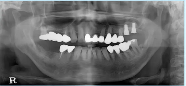
Fig. 7. 방사선 사진(파노라마, 수술 당일)
Jin-Hyuk Yang et al: Sinus Floor Grafting with Simultaneous Implant Placement on Severe Artrophied Maxillary Ridge. Implantology 2009
Fig. 5. 외측창 골 절제술과 상악동막의 거상
Jin-Hyuk Yang et al: Sinus Floor Grafting with Simultaneous Implant Placement on Severe Artrophied Maxillary Ridge. Implantology 2009
Fig. 6. 임프란트 매식체 식립
Jin-Hyuk Yang et al: Sinus Floor Grafting with Simultaneous Implant Placement on Severe Artrophied Maxillary Ridge. Implantology 2009
Nantes, France)을 혼합한 뒤 거상된 상악동막 하방에 채워 넣었다. 이 후 치조정으로 단계적 drilling을 시행하고 Osteotome을 통해 골밀도를 증가시켰다(Fig. 5). 그 후 길이 10mm를 가지고 하부직경 4.8mm와 상부 직경 6.5mm로 상 부가 넓어지는 형태의 임프란트(ITI TE type, StraumannⓇ, Basel, Switzerland)을 식립하였다(Fig. 6).
#26번은 50N, #27은 30N의 최종 토크로 고정을 얻었다.
연결지대주(Healing abutement)를 연결시켰는데, #27의 경 우 연결 시 약간의 움직임(rotational movement)이 있었다.
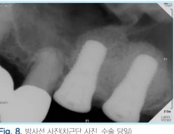
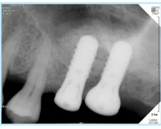
4-0 단섬유봉합사(MonosynⓇAesculap, Tuttlingen, Germany)로 봉합하였으며 10일 후 발사하였다. 수술 당일 방사선 사진으로 식립된 임프란트를 평가하였는데, 처음 계 획된 위치에 잘 식립되었으며 골이식재도 거상된 상악동 막 아래 잘 집결되어 있었다(Fig. 7, 8). 술 후 1주간 항생제, 소 염진통제 및 소화제를 처방하였으며 구강세척액으로 소독을 지시하였다. 6주 후 내원하여 임상 및 방사선학적 평가를 시 행하였다. 검사 결과 상 좋은 치유상태를 보였으며 고정검사 측정기구(PeriotestⓇ, Siemens Dental, Bensheim, Germany)로 측정결과 -1값을 보이며 방사선학적으로 골이 식재의 유지도 양호하였다(Fig. 9). 술 후 5개월에 임상, 방 사선 평가가 이루어졌는데 고정검사측정기구(PeriotestⓇ)로 측정결과 #26번은 -5, #27번은 -6 보여 고정이 잘 유지되고 있음을 알 수 있었다(Fig. 9). 술 후 8개월에 보철물이 완성되 어, 현재 9개월간 보철부하가 가해진 상태로 환자는 큰 불편 감 없으며 임상, 방사선적으로도 좋은 결과를 유지하고 있다 (Fig. 11).
Fig. 9. 방사선 사진(파노라마, 술 후 6주)
Jin-Hyuk Yang et al: Sinus Floor Grafting with Simultaneous Implant Placement on Severe Artrophied Maxillary Ridge. Implantology 2009
Fig. 8. 방사선 사진(치근단 사진, 수술 당일)
Jin-Hyuk Yang et al: Sinus Floor Grafting with Simultaneous Implant Placement on Severe Artrophied Maxillary Ridge. Implantology 2009
Fig 10. 방사선 사진(치근단 사진, 술 후 5주)
Jin-Hyuk Yang et al: Sinus Floor Grafting with Simultaneous Implant Placement on Severe Artrophied Maxillary Ridge. Implantology 2009
Fig. 11. 임상사진(술 후 8개월, AA:: 협측면, BB:: 교합면)
Jin-Hyuk Yang et al: Sinus Floor Grafting with Simultaneous Implant Placement on Severe Artrophied Maxillary Ridge.
Implantology 2009
A B
증례 2
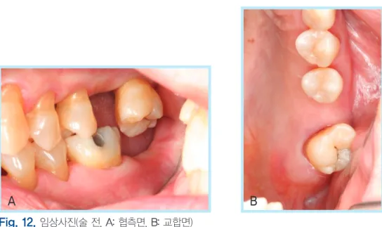
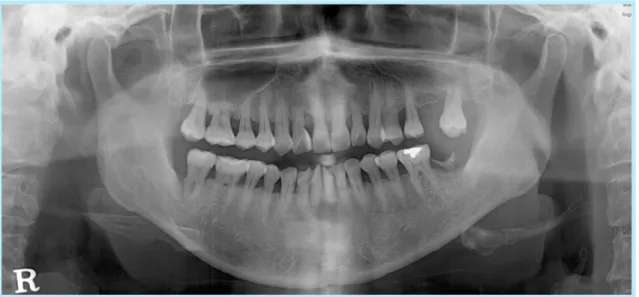
2007년 1월 55세 여자환자가 오른쪽 위, 아래 잇몸이 붓고 아프다는 주소로 내원하였다. Aspirin allergy 이외에 전신 적으로 특이한 사항이 없었으며 상악 좌측의 경우 #26은 상 실된 상태로 오래있었으며 #27은 내원 당시 동요도 2도로 이 개부병소가 이환되어 있으며 깊은 치주낭 깊이를 보이고 있 었다. #27은 예후가 매우 불량하여 발치하였으며, 무치악부 위의 기능적 회복을 위해 임프란트 시술을 계획하였다. 임상 적 평가 결과, 상악 좌측 구치부 치조골의 수직적, 수평적 골 소실이 다른부위와 비슷한 정도로만 일어난 상태였으며, 방 사선적 평가로는 상악동 함기화(maxillary sinus pneuma- tization)가 일어나 잔존골의 두께는 3~5mm로 얇은 상태임 을 알 수 있었다(Fig. 12, 13, 14). 정확한 잔존골의 양과 위치 를 확인하고자 전산화 단층 촬영(computed tomography)를 시행하였는데, 1mm 이상의 피질골(cortical bone)이 존재함 을 확인할 수 있다(Fig. 15).
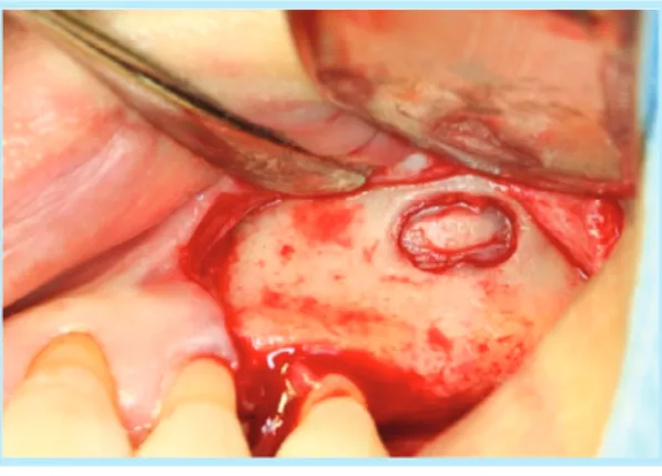
앞선 증례와 마찬가지로 좌측 상악동 거상술과 골이식을 한 후 #26, 27 부위에 임프란트 식립을 계획하였다. 잔존골이 3~5mm로 단계적 식립 또는 동시 식립을 모두 고려할 수 있 으나 수술의 횟수를 줄여 환자의 불편감을 최소화하는 술식 을 위해 측방접근법을 통한 상악동 거상술과 임프란트 동시 식립을 시행하기로 하였다. 골이식 재료로는 동종골(Freeze dried bone allograft-FDBA, Oragraft, Lifenet health, Virginia Beach, VA, USA)과 합성골(Macroporous bipha- sic calcium phosphate-MBCPⓇ, 60% HydroxyApatite + 40% β-TriCalciumPhosphate, Biomatlante, Sarl, Nantes, France)을 사용하기로 하였다. 통상적 방법대로 술 부인 좌측 상악 구치부에 국소마취를 시행한 후 수평절개와 수직절개를 한 후 전층판막을 거상하였다. 상악동 기저부로 부터 최소 2mm 상방, 치조정으로부터 최소 4mm 상방에 외 측창 골절제술(window ostectomy)을 충분한 주수 하(irri- gation)에 Piezo-surgery kit bur로 시행하였다. 외측창 (window)은 가로 세로 각각 8×4 mm 크기로 형성하였다 (Fig. 16).
Fig. 12. 임상사진(술 전, AA:: 협측면, BB:: 교합면)
Jin-Hyuk Yang et al: Sinus Floor Grafting with Simultaneous Implant Placement on Severe Artrophied Maxillary Ridge.
Implantology 2009
A B
Fig. 13. 방사선 사진(파노라마, 술 전)
Jin-Hyuk Yang et al: Sinus Floor Grafting with Simultaneous Implant Placement on Severe Artrophied Maxillary Ridge. Implantology 2009
Fig. 14. 방사선 사진(치근단 사진, 술 전)
Jin-Hyuk Yang et al: Sinus Floor Grafting with Simultaneous Implant Placement on Severe Artrophied Maxillary Ridge. Implantology 2009
Fig. 15. 방사선 사진(전산화 단층 사진, 술 전)
Jin-Hyuk Yang et al: Sinus Floor Grafting with Simultaneous Implant Placement on Severe Artrophied Maxillary Ridge. Implantology 2009
골절편을 주위골로부터 분리하고 거상기구로 상악동막 (sinus membrane)을 거상한 후 준비한 동종골과 합성골의 혼합 이식재를 막 아래에 채워넣었다. 이 후 치조정으로 단계 적으로 drilling을 한 후 Osteotome을 통해 골밀도를 증가시 켰다. 그 후 #26 부위는 길이 10mm에 4.8mm 직경을 가진 임프란트(ITI, SrtaumannⓇ, Basel, Switzerland)를, #27
부위는 같은 길이에 4.8mm 직경과 넓은 상부목구조 형태의 임프란트(Wide neck, StaumannⓇ, Basel, Switzerland)를 식립하였다. #26번은 20N, #27은 30N의 최종 토크로 고정 을 얻었다. 연결지대주(Healing abutement)를 연결시켰으 며 4-0 단섬유봉합사(MonosynⓇ Aesculap, Tuttlingen, Germany)로 봉합하였으며 10일 후 발사하였다. 이번 증례에 서도 수술 당일 방사선 사진을 촬영하였는데 처음 계획된 위 치에 잘 식립되었으며 골이식재도 거상된 상악동 막 아래 잘 집결되어 있었다(Fig. 17, 18). 술 후 1주간 항생제, 소염진통 제 및 소화제를 처방하였으며 구강세척액으로 소독을 지시하 였다. 4주 후 내원 시 임상 및 방사선학적 평가를 시행하였다.
결과 좋은 치유상태를 보였다. 술 후 6개월에 임상, 방사선 평 가가 이루어졌는데 고정검사측정기구(PeriotestⓇ)로 측정결 과 #26, 27 모두 -5 보여 고정이 잘 유지되고 있음을 알 수 있 었다(Fig. 9). 술 후 7개월에 보철물이 완성되어 현재 2개월간 보철부하가 가해진 상태로 환자는 큰 불편감 없으며 임상, 방 사선적으로도 좋은 결과를 유지하고 있다(Fig. 19).
Fig. 17. 방사선 사진(파노라마, 수술 당일)
Jin-Hyuk Yang et al: Sinus Floor Grafting with Simultaneous Implant Placement on Severe Artrophied Maxillary Ridge. Implantology 2009
Fig. 16. Piezo-surgery bur를 사용한 외측창 골 절제술
Jin-Hyuk Yang et al: Sinus Floor Grafting with Simultaneous Implant Placement on Severe Artrophied Maxillary Ridge. Implantology 2009
환자의 상악 무치악 부위는 생리적, 기능적 회복을 위한 시 술에 해부학점 취약점과 제약점이 있는 곳이다. 골질이 약하 여 치아가 상실되면 치조제 흡수가 다른 곳에 비해 심하게 일 어나고 상악동과 인접해있어, 상악동의 함기화까지 일어나면 임프란트를 시술하기에 치조골 고경이 부족해진다. 이러한 제약을 극복할 수 있는 예지성 높은 술식으로 상악동 거상술 이이용되고 있다. Wallace15) 등은 상악동 거상술에 관한 review연구에서 상악동 거상술 후 임프란트를 식립했을 때 일반적인 성공률과 차이가 없다고 보고하였다. Chiapasco7,8) 등은 상악구치부의 상태에 따라 다른 술식을 해야 한다는 기 준을 제시하였으며, Zitzmann과 Sharer18) 등도 잔존골양에 따라 Osteotome 방법이나 측방접근법을 통해 상악동거상술 을 선택적으로 시행해야 한다고 하였다. 그러나 최근 연구들 에 따르면 기준에 따른 분류와 상관없다는 연구결과들이 많 다. Mardinger19)와 Peleg16)등은 상악동 거상술과 임프란트 를 동시식립한 연구에서 각각 92%와 97.9%로 높은 성공률을 보고하였으며 Jurisic20)등도 동시식립한 경우 단계적 식립한
경우와 임프란트 성공률의 차이를 보이지 않는다고 하였다.
골이식재의 경우도 block bone이나 분쇄된 자가골 단독으 로의 사용보다는 동종골, 이종골, 합성골 등을 혼합하여 사용 하는 경우에 좋은 결과를 보이고 있다13). 또한 Valentini21)의 관찰 비교 연구에서 순수 자가골만을 이식한 경우보다 이종 골 이식재만 이식한 경우 높은 임프란트 성공률을 보인다고 하였다. Buser22)와 Wennerberr23)등의 연구에서 SLA 처리 한 임프란트는 세포활성을 촉진시켜 골과 임프란트 접촉을 신속하게 일으키기 때문에 높은 성공률을 보인다고 보고하였 다. Lai24)와 Ferrigno25)등이 연구에서 SLA 표면처리된 ITI 임프란트를 osteotome을 시행한 부위에서 높은 성공률을 나 타냈으며 상악동 거상술을 시행한 부위에 식립한 ITI 임프란 트에 관한 Nedir26)의 연구에서도 높은 성공률을 가진다고 하 였다.
Chiapasco7,8)등이 제시한 기준에 따르면 본 증례에서는 단계적 식립을 하여야 하지만, 동시 식립으로 수술횟수를 줄 여 술식을 간략하게 하고 수술에 대한 환자의 심리적 불안감 을 줄여 결과적으로 환자에게 최소한의 불편감을 주었다. 또 한 임프란트에 적절한 부하가 가해질 때 임프란트 주위골이 치밀화되고 이식골의 안정성이 증진된다는 연구27)등은 동시
Ⅲ
Fig. 18. 방사선 사진(치근단 사진, 수술 당일)
Jin-Hyuk Yang et al: Sinus Floor Grafting with Simultaneous Implant Placement on Severe Artrophied Maxillary Ridge. Implantology 2009
Fig. 19. 방사선 사진(치근단 사진, 술 후 4주)
Jin-Hyuk Yang et al: Sinus Floor Grafting with Simultaneous Implant Placement on Severe Artrophied Maxillary Ridge. Implantology 2009
식립을 하였을 때에도 임프란트가 안정성을 가진다는 것을 뒷받침해 준다.
잔존골의 양이 충분하지 않아 임프란트의 초기고정을 얻기 힘들 수도 있으나, 본 증례에서는 30N 이상으로 양호한 고정 을 얻을 수 있었다. 전산화 단층 촬영에서 두 증례 모두에서 피질골(cortical bone)의 두께가 1mm 이상을 가진 것을 확인 할 수 있는데, 충분한 피질골 두께가 임프란트 식립 시 양호한 초기 고정을 얻을 수게 한 이유라 사료된다.
본 증례에서 자가골과 합성골의 혼합 또는 동종골과 합성골 의 혼합된 이식재를 사용하여 상악동 거상술을 시행한 후 SLA 표면을 가지는 ITI 임프란트로 동시식립하였다. 식립된 임프란트들은 이식된 골과 잔존골의 밀도 증가를 통해 초기 고정을 얻은 후 합병증 없이 보철물이 완성되어 부하 후에도 임상, 방사선적 문제없이 잘 유지되고 있다. 각각 술 후 17개 월, 9개월인 현재에도 환자들은 불편감없이 기능적, 생리적 회복에 만족감을 나타내고 있다. 많은 증례가 없기에 일반화 할 수 없지만, 잔존골의 흡수가 많은 상악 무치악 부위에서 환 자의 불편감을 최소한으로 줄이며 기능을 회복시킬 수 있는 방법으로 혼합된 골이식재를 사용하여 측방접근법을 통한 상 악동 거상술을 동반한 SLA 표면의 임프란트 동시 식립방법 은 가능한 술식이라 할 수 있다.
1. Khoury F. Augmentation of the sinus floor with mandibular bone block and simultaneous implantation: a 6-year clinical investigation. Int J Oral Maxillofac Implants 1999;14:557-564.
2. Smiler DG, Johnson PW, Lozada JL, et al. Sinus lift grafts and endosseous implants. Treatment of the atrophic posterior maxilla. Dent Clin North Am 1992;36:151-186; discussion 187-158.
3. Boyne PJ. US Navy Dental School lectures to postgraduates. 1965- 1968.
4. Boyne PJ, James RA. Grafting of the maxillary sinus floor with autoge-
nous marrow and bone. J Oral Surg 1980;38:613-616.
5. Jensen OT, Shulman LB, Block MS, et al. Report of the Sinus Consensus Conference of 1996. Int J Oral Maxillofac Implants 1998;13 Suppl:11-45.
6. Misch CE. Maxillary sinus augmentation for endosteal implants: orga- nized alternative treatment plans. Int J Oral Implantol 1987;4:49-58.
7. Chiapasco M, Zaniboni M, Rimondini L. Dental implants placed in grafted maxillary sinuses: a retrospective analysis of clinical outcome according to the initial clinical situation and a proposal of defect classifi- cation. Clin Oral Implants Res 2008;19:416-428.
8. Chiapasco M. Tecniche ricostruttive con innesti e/o osteotomie in:
Chiapasco M, Romeo E (eds). Torino: UTET 2003:225-303.
9. Tatum H, Jr. Maxillary and sinus implant reconstructions. Dent Clin North Am 1986;30:207-229.
10. Jensen J, Simonsen EK, Sindet-Pedersen S. Reconstruction of the severely resorbed maxilla with bone grafting and osseointegrated implants: a preliminary report. J Oral Maxillofac Surg 1990;48:27-32;
discussion 33.
11. Summers RB. A new concept in maxillary implant surgery: the osteotome technique. Compendium 1994;15:152, 154-156, 158 passim;
quiz 162.
12. Summers RB. The osteotome technique: Part 3--Less invasive methods of elevating the sinus floor. Compendium 1994;15:698, 700, 702-694 passim; quiz 710.
13. Fugazzotto PA, Vlassis J. Long-term success of sinus augmentation using various surgical approaches and grafting materials. Int J Oral Maxillofac Implants 1998;13:52-58.
14. Hurzeler MB, Kirsch A, Ackermann KL, Quinones CR. Reconstruction of the severely resorbed maxilla with dental implants in the augmented maxillary sinus: a 5-year clinical investigation. Int J Oral Maxillofac Implants 1996;11:466-475.
15. Wallace SS, Froum SJ. Effect of maxillary sinus augmentation on the survival of endosseous dental implants. A systematic review. Ann Periodontol 2003;8:328-343.
16. Peleg M, Garg AK, Mazor Z. Predictability of simultaneous implant placement in the severely atrophic posterior maxilla: A 9-year longitudi- nal experience study of 2132 implants placed into 731 human sinus grafts. Int J Oral Maxillofac Implants 2006;21:94-102.
17. Ioannidou E, Dean JW. Osteotome sinus floor elevation and simultane- ous, non-submerged implant placement: case report and literature review. J Periodontol 2000;71:1613-1619.
18. Zitzmann NU, Scharer P. Sinus elevation procedures in the resorbed
posterior maxilla. Comparison of the crestal and lateral approaches. Oral Surg Oral Med Oral Pathol Oral Radiol Endod 1998;85:8-17.
19. Mardinger O, Nissan J, Chaushu G. Sinus floor augmentation with simultaneous implant placement in the severely atrophic maxilla: tech- nical problems and complications. J Periodontol 2007;78:1872-1877.
20. Jurisic M, Markovic A, Radulovic M, et al. Maxillary sinus floor aug- mentation: comparing osteotome with lateral window immediate and delayed implant placements. An interim report. Oral Surg Oral Med Oral Pathol Oral Radiol Endod 2008.
21. Valentini P, Abensur DJ. Maxillary sinus grafting with anorganic bovine bone: a clinical report of long-term results. Int J Oral Maxillofac Implants 2003;18:556-560.
22. Buser D, Schenk RK, Steinemann S, et al. Influence of surface charac- teristics on bone integration of titanium implants. A histomorphometric study in miniature pigs. J Biomed Mater Res 1991;25:889-902.
23. Wennerberg A, Albrektsson T, Andersson B, et al. A histomorphomet-
ric and removal torque study of screw-shaped titanium implants with three different surface topographies. Clin Oral Implants Res 1995;6:24- 30.
24. Lai HC, Zhang ZY, Wang F, et al. Resonance frequency analysis of sta- bility on ITI implants with osteotome sinus floor elevation technique without grafting: a 5-month prospective study. Clin Oral Implants Res 2008;19:469-475.
25. Ferrigno N, Laureti M, Fanali S. Dental implants placement in conjunc- tion with osteotome sinus floor elevation: a 12-year life-table analysis from a prospective study on 588 ITI implants. Clin Oral Implants Res 2006;17:194-205.
26. Nedir R, Bischof M, Vazquez L, et al. Osteotome sinus floor elevation without grafting material: a 1-year prospective pilot study with ITI implants. Clin Oral Implants Res 2006;17:679-686.
27. Listrom RD, Symington JM. Osseointegrated dental implants in con- junction with bone grafts. Int J Oral Maxillofac Surg 1988;17:116-118.
교신저자 : 최성호
우편번호 : 120-752, 서울특별시 서대문구 신촌동 134 연세대학교 치과대학 치주과학교실
전자우편 : [email protected]