저작자표시-비영리-변경금지 2.0 대한민국 이용자는 아래의 조건을 따르는 경우에 한하여 자유롭게 l 이 저작물을 복제, 배포, 전송, 전시, 공연 및 방송할 수 있습니다. 다음과 같은 조건을 따라야 합니다: l 귀하는, 이 저작물의 재이용이나 배포의 경우, 이 저작물에 적용된 이용허락조건 을 명확하게 나타내어야 합니다. l 저작권자로부터 별도의 허가를 받으면 이러한 조건들은 적용되지 않습니다. 저작권법에 따른 이용자의 권리는 위의 내용에 의하여 영향을 받지 않습니다. 이것은 이용허락규약(Legal Code)을 이해하기 쉽게 요약한 것입니다. Disclaimer 저작자표시. 귀하는 원저작자를 표시하여야 합니다. 비영리. 귀하는 이 저작물을 영리 목적으로 이용할 수 없습니다. 변경금지. 귀하는 이 저작물을 개작, 변형 또는 가공할 수 없습니다.
Sinus augmentation using
rhBMP-2-loaded synthetic bone substitute with
simultaneous implant placement in rabbits
Myung-Jae Joo
Department of Dentistry
Sinus augmentation using
rhBMP-2-loaded synthetic bone substitute with
simultaneous implant placement in rabbits
Directed by Professor Ui-Won Jung
The Doctoral Dissertation
submitted to the Department of Dentistry
the Graduate School of Yonsei University
in partial fulfillment of the requirements for the degree of
Ph.D. in Dental Science
Myung-Jae Joo
감사의 글
이 논문이 완성되기까지 부족한 저를 격려해 주시고 연구에 매진할 수 있도록 끊임없는 지도와 관심으로 이끌어 주신 정의원 지도교수님께 진심으로 감사 드립니다. 학문에 대한 열정을 알려주신 김종관 교수님, 언제나 따뜻한 관심과 격려를 아끼지 않으신 채중규 교수님, 올바른 자세와 태도를 가질 수 있게 해주신 조규성 교수님, 작은 부분까지 조언해 주시고 마지막까지 큰 힘이 되어주신 최성호 교수님, 연구에 있어 폭넓은 시야를 보여주신 김창성 교수님, 연구 초기 힘든 시기에 많은 격려와 조언을 해주신 이중석 교수님, 세심한 부분까지 성심껏 지도해주신 차재국 교수님께 깊이 감사 드립니다. 바쁘신 와중에도 심사를 맡아주시고 조언을 해주신 조성원 교수님께도 감사 드립니다. 연구가 무사히 완료될 수 있도록 도움을 준 치주과의 모든 의국원들과 연구원들, 학생에게도 고마움을 전합니다. 항상 바른길로 인도해주시고 무한한 신뢰로 저를 지켜봐 주시는 부모님, 언제나 저에게 의지가 되어주는 누나에게 사랑과 감사의 마음을 전합니다. 모든 분들께 진심으로 감사 드립니다. 2016년 12월Table of Contents
List of figures
··· ii
List of tables
··· iii
Abstract (English)
··· iv
I. Introduction
··· 1
II. Materials & Methods
··· 4
1. Experimental Animals ··· 4
2. Experimental Materials ··· 4
3. Experimental Design ··· 5
4. Surgical Procedures ··· 6
5. Radiographic Analysis
··· 7
6. Histologic and Histomorphometric Analyses
··· 7
7. Statistical Analysis
··· 8
III. Results
··· 9
1. Clinical Findings
··· 9
2. Radiographic Analysis
··· 9
3. Histologic and Histomorphometric Analyses
··· 10
IV. Discussion
··· 12
References
··· 16
Figure Legends
··· 20
Tables
··· 22
Figures
··· 25
Abstract (Korean)
··· 30
List of figures
Figure 1.
Photographs of implant and surgical sites.Figure 2.
Schematic illustration of histometric measurements.Figure 3.
Radiographic images in micro computed tomography.Figure 4.
Histological photomicrographs of the total augmented area.List of tables
Table 1.
Volumetric measurements from micro-CT data.Table 2.
The histomorphometric linear measurements in the augmented pouch.Abstract
Sinus augmentation using rhBMP-2-loaded synthetic bone
substitute with simultaneous implant placement in rabbits
Myung-Jae Joo, D.D.S., M.S.D
Department of Dentistry
The Graduate School, Yonsei University
(Directed by Professor Ui-Won Jung, D.D.S., M.S.D., PhD.)
Objective: The aim of this study was to determine the effect of recombinant human
bone morphogenetic protein-2 (rhBMP-2) loaded synthetic bone substitute on implants that are simultaneously placed with sinus augmentation in rabbits.
Materials and methods: A circular access window was prepared in the maxillary
sinus of rabbits (n = 5) for a bone graft around an implant (ø3 x 6 mm) that was simultaneously placed anterior to the window. RhBMP-2(0.1mg/ml) loaded synthetic bone substitute was placed on one side of the sinus for the experimental group, and saline-soaked synthetic bone substitute was placed on the other side of the sinus for the control group. After 4 weeks, block sections containing the augmented sinus and implants were obtained for analyses by micro computed tomography (CT) and
Results: Osseointegration occurred between the implant and cortical bone, and newly
formed bone lining the implant surface was observed in both micro CT and light microscopy. Volumetric analysis showed that the median amount of newly formed bone was significantly greater in the BMP group than the control group (51.6 mm3
and 46.6 mm3, respectively; P = 0.019). In the histometric analysis, the
osseointegration height was also significantly greater in the BMP group at the medial surface of the implant (5.2 mm and 4.3 mm, respectively; P = 0.037).
Conclusion: Within the limitations of the study, the implant simultaneously placed
with sinus augmentation using rhBMP-2 loaded synthetic bone substitute can be successfully osseointegrated even when only a limited bone height is available during the early stage of healing.
Keywords: Sinus floor augmentation, dental implants, bone substitute, collagen,
Sinus augmentation using rhBMP-2-loaded synthetic bone
substitute with simultaneous implant placement in rabbits
Myung-Jae Joo, D.D.S., M.S.D
Department of Dentistry
The Graduate School, Yonsei University
(Directed by Professor Ui-Won Jung, D.D.S., M.S.D., PhD.)
I. Introduction
A dental implant can be either simultaneously placed (in a one-stage procedure) or placed at the second stage when the atrophic posterior maxilla is to be augmented by sinus floor elevation.1,2If primary stability of the implant is achievable,
the one-stage technique might be preferred for shortening the overall treatment period and skipping the second operation to place the implant.3,4 A randomized controlled
clinical study found that the survival rate and peri-implant bone-level changes at 1 year after loading did not differ significantly between implants placed using one-and two-stage sinus graft procedures.5
While there are obvious advantages when using one-stage surgery, it might be very difficult to achieve primary stability of implants with this approach when there is a minimal bone height (< 3 mm) with poor bone quality in the posterior maxilla. In some cases the implants could be displaced into the maxillary sinus, causing maxillary sinusitis.6However, improvements in surgical techniques as well as
in technologies such as the characteristics of the implant surface, thread design and surgical devices make it easier for clinicians to achieve initial stability of the implant.7Also, the development of bone substitute to promote bone regeneration and
osseointegration provides a more predictable procedure along with allowing the extended application of the one-stage approach.8 Furthermore, many attempts have
been made using growth factors (e.g., bone morphogenetic protein), which are known to induce rapid bone formation in the maxillary sinus.9
Bone morphogenetic proteins require a carrier material that serves as a scaffold for cellular growth and attachment.10 There have been many reports on the
osteopromotive effects of different carriers, with collagenated biphasic calcium phosphate (CBCP) having been used recently as a carrier material in a sinus augmentation model showing excellent osteoconductive properties.11,12Brodie et al13
reported that CBCP increased the proliferation and survival rate of osteoblasts. Our previous quantitative and qualitative analyses using micro computed tomography (CT) and histology found that CBCP could be an appropriate carrier system.14
Several previous studies have focused on the augmented area without implant placement in a rabbit sinus model15-17, with sinus augmentation with
simultaneous implant placement in a rabbit sinus model rarely being studied.18 We
considered it important to determine whether implant osseointegration is achieved with and without recombinant human bone morphogenetic protein-2 (rhBMP-2) loading during the early stage of healing. The aim of this study was therefore to determine the effect of rhBMP-2-loaded CBCP on simultaneous implant placement with sinus augmentation in rabbits.
II. Materials and methods
Experimental Animals
Five male New Zealand white rabbits weighing between 2.5 kg and 3.0 kg were selected as the experimental model. The number of animals was determined from newly formed bone volume data of our previous study14 and required to have
95% chance of detecting, as significant at the 5% level. Animals were kept in separate cages under standard laboratory conditions with ad libitum access to a diet of standard laboratory pellets and water. Animal selection and care, the preparation procedures, and the surgical protocols were certified by the Institutional Animal Care and Use Committee, Yonsei Medical Center, Seoul, Korea (approval no. 2011-0262).
Experimental Materials
Experimental implants
The acid-etched and sandblasted rough-surface implants used in the study (Dentium, Seoul, Korea) had a cylindrical shape with dimensions of ø3 ´ 6 mm (Figure 1A).
Preparation of rhBMP-2 and assessment of bioactivity
RhBMP-2 supplied by the Genoss Institute (Suwon, Korea) was extracted from inclusion bodies at room temperature, refolded and concentrated. The protein was ultimately purified by heparin-affinity chromatography, filtered and then freeze-dried. Mouse bone-marrow stromal cells and fetal bovine serum were incubated. RhBMP-2 was added at various concentrations to 1 ml of fresh medium after removing the supernatant. RhBMP-2 activity was determined using the Alkaline Phosphatase Assay Kit (BioVision, Milpitas, CA, USA) after further cultivation. The absorbance at 405 nm was recorded after 20 min of incubation at 37°C. The activity was related to the protein content in each sample using a BCA protein assay (Pierce, Rockford, IL, USA).
Preparation of rhBMP-2-loaded CBCP
CBCP (Osteon Collagen, Genoss Institute) with a particle size of 0.3–0.5 mm was used as the carrier of rhBMP-2 in this study. This carrier was a cylindrical shaped bone filler (ø6.0 ´ 5.0 mm) composed of synthetic bone (70% hydroxyapatite, 30% β-tricalcium phosphate and a natural type I collagen). RhBMP-2 (0.1 mg/ml) was diluted in a buffer, and 0.2 ml of this rhBMP-2 solution was loaded onto particles of CBCP for the experimental (BMP) group, while CBCP was soaked in saline only for the control group.
Experimental Design
Two groups were allocated into both sinuses of rabbit, respectively. In each animal, CBCP soaked with saline was placed on one side of the maxillary sinus for the control group, while CBCP loaded with rhBMP-2 was inserted on the other side for the BMP group. The BMP and control groups were placed at random. After bone grafting, two implants were placed at 3 mm anterior to the holes in the left and right lateral walls, respectively. The sample size per group was five.
Surgical Procedures
All surgeries were performed under general anesthesia with additional infiltration anesthesia being applied to the nasal dorsum. Protocols reported by Kim et al14 were used to prepare the access window and elevate the sinus membrane (SM).
After the nasal dorsum of each rabbit was shaved, the surgical field was disinfected with iodine solution. The dorsal surface of the nasal bone was exposed by making a midline incision on the skin and periosteum. A trephine bur (C-reamer, Neobiotech, Seoul, Korea) was used to form two circular holes with diameters of 5.5 mm on the two sides of the nasal bone. The SM was elevated to the hole approximately 10 mm anteriorly. While protecting the SM using a surgical curette, the implant sites were drilled to a diameter of 3 mm in front of the holes using a pilot drill followed by a
final drill (2.7 mm in diameter). CBCP with rhBMP-2 or saline was packed into the hole anteriorly towards the implant site before inserting the implants. Two implants were inserted manually into the cortical bone until they were seated up to their shoulder (Figure 1B). The holes were covered with periosteum. The skin and periosteum were sutured with glyconate absorbable monofilament (4-0 Monosyn, B-Braun, Aesculap, PA, USA). At 4 weeks after surgery, the rabbits were sacrificed using an overdose of anesthesic.
Radiographic Analysis
Including the implant and surrounding tissues, all specimens were fixed in 10% formalin for 10 days, and then images were obtained using a high-resolution micro CT system (SkyScan 1173, SkyScan, Aartselaar, Belgium) at a resolution of 35 μm (achieved using 100 kV and 100 μA). The specimens were analyzed for the remaining bone substitute material and newly formed bone according to 8-bit greyscale values from 100 to 255 and from 70 to 100, respectively. The On-Demand three-dimensional (3D) software (Cybermed, Seoul, Korea) was utilized to obtain 3D images for making volume measurements of the overall augmented region, newly formed bone, implant and remaining particles of graft materials within the entire region of interest.
Histologic and Histomorphometric Analyses
The fixed specimens were dehydrated in ethanol, embedded in methacrylate and sectioned in the centre of the augmented sinus at the sagittal plane using a diamond saw (Exakt, Apparatebau, Norderstedt, Germany). The final thickness of the reduced central section was about 20 μm. Each section was stained with hematoxylin-eosin. The histological slides were observed and captured digitally under a light microscope (BX50, Olympus, Tokyo, Japan).
Histomorphometric measurements were made with the aid of an automated image-analysis system (ImagePro Plus, Media Cybernetics, Silver Spring, MD, USA). Linear measurements including the augmented height (AH) and protruding height (PH) into the sinus pouch of the implant were made on each section. The cortical bone height (CBH) and the distance from the basal cortical bone to the highest point of osseointegration (OH) were measured on both the medial and lateral sides of the implant (Figure 2). Characteristics of the total augmented area such as the areas of newly formed bone, residual materials, non-mineralized tissue and implant were separately and manually traced and calculated. The ratios of the total length of bone contact to the implant thread length (also known as BIC) in the section were also obtained.
Statistical Analysis
The statistical analysis was performed using the R statistical software (version 3.2.2, http://www.r-project.org). The non-parametric mixed model was used for comparing radiographic and histomorphometric parameters between two groups.19
The cut-off for statistical significance was set at P < 0.05. Bonferroni correction was used for multiple comparisons.
III. Results
Clinical Findings
During the surgical procedure, rigid fixation was achieved for all implants by manual insertion into the cortical bone. The sinus augmentation surgery with simultaneous implant placement was performed without any significant problems such as SM perforation in all of the experimental animals. All of the animals survived until the planned time and their wounds healed without any specific events.
Radiographic Analysis : micro CT
At 4 weeks there were no instances of perforation in the augmented pouches in the BMP and control groups, and they were filled evenly with the remaining bone substitute and newly formed bone in both coronal and sagittal micro CT sectional views. In both groups the inserted part of the implant was thoroughly enveloped by newly formed bone and bone substitutes in cross-sectional views (Figure 3). The amount of newly formed bone on the apex of the implant was greater in the BMP group than the control group. The remaining bone substitute was distributed more laterally than medially in sagittal views and more anteriorly than posteriorly in
coronal views in both groups. The median augmented volume was significantly greater in the BMP group than the control group (153.5 mm3 and 116.1 mm3,
respectively; P = 0.034), as was the median newly formed bone volume (51.6 mm3
and 46.6 mm3, P = 0.019; Table 1).
Histologic and Histomorphometric Analyses
The maxillary sinus pouch was enveloped by a thin cortical layer and respiratory mucosa. The SM was intact, and there were no signs of inflammation. Osseointegration of the implant was evident throughout the cortical bone area in all specimens. The newly formed bone appeared to form from the cortical bone towards the implant apex along the implant surface and around the residual bone substitute. Newly formed bone was observed evenly in the hole to the SM in the BMP group, but mainly in the hole to middle parts of the augmented pouch in the control group (Figure 4). The contact between newly formed bone and implant medial surface was positioned more apically in the BMP group. The trabecular pattern of bone formation did not show any significant histological differences between the BMP and control groups. The intertrabecular space was filled with fibrovascular tissue and bone marrow in both groups (Figure 5).
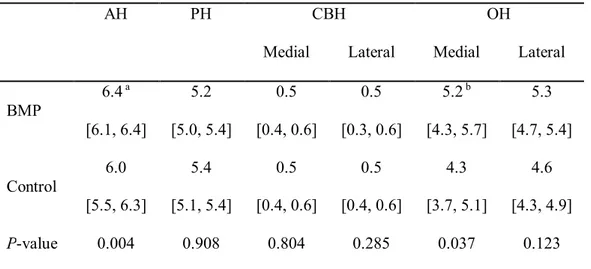
AH of the maxillary sinus pouch was significantly greater in the BMP group than the control group (6.4 mm and 6.0 mm, respectively; P = 0.004). The OH was significantly greater in the BMP group than the control group at the medial surface of the implant (5.2 mm and 4.3 mm, respectively; P = 0.037), but it did not differ significantly at the lateral surface of the implant (5.3 mm and 4.6 mm, P = 0.123; Table 2).
None of the measured areas differed significantly between the BMP and control groups (P > 0.05, Table 3); this was also the case for BIC (25.3% and 24.7%, respectively; P > 0.05).
IV. Discussion
The present study evaluated the effect of rhBMP-2-loaded CBCP on bone formation of sinus augmentation with simultaneous implant placement in rabbits. In the surgical phase, rigid fixation of the implants was achieved in rabbit sinuses with a median CBH of 0.5 mm. At 4 weeks after the surgery, augmented bone substitute and implants were localized in sinus pouches without any complications, and newly formed bone had formed from cortical bone to the apical portion of the implants. The augmented volume, newly formed bone volume, AH and OH were greater in the BMP than the control group, but there was no intergroup difference in BIC. These results support that the use of rhBMP-2-loaded CBCP allows successful implantation with sinus augmentation even when only a minimal bone height is available during the early stage of healing.
The previous study was performed to graft 0.1 mg/ml rhBMP-2-loaded CBCP into rabbit sinus, and significantly larger volumes of augmented and newly formed bone were founded in the experimental group than the control group 4 weeks later.14 It was speculated that postoperative swelling induced by rhBMP-2 would
result in a larger initial augmented volume, which could subsequently be replaced by accelerated bone formation. Such early corticalization of the SM surrounding area can resist the positive respiratory pressure and thereby reduce volumetric shrinkage. As a
augmentation. Despite the same rhBMP-2 concentration and dosage being applied in the present study, the differences in volume appeared to be smaller than in the previous study. This was attributed to the effects of rhBMP-2 not only on early corticalization of the SM but also on new bone formation in the peri-implant area. Several studies have found that rhBMP-2 contributes to new bone formation around the implant, thereby supporting this role of rhBMP-2.20-22 Therefore, a higher
concentration and dosage of rhBMP-2 might be needed in order to achieve results similar to those in the previous study.
The early stability of the implant is determined by the peri-implant bone quantity and maturity23, which could be predictive of the long-term prognosis of the
implant. When an implant is placed, the new bone formation appears to start from the original cortical bone of the sinus wall and progress towards the center in the apical direction.9 Newly formed cortical bone could play a similar role to the original
cortical bone. In the present study, the OH-to-PH ratios were 103.5% and 97.3% medially and laterally, respectively, in the BMP group, and 83.7% and 89.3% in the control group. This indicates that almost the entire height of implant was covered by newly formed bone in the BMP group. RhBMP-2 was effective in bone formation on the medial surface of the implant, which was closer to the axial wall24, resulting in
rhBMP-2 stimulating angiogenesis through the chemotaxis of endothelial cells and osteoblasts that exist in cortical bone and around the SM.25 It is therefore suggested
that rhBMP-2 accelerates peri-implant bone formation and thereby enhances the early stability of the implant.
Comparing with the BIC values of Kim et al18(11.3% for blood clots, 23.0%
for autogenous bone and 29.0% for bovine-derived hydroxyapatite), BIC of CBCP grafting was similar to autogenous bone grafting regardless of whether or not rhBMP-2 was used (rhBMP-25.3% for BMP and rhBMP-24.6% for control). It seems that rhBMP-rhBMP-2 itself was not effective in directing newly formed bone to contact the implant surface. Wikesjo et al20found no significant difference in BIC when rhBMP-2 and absorbable collagen
sponge were applied to supra-alveolar defects of mongrel dogs for 8 weeks. However, a histological analysis revealed that rhBMP-2 affected new bone formation more widely on the implant surface. A wider area of newly formed bone means that more of the implant surface will be covered, which can increase the implant stability. Fixation at the cortical bone is always important for initial stability of an implant.26
However, one of the main disadvantages of one-stage implantation is the possibility of fixation failure leading to displacement of the fixture into the maxillary sinus. In the present study model, the implant was fixed to thin cortical bone (less than 0.5 mm thick, representing less than 10% of the fixture length) by manipulation and modification of the implant design such as the inclusion of a crestal shoulder and microthreading in the cortical contact area. Also, adding rhBMP-2 to CBCP can achieve secondary stability via new bone formation around the implant surface, as discussed above.
The volume but not the area differed significantly between the two study groups. This could have been due to the smallness of the sample and variations in the
of the sample and performing measurements at a single point in time. Therefore, future studies should include larger samples and obtain results at various time points. Although the rabbit sinus model has a strong osteogenic potential for observing the effects of rhBMP-2 on a sinus graft after 4 weeks, the bone contact around the implant should also be observed during the later stage of healing; that is, after more than 4 weeks.
In conclusion, an implant simultaneously placed with sinus augmentation using rhBMP-2-loaded synthetic bone substitute can be successfully osseointegrated even when there is a limited height of bone available during the early stage of healing, thereby achieving a reduced healing time when cortical fixation is obtained.
References
1. Jensen OT, Shulman LB, Block MS, Iacono VJ. Report of the Sinus Consensus Conference of 1996. Int J Oral Maxillofac Implants 1998;13 Suppl:11-45. 2. Wallace SS, Froum SJ. Effect of maxillary sinus augmentation on the survival of
endosseous dental implants. A systematic review. Ann Periodontol 2003;8(1):328-343. 3. Peleg M, Mazor Z, Garg AK. Augmentation grafting of the maxillary sinus and
simultaneous implant placement in patients with 3 to 5 mm of residual alveolar bone height. Int J Oral Maxillofac Implants 1999;14(4):549-556.
4. Peleg M, Mazor Z, Chaushu G, Garg AK. Sinus floor augmentation with simultaneous implant placement in the severely atrophic maxilla. J Periodontol 1998;69(12):1397-1403.
5. Felice P, Pistilli R, Piattelli M, Soardi E, Barausse C, Esposito M. 1-stage versus 2-stage lateral sinus lift procedures: 1-year post-loading results of a multicentre randomised controlled trial. Eur J Oral Implantol 2014;7(1):65-75.
6. Zitzmann NU, Scharer P. Sinus elevation procedures in the resorbed posterior maxilla. Comparison of the crestal and lateral approaches. Oral Surg Oral Med Oral Pathol Oral Radiol Endod 1998;85(1):8-17.
7. Jung UW, Hong JY, Lee JS, Kim CS, Cho KS, Choi SH. A hybrid technique for sinus floor elevation in the severely resorbed posterior maxilla. J Periodontal Implant Sci 2010;40(2):76-85.
approach: rigid synthetic resorbable barriers versus anorganic bovine bone. Five-month post-loading clinical and histological results of a pilot randomised controlled clinical trial. Eur J Oral Implantol 2009;2(4):293-306.
9. Choi Y, Lee JS, Kim YJ, Kim MS, Choi SH, Cho KS, Jung UW. Recombinant human bone morphogenetic protein-2 stimulates the osteogenic potential of the Schneiderian membrane: a histometric analysis in rabbits. Tissue Eng Part A 2013;19(17-18):1994-2004.
10. Seeherman H, Wozney J, Li R. Bone morphogenetic protein delivery systems. Spine (Phila Pa 1976) 2002;27(16 Suppl 1):S16-23.
11. Jung IH, Lim HC, Lee EU, Lee JS, Jung UW, Choi SH. Comparative analysis of carrier systems for delivering bone morphogenetic proteins. J Periodontal Implant Sci
2015;45(4):136-144.
12. Alam I, Asahina I, Ohmamiuda K, Enomoto S. Comparative study of biphasic calcium phosphate ceramics impregnated with rhBMP-2 as bone substitutes. J Biomed Mater Res 2001;54(1):129-138.
13. Brodie JC, Goldie E, Connel G, Merry J, Grant MH. Osteoblast interactions with calcium phosphate ceramics modified by coating with type I collagen. J Biomed Mater Res A 2005;73(4):409-421.
14. Kim JS, Cha JK, Cho AR, Kim MS, Lee JS, Hong JY, Choi SH, Jung UW. Acceleration of Bone Regeneration by BMP-2-Loaded Collagenated Biphasic Calcium Phosphate in Rabbit Sinus. Clin Implant Dent Relat Res 2015;17(6):1103-1113.
15. Watanabe K, Niimi A, Ueda M. Autogenous bone grafts in the rabbit maxillary sinus. Oral Surg Oral Med Oral Pathol Oral Radiol Endod 1999;88(1):26-32.
16. Wada K, Niimi A, Watanabe K, Sawai T, Ueda M. Maxillary sinus floor augmentation in rabbits: a comparative histologic-histomorphometric study between rhBMP-2 and autogenous bone. Int J Periodontics Restorative Dent 2001;21(3):252-263.
17. Sun XJ, Zhang ZY, Wang SY, Gittens SA, Jiang XQ, Chou LL. Maxillary sinus floor elevation using a tissue-engineered bone complex with OsteoBone and bMSCs in rabbits. Clin Oral Implants Res 2008;19(8):804-813.
18. Kim YS, Kim SH, Kim KH, Jhin MJ, Kim WK, Lee YK, Seol YJ, Lee YM. Rabbit maxillary sinus augmentation model with simultaneous implant placement: differential responses to the graft materials. J Periodontal Implant Sci 2012;42(6):204-211. 19. Brunner E, Langer F. Nonparametric analysis of ordered categorical data in designs
with longitudinal observations and small sample sizes. Biom J 2000;42(6):663-675. 20. Wikesjo UM, Qahash M, Thomson RC, Cook AD, Rohrer MD, Wozney JM, Hardwick
WR. rhBMP-2 significantly enhances guided bone regeneration. Clin Oral Implants Res 2004;15(2):194-204.
21. Yasko AW, Lane JM, Fellinger EJ, Rosen V, Wozney JM, Wang EA. The healing of segmental bone defects, induced by recombinant human bone morphogenetic protein (rhBMP-2). A radiographic, histological, and biomechanical study in rats. J Bone Joint Surg Am 1992;74(5):659-670.
22. Sigurdsson TJ, Fu E, Tatakis DN, Rohrer MD, Wikesjo UM. Bone morphogenetic protein-2 for peri-implant bone regeneration and osseointegration. Clin Oral Implants Res 1997;8(5):367-374.
23. Guillot R, Gilde F, Becquart P, Sailhan F, Lapeyrere A, Logeart-Avramoglou D, Picart C. The stability of BMP loaded polyelectrolyte multilayer coatings on titanium.
24. Jung UW, Unursaikhan O, Park JY, Lee JS, Otgonbold J, Choi SH. Tenting effect of the elevated sinus membrane over an implant with adjunctive use of a hydroxyapatite-powdered collagen membrane in rabbits. Clin Oral Implants Res 2015;26(6):663-670. 25. Chen D, Zhao M, Harris SE, Mi Z. Signal transduction and biological functions of bone
morphogenetic proteins. Front Biosci 2004;9:349-358.
26. Khoury F. Augmentation of the sinus floor with mandibular bone block and simultaneous implantation: a 6-year clinical investigation. Int J Oral Maxillofac Implants 1999;14(4):557-564.
Figure Legends
Figure 1.
(A) Photograph of the implant used in this study. (B) Two implant sites and holes were prepared bilaterally. Venous-blood-filled parts of the sinus after placing the implants. (Ant, anterior; Post, posterior).Figure 2.
Schematic showing the measured parameters. (Med, medial; Lat, lateral; CBH, cortical bone height; AH, augmented height; PH, protruding height; OH, distance from the basal cortical bone to the highest point of osseointegration).Figure 3.
Radiographic findings in micro computed tomography. False-colorrepresentation of radiographic findings in a cross-sectional view. Note that the augmented bone substitutes (green) are well maintained within the maxillary sinus, and that the implants are thoroughly enveloped by newly formed bone (purple) and augmented bone substitutes. (A and B) BMP group; (C and D) Control group. (A and C) are coronal views, while (B and D) are sagittal views.
Figure 4.
Histological photomicrographs of the total augmented area after 4 weeksof healing. (A) BMP group. (B) Control group. The osseointegration of the implant was evident throughout the cortical bone area in both groups. (CB, cortical bone; RBS,
Figure 5.
Histological photomicrographs of the apical area after 4 weeks of healing. (A) BMP group. (B) Control group. More new bone formed around the apex of the implant in the BMP group than the control group. (SM, sinus membrane; NB, newly formed bone; H&E, the scale bar = 500 μm).Tables
Table 1. Volumetric measurements from micro-CT data (Median [min, max]; mm3)
NB RBS Implant Augmented Total BMP 51.6 a [43.9, 59.2] 30.0 [27.4, 37.4] 95.7 [93.4, 101.7] 153.5b [98.9, 181.3] 249.2 [200.7, 276.5] Control 46.6 [34.5, 48.7] 33.4 [28.2, 37.1] 101.5 [97.8, 102.5] 116.1 [104.6, 121.1] 218.6 [208.0, 249.8] P-value 0.019 0.476 0.127 0.034 0.095 Note : NB, newly formed bone; RBS, remaining bone substitute.
Table 2. The histomorphometric linear measurements in the augmented pouch
(Median [min, max]; mm)
AH PH CBH OH
Medial Lateral Medial Lateral
BMP 6.4a [6.1, 6.4] 5.2 [5.0, 5.4] 0.5 [0.4, 0.6] 0.5 [0.3, 0.6] 5.2b [4.3, 5.7] 5.3 [4.7, 5.4] Control 6.0 [5.5, 6.3] 5.4 [5.1, 5.4] 0.5 [0.4, 0.6] 0.5 [0.4, 0.6] 4.3 [3.7, 5.1] 4.6 [4.3, 4.9] P-value 0.004 0.908 0.804 0.285 0.037 0.123 Note: AH, augmented height; PH, protruding height; CBH, cortical bone height; OH, the most highest osseointegrated point to cortical bone base length.
aSignificantly greater than control group (P < 0.01)
Table 3. The histomorphometric area measurements in the augmented pouch (Median
[min, max]; mm2)
NB RBS Soft tissue Implant Total
BMP 2.0 [1.2, 3.5] 4.4 [0.6, 4.8] 10.9 [10.3, 12.1] 13.4 [10.4, 14.1] 30.2 [25.5, 34.5] Control 1.9 [0.9, 2.9] 3.0 [2.7, 5.8] 10.5 [7.0. 12.4] 13.6 [13.4, 13.8] 29.8 [26.4, 32.6] P-value 0.794 0.908 0.598 0.270 0.587 Note: NB, newly formed bone; RBS, remaining bone substitute.
Figures
국문요약
토끼에서 rhBMP-2 동반하여 합성골 적용한
상악동 증대술과 임플란트 동시식립
<지도교수 정 의 원> 연세대학교 대학원 치의학과주 명 재
임플란트 식립에 있어 상악 후방 무치악 부위는 상악동의 함기화, 치조골의 흡수 등으로 인하여 수직적인 공간의 부족을 흔하게 보인다. 수직적인 공간을 확보하기 위하여 임플란트 식립 전 상악동 증대술을 주로 시행하며 대개 식립 전까지 4 개월 이상의 치료기간이 추가로 필요하다. 추가로 필요한 기간을 단축하기 위하여 상악동 증대술과 동시에 임플란트를 식립하는 방법이 있으며 동시 식립 후 안정적인 초기치유가 중요하다. 본 연구는 토끼 상악동에 임플란트 식립 시 골형성유도단백질-2 (rhBMP-2)가 적용된 collagenated biphasic calcium phosphate (CBCP)를총 5 마리의 토끼을 대상으로 상악동 양측에 골이식을 위한 창을 형성하고 전방으로 임플란트(ø3 x 6 mm)를 수조작을 통해 고정을 확인하며 식립하였다. 상악동 좌우로 실험군에는 rhBMP-2 가 적용된 CBCP 를, 대조군에는 생리식염수가 적용된 CBCP 를 각각 이식하였다. 4 주 후, 방사선학적으로 2 차원적인 단면영상과 3 차원적인 재건영상을 통해 증대된 부피, 신생골의 부피 등을 분석하고, 조직계측학적으로는 선형분석으로 피질골의 두께, 골-임플란트 접촉, 임플란트 플랫폼으로부터 신생골 형성 근단부까지의 거리(내측 및 외측), 임플란트 플랫폼으로부터 증대된 상악동 경계까지의 거리를 측정하였으며, 면적분석으로 신생골양, 잔존 골대체제양, 광화조직양, 섬유혈관조직양을 분석하였다. 방사선 및 조직학적 관찰에서 양쪽 군 모두 임플란트와 피질골과의 골유착이 이루어졌으며 임플란트 근단부를 향하여 신생골이 선상으로 형성되는 것을 확인 할 수 있었다. 부피 분석에서 실험군(51.6 mm3)이 대조군(46.6 mm3)보다 신생골 형성이 더 많았다. 골-임플란트 접촉은 통계학적으로 유의한 차이가 나지 않았으나 임플란트 플랫폼으로부터 신생골 형성 근단부까지의 거리는 내측에서 실험군(5.2 mm)이 대조군(4.3 mm)보다 통계적으로 유의하게 크게 나타났다. 이상의 연구를 통해, 상악 후방 무치악에서 수직적 공간이 부족한 부위의 rhBMP-2 적용 합성골의 상악동 거상술과 동시에 임플란트 식립을
시행하는 것은 초기 회복 시기에서 성공적인 골유착을 가능하게 함을 확인할 수 있었다.
_____________________________________________________________________ 핵심되는 말 : 상악동 증대술, 치과 임플란트, 골대체제, 교원질,
![Table 1. Volumetric measurements from micro-CT data (Median [min, max]; mm 3 )](https://thumb-ap.123doks.com/thumbv2/123dokinfo/5099332.78781/33.892.155.741.343.558/table-volumetric-measurements-micro-ct-data-median-min.webp)