: Vol. 35, No. 3, 2005
Bone Added Osteotome Sinus Floor Elevation with Simultaneous Placement of Branemark
Ti-Unite and ITI SLA implants
Nam-Won Kang1·Ui-Won Jung1,2·Seong-Ho Choi1,2,3·
Kyoo-Sung Cho1,2,3·Jung-Kiu Chai1,2·Chong-Kwan Kim1,2,3·Chang-Sung Kim1,2,3
1Department of Periodontology
2Oral Science Research Center, College of Dentistry
3Brain Korea 21 Project for Medical Science, Yonsei University
Ⅰ. Introduction
The placement of implants in the posterior maxilla is occasionally limited by insufficient bone volume as a result of alveolar atrophy or pneumatization of the maxillary sinus.
This clinical problem can be resolved by si- nus augmentation using various surgical pro- cedures, including an onlay augmentation of the alveolar crest1,2, Le Fort I osteotomies with an interpositional bone graft3,4, lateral approach sinus augmentation 5-7 and osteo- tome sinus augmentation8-11. The placement of the implants in a bone- grafted maxilla has been reported to be successful as a 1-step approach with sinus augmentation or
in a 2-step approach after sinus augmen- tation. However, when placed in the bone- grafted maxilla, a lower survival rate of ma- chined surface implants compared with rough surface implants has been reported.1)
In 1994, a less invasive sinus floor eleva- tion procedure with simultaneous grafting and the immediate placement of implants was introduced by Summers8. Using the Summers osteotome kit8,9, which was specifically de- signed for this procedure, the pre-existing crestal bone is displaced toward the sinus floor as the osteotomes are inserted. Various types of graft materials and implants can be used in this surgical procedure. Clinical case reports and studies on the BAOSFE proce-
This work was supported in part by Yonsei University, College of Dentistry Research Fund of 2004.
Correspondence author : Chang-Sung Kim, Department of Periodontology, Oral Science Research Center, College of Dentistry, Brain Korea 21 Project for Medical Science, Yonsei University. 134 Shinchon-Dong, Seodaemun-gu, Seoul, Korea.
Phone: +82-2-2228-3186 Telefax: +82-2-392-0398 E-mail: [email protected]
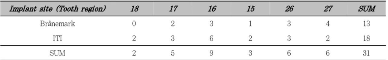
Table 1. Distribution of Implant According to the Implant Systems (n=31)
Implant site (Tooth region) 18 17 16 15 26 27 SUM
Brånemark 0 2 3 1 3 4 13
ITI 2 3 6 2 3 2 18
SUM 2 5 9 3 6 6 31
dure with the simultaneous placement of im- plants showing a relatively high survival rate in both the Brånemark(91.4 to 100%) and ITI SLA implants (94 to 98 %) have been pub- lished10-15. However, no comparative clinical study was available on the Brånemark Ti- Unite and ITI SLA implant.
Clinical and radiographic studies on the dimensional change in the grafted bone have also been reported16,17. It was reported that all the graft materials resulted in a radio- graphic reduction ranging from 0.79 to 2.09 mm over a 3-year follow-up. However, it was not determined whether this reduction in graft height occurred during the initial healing period or was an ongoing process.
Recently, Hatano et al. assessed the long- term changes in the sinus-graft height after a maxillary sinus floor augmentation with simultaneous placement of 18 implants. The results showed that the graft height de- creased during the first 2~3 years after augmentation, but all subsequent changes were minimal.
The aim of this study was to evaluate and compare the clinical results of the Brånemark Ti-Unite and ITI SLA implants placed simul- taneously using BAOSFE procedure and to assess the change in the graft height radio- graphically in these two different implant systems after the BAOSFE procedure during
the initial healing period.
Ⅱ. Materials and Methods
1. Patients
Twenty two patients(10 women and 12 men, mean age of 50 years, age range of 20 to 65 years) with severe atrophy of the al- veolar process in the posterior maxilla were treated at the Department of Periodontology, College of Dentistry, Yonsei University. None of the patients showed signs and symptoms of sinus and intraoral disease. The patients were provided informed consent to partic- ipate in this clinical study. None of the sub- jects had systemic diseases or had undergone drug therapy in the previous 12 months.
Eleven patients underwent the BAOSFE pro- cedure with the simultaneous placement of 13 Brånemark Ti- Unite implants(Nobel Biocare, Sweden). The other 11 patients un- derwent the BAOSFE procedure with the si- multaneous placement of 18 ITI SLA im- plants(Institut Straumann AG, Switzerland) (Table 1). There was no case of sinus mem- brane perforation during surgery.
2. Operative technique
On the initial examination, the patients'
medical histories were reviewed in order to rule out any local or systemic diseases that might contraindicate the surgical proce- dures. The patients received oral hygiene instructions and whole-mouth scaling prior to the surgery.
The BAOSFE procedure was performed using a Summers Osteotome kit†, as de- scribed by Summers8,9. Briefly, an incision was made under local anesthesia(Lidocaine 2% with 1:80.000 epinephrine¶) at the eden- tulous area to be treated. After the crestal incision had been made, full thickness buc- cal and palatal flaps were reflected. The site preparation began using the Summers #1 and #2 osteotomes. When the bone was too dense for hand instrumentation, 2mm twist drilling was used to reach the cancellous bone. The drilling remained 1mm below the floor of the sinus. The preparation site was widened using #2 and #3 Summers osteo- tomes. No instrument penetrated the cavity of the sinus at any time. A prepared various bone mix, which acts as a shock absorber, was added to the preparation site with a carrier. Elevation of the maxillary sinus membrane was achieved using the #3 osteo- tome that was used previously to force the graft ahead of its tip to achieve the sinus floor up-fracture. At this stage, the in- tegrity of the sinus membrane was con- firmed by the Valsalva manuever. Finally, each patient received the Brånemark Ti- Unite implants or the ITI SLA implants into the osteotomy site. The primary stability was achieved in all implants. Primary clo- sure was achieved by using monofilament*
suture material.
Postoperatively, the patients were in- structed to rinse their mouth twice a day with a 0.12% chlorhexidine solution‡ during the first 2 weeks after surgery. Antibiotic regimens were prescribed for 7 days, and the sutures were removed after 10 days.
3. Prosthetic procedures
After a mean healing period of 9 months for the Brånemark implants and 8 months for the ITI implants, all the patients were rehabilitated with fixed crown or bridges.
4. Follow-up
After inserting the implants, the patients were followed-up 1 and 2 weeks, 3, 6, 9 and 12 months. A radiological evaluation was performed using minimum of three panoramic radiographs according to the following sched- ule: prior to surgery, immediately after sur- gery, and 6 months after surgery(Figure 1).
5. Analysis of radiographs
Using a scanner, the panoramic radio- graphs were digitalized. The Digital image analysis program# was used for the linear analysis of the panoramic radiographs. The magnification of panoramic radiograph was corrected using the known actual length of the inserted implants and an accurate graft height could be obtained. This was under- taken by one investigator. The radiographs from the same patient were blinded to the time. The following radiographic parameters from each radiograph were measured(Fig.2):
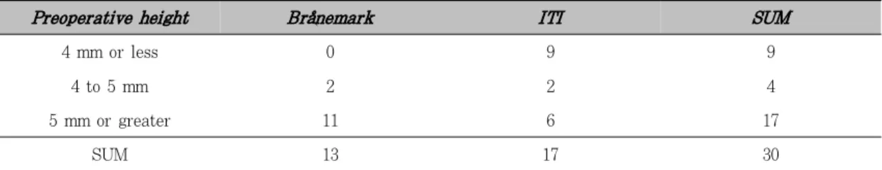
Table 2. Native Bone Height and Implant Distribution
Preoperative height Brånemark ITI SUM
4 mm or less 0 9 9
4 to 5 mm 2 2 4
5 mm or greater 11 6 17
SUM 13 17 30
▶ The native bone height ; the distance from the alveolar crest to the floor of the maxillary sinus at the implant site, which is represented as a mean of the mesial and distal native bone heights.
▶ The grafted bone height; the distance from the floor of the maxillary sinus to the border of the grafted bone at the implant site, which is represented as a mean of mesial and distal grafted bone height.
▶ The implant height; the distance from the apex to the head of the fixture.
6. Statistical analysis
The survival rate of each implant system was calculated. A paired t-test was used to calculate the statistical differences of the changes in the grafted bone height during the observation period within the each im- plant system. Unpaired t-test was used to calculate the statistical differences in graft- ed bone height change between the two im-
plant systems. A P value < 0.05 was consid- ered to be significant.2)
Ⅲ. Results
Clinical and radiographic healing was un- eventful during the observation periods of 12 months. Table 1 shows the distribution of the implants. The 31 osseointegrated im- plants represent a survival rate of 96.8%.
The Brånemark Ti-Unite surface implants showed 100%(13/13) survival rate and the ITI SLA surface implants showed 94.4%
(17/18) survival rate. One of the 18 ITI im- plants was lost during the observation period. A lateral force or overload induced by the temporary denture after placing the implant might be responsible for the failure.
The native bone height of the Brånemark Ti-Unite surface implant was significantly larger than that of the ITI SLA surface im- plant(Table 2). The patients' details are documented in tables 3 and 4 according to the implant systems.
¶ 2% lidocaine, 1:100,000 epinephrine, Kwangmyung Pharm., Seoul, Korea
† 3i, Implant Innovations, Palm Beach Garden, FL, USA
* Ethilon, Ethicon, Johnson & Johnson Int., Edinburgh, UK
‡ Hexamedin, Bukwang Pharmaceutical Co., Korea.
§ HP scanjet 7400c , Hewlett Packard, USA
# Image-Pro Plus, Media Cybernetics, Silver Spring, M.D., USA
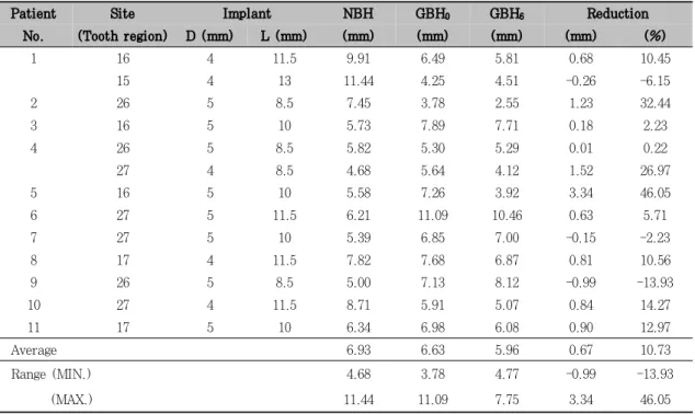
Table 3. Native, Grafted bone height and Reduction of the grafted bone height of the Brånemark Ti-Unite System
Patient Site Implant NBH GBH0 GBH6 Reduction
No. (Tooth region) D (mm) L (mm) (mm) (mm) (mm) (mm) (%)
1 16 4 11.5 9.91 6.49 5.81 0.68 10.45
15 4 13 11.44 4.25 4.51 -0.26 -6.15
2 26 5 8.5 7.45 3.78 2.55 1.23 32.44
3 16 5 10 5.73 7.89 7.71 0.18 2.23
4 26 5 8.5 5.82 5.30 5.29 0.01 0.22
27 4 8.5 4.68 5.64 4.12 1.52 26.97
5 16 5 10 5.58 7.26 3.92 3.34 46.05
6 27 5 11.5 6.21 11.09 10.46 0.63 5.71
7 27 5 10 5.39 6.85 7.00 -0.15 -2.23
8 17 4 11.5 7.82 7.68 6.87 0.81 10.56
9 26 5 8.5 5.00 7.13 8.12 -0.99 -13.93
10 27 4 11.5 8.71 5.91 5.07 0.84 14.27
11 17 5 10 6.34 6.98 6.08 0.90 12.97
Average 6.93 6.63 5.96 0.67 10.73
Range (MIN.) 4.68 3.78 4.77 -0.99 -13.93
(MAX.) 11.44 11.09 7.75 3.34 46.05
D : Distal M : Mesial
NBH : Native bone height
GBH0 : Grafted bone height(Baseline) GBH6 : Grafted bone height(6 Months) MIN : Minimum
MAX : Maximum
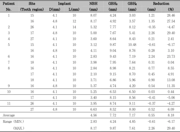
The gain in the grafted bone height of the Brånemark Ti-Unite implants was 6.63mm ranging from 3.78mm to 11.09mm, and that of the ITI SLA implants was 7.72mm, rang- ing from 4.24mm to 9.87mm. A statistically significant difference between the pre-surgi- cal and post-surgical bone height existed in both implant systems(P<0.05). However, there was no significant difference in the gain of the grafted bone height between the implant systems.
The total mean reduction in the grafted bone height was 0.6mm(9.29%) of the graft-
ed bone 6 months after surgery. There was a statistically significant reduction in the grafted bone height between that observed immediately after surgery and 6 months af- ter surgery(p<0.05). The mean reduction in the grafted bone height of the Brånemark Ti-Unite implants was 0.67mm(10.73%) ranging from -0.99mm to 3.34mm. Regarding the ITI SLA implants, the mean reduction in the grafted bone height was 0.55mm(8.18%) ranging from -0.61mm to 2.26mm(Table 4).
However, there was no statistically sig- nificant difference between the two systems.
Table 4. Native, Grafted bone height and Reduction of the bone height of the ITI SLA System
Patient Site Implant NBH GBH0 GBH6 Reduction
No. (Tooth region) D(mm) L(mm) (mm) (mm) (mm) (mm) (%)
1 15 4.1 10 8.07 4.24 3.03 1.21 28.46
16 4.8 12 8.17 4.92 3.57 1.35 27.54
2 26 4.1 14 5.32 7.77 8.12 -0.35 -4.47
3 17 4.8 10 5.00 7.67 5.41 2.26 29.40
4 27 4.1 10 3.60 8.64 8.43 0.21 2.41
5 15 4.1 10 3.12 9.87 10.48 -0.61 -6.17
16 4.8 10 4.11 9.04 8.76 0.28 3.10
6 16 4.1 10 2.83 9.43 7.19 2.24 23.73
7 18 4.1 10 3.98 7.95 7.64 0.31 0.04
8 16 4.1 10 2.84 8.98 8.21 0.77 8.55
17 4.1 10 2.10 9.15 8.70 0.45 4.91
18 4.1 10 3.71 6.86 5.96 0.90 13.08
9 16 4.8 10 5.37 4.74 4.20 0.54 11.35
10 16 4.1 10 5.25 6.53 6.50 0.03 0.44
17 4.1 10 3.40 8.15 8.56 -0.41 -5.06
11 26 4.1 10 3.95 8.74 9.11 -0.37 -4.27
27 4.8 10 6.63 8.52 8.00 0.52 6.09
Average 4.56 7.72 7.17 0.55 8.18
Range (MIN.) 2.83 4.24 4.85 -0.61 -6.17
(MAX.) 8.17 9.87 7.61 2.26 29.40
D : Distal M : Mesial
NBH : Native bone height
GBH0 : Grafted bone height (Baseline) GBH6 : Grafted bone height (6 Months) MIN : Minimum
MAX : Maximum
Ⅳ. Discussion
The aim of this study was to evaluate and compare the clinical results of the Bråne- mark Ti-Unite and ITI SLA implants placed simultaneously using BAOSFE procedure, and to assess the change in the graft height radiographically in these two different im- plant systems after the BAOSFE procedure during the initial healing period. The results
indicated that the simultaneous placement of the Brånemark Ti-Unite as well as the ITI SLA implant using the BAOSFE procedure is a feasible treatment option for patients with atrophic posterior maxilla. In addition, radiographic reduction of the grafted bone height was found during the initial healing period of 6 months in similar pattern at these two different implant systems.
Although there were various results with
different follow-up periods, inclusion cri- teria, surgical and prosthetic techniques, and other factors, the BAOSFE procedure with the simultaneous placement of an im- plant showed a predictable survival rate ranging from 95% to 100%6,10,11. The 1-step approach to the atrophic posterior maxilla using the BAOSFE procedure has the advan- tages of being less invasive. This technique can also enhance the bone quality of the im- plant site from type III or IV to type II.
Reduction of surgical and healing time can be achieved because a coordinated con- solidation of the graft around the implants during the healing period is expected. More- over, there has been little difference re- ported between the survival rate of the im- plants placed immediately at the time of the grafting or those placed after a delay19. It has been reported that the differences in the implant designs and surface characteristics may influence the survival rate of the differ- ent types of implants. Regarding the extent of bone retention, some studies have re- ported that the SLA surface is superior to the machined surface implant20,21. Moreover, it was reported that the survival rate of the SLA surfaced implants in the sinus-aug- mented maxilla was significantly higher than that of the machined surface implants22.
It was reported that the survival rate of the implants was also influenced by the quality and quantity of the native bone
11,12,23. In particular, the survival rate is markedly reduced when the native bone height in a implant site was 4mm or less11 because it is difficult to achieve primary sta- bility of the implant, and there is a higher
possibility of the Schneiderian membrane tearing24. Therefore, at least 5mm of the na- tive bone was recommended for the 1 step approach. In this study, the mean height of the native bone was 5.58mm with a dis- tribution of 6.93mm for the Brånemark and 4.56mm for the ITI SLA implant. Thirteen of 30(43%) sites were < 5mm in the native bone height and 9 out of ITI SLA implant were 4mm or less. Nevertheless, a predictable high survival rate could be obtained at both implant systems. Peleg et al.(1999) eval- uated the efficacy of the augmentation of the maxillary sinus using the lateral approach with the simultaneous placement of hydrox- yapatite surface implants in patients with 3 to 5 mm of the residual bone height 25. All the 160 implants in the 63 patients were sta- ble during 2 to 4 year follow-up periods.
Together with previous studies, these results showed that the rough surface implants used in the augmented sinus area could provide a predictable prognosis. Therefore, a 1-step procedure of grafting the maxillary sinus and the simultaneous placement of rough surface implants might be selected as a feasible treatment option for patients with as little as 5mm of the native bone height.
The dimensional changes in the height of the graft augmented in the sinus have been documented. At the Sinus Consensus Con- ference of 1996, 100 patients, 145 sinus- grafting sites were evaluated using pan- oramic radiographs over a 3-year period. It was reported that all graft materials re- sulted in a radiographic reduction ranging from 0.79 to 2.09mm. However, it was not determined whether this reduction in the
graft height occurred in an initial healing period or was a part of an ongoing healing process. Hallman et al. analyzed 30 maxil- lary sinuses in 20 patients who were grafted with a mixture of autogenous bone and bo- vine hydroxyapatite, and reported that a small(<10%) but statistically significant di- mensional reduction was observed 12 months after surgery and after 1 year of loading26. Other studies on the reduction of sinus grafts using X-rays were also available27. In this study, it was demonstrated that during the course of the initial healing periods of 6 months, the height of the grafted bone was reduced by an overall mean of 0.6mm (9.29%), which comprised of a mean of 0.67 mm(10.73%) for the Brånemark Ti-unite im- plants and 0.55mm(8.18%) for the ITI SLA implants. However, the difference between two implant systems was not statistically significant. Therefore, it appears that adi- mensional healing response of the grafted bone may occur with a similar pattern in the Brånemark Ti-Unite and the ITI SLA implants. The reduction of the grafted mate- rial was influenced more by the host healing response than by submergence or implant characteristics. The radiographic evaluations in this study could not fully characterize the nature of the graft materials in the sinus. A histological finding will be essential for as- sessing the healing event in augmented sinus. Longer follow-up periods will be also be needed to determine if the reduction ob- served in this study is an ongoing process or occurs only in the initial healing period.
However, together with other studies, it can be concluded that a major volumetric reduc-
tion of the grafted materials in sinus occurs during initial healing period.
Ⅴ. Conclusion
The simultaneous placement of the Bråne- mark Ti-Unite and ITI SLA implants with BAOSFE procedure showed predictable clin- ical results. In addition, radiographic reduc- tion of the grafted bone height was found during the initial healing period of 6 months in similar pattern at these two different im- plant systems.
Ⅵ. References
1. Jenson J, Krantz Simonsen E, Sindet Pedersen S. Reconstruction of the se- verely resorbed maxilla with bone graft- ing and osseointegrated implants: A preliminary report. J Oral Maxillofac Surg 1990;48:27-32.
2. Adell R, Lekholm U, Grndahl K, Bråne- markP-I, Lindstrm J, Jacobsson M. Re- construction of severely resorbed eden- tulous maxillae using osseointegrated fixtures in immediate autogenous bone grafts. Int J Oral Maxillofac Implants 1990;5:233-246.
3. Isaksson S. Evaluation of three bone grafting techniques for severely resorbed maxillae in conjuction with immediated endosseous implants. Int J Oral Maxillo- fac Implants 1994;9:679-688.
4. Kahnberg K-E, Nystrm E, Bartholdsson L. Combined use of bone grafts and Brånemark fixtures in the treatment of severely resorbed maxillae. Int J Oral
Maxillofac Implants 1989;4:297-304.
5. Fugazzotto PA. Maxillary sinus grafting with and without simultaneous implant placement: Technical considerations and case reports. Int J Periodont Rest Dent 1994;14(6):545-551.
6. Fugazzotto PA, Vlassis J. Long-term success of sinus augmentation using various surgical approaches and grafting materials. Int J Oral Maxillofac Im- plants 1998;13:52-58.
7. Blomqvist JE, Alberius P, Isaksson S.
Two stage maxillary sinus reconstruc- tion with endosseous Implants: A pro- spective study. Int J Oral Maxillofac Implants 1998;13:758-766.
8. Summers RB. Maxillary implant sur- gery: The osteotome technique; Part 1.
Compend Contin Educ Dent 1994;15 (2):152-162.
9. Summers RB. The osteotome technique;
Part 3. Less invasive methods of elevat- ing the sinus floor. Compend Contin Educ Dent 1994;15(6):698-708.
10. Zitzmann N, Scharer P. Sinus elevation procedures in the resorbed posterior maxilla: Comparison of the crestal and lateral approaches. Oral Surg Oral Med Oral Pathol Oral Radiol Endodontol 1998;85:8-17
11. Rosen PS, Summers R, Mellado JR, et al. The bone added osteotome sinus floor elevation technique: multicenter retro- spective report of consecutively treated patients. Int J Oral Maxillofac Implants 1999;14:853-858
12. Bruschi GB, Scipioni A, Calesini G, Bruschi E. Localized management of si-
nus floor with simultaneous implant placement: A clinical report. Int J Oral Maxillofac Implants 1998;13:219-226 13. Winter AA, Pollack AS, Odrich RB.
Placement of implants in the severely trophic posterior maxilla using localized management of the sinus floor: A pre- liminary study. Int J Oral Maxillofac Implants 2002;17:687-695
14. Horowitz RA. The use of osteotomes for sinus augmentation at the time of im- plant placement. Compend Cont Educ Dent 1997;18:441-452.
15. Komarnyckyj OG, London RM. Osteo- tome single stage dental implant place- ment with and without sinus elevation:
A clinical report. Int J Oral Maxillofac Implants 1998;13:799-804.
16. Buchmann R, Khoury F, Faust C, E.
Lange. Peri-implant conditions in perio- dontally compromised patients following maxillary sinus augmentation. A long- term post-therapy. Clinical Oral Im- plants Research 1999;10:103
17. Raghoebar, G.M., Timmenga, N.M., Reintsema, H., Stegenga, B. & Vissink, A. Maxillary bone grafting for insertion of endosseous implants: results after 12-24 months. Clinical Oral Implants Research 2001;12:279-286.
18. Naoki Hatano, Yoshinaka Shimizu, Kiyo- shi Ooya. A clinical long-term radio- graphic evaluation of graft height changes after maxillary sinus floor aug- mentation with a 2autogenous bone/xen- ograft mixture and simultaneous place- ment of dental implants. Clinical Oral Implants Research 2004;15:339-345.
19. Tong DC, Drangsholt M, Beine OR. A review of survival rates for implants placed in grafted maxillary sinuses us- ing metaanalysis. Int J Oral Maxillofac Implants 1998;13:175-182
20. Wennerberg A, Albrektsson T, Johans- son C, Andersson B. Experimental study of turned and grit-blasted screw-shaped implants with special emphasis on ef- fects of blasting material and surface topography Biomaterials. 199617:15-22.
21. Ogawa T, Ozawa S, Shih JH, Ryu KH, Sukotjo C, Yang JM, Nishimura I.
Biomechanical evaluation of osseous im- plants having different surface top- ographies in rats. J Dent Res 2000;79 :1857-63.
22. Pinholt EM. Branemark and ITI dental implants in the human bone-grafted maxilla: a comparative evaluation. Clin Oral Implants Res 2003;14:584-92.
23. Cavicchia F, Brevi F, Petrelli G. Loca- lized augmentation of the maxillaru si- nus floor to a coronal approach for the placement of implants. Int J Periodon- tics Restorative Dent 2001;21:475-485.
24. Fugazzotto PA. Augmentation of the posterior maxilla: a proposed hierarchy of treatment selection. J Periodontol 2003;74:1682-1691.
25. Peleg M, Mazor Z, Garg AK. Augmen- tation grafting of the maxillary sinus and simultaneous implant placement in patients with 3 to 5 mm of residual al- veolar bone height. Int J Oral Maxillofac Implants 1999;14:549-56.
26. Mats Hallman, Mans Hedin, Lars Sen- nerby, Stefan Lundgren. A Prospective 1-Year Clinical and Radiographic Study of Implants Placed After Maxillary Sinus Floor Augmentation With Bovine Hydro- xyapatite and Autogenous Bone. J Oral Maxillofac Surg 2002;60:277-284
27. Nicolaas C.Geurs, I. Chung Wang, Leonard B. Shulman, Marjorie K. Jeff- coat. Retrospective Radiographic Analysis of Sinus Graft and Implant Placement Procedures from the Academy of Osseo- integration Consensus Conference on Sinus Grafts. Int J Periodontics Resto- rative Dent 2001;21:517-523
설명
Figure 1a. Taking panoramic radiographs(Brånemark Ti-Unite implant)
(1) Prior to surgery (2) Immediately after surgery (3) 6 months after sur- gery
Figure 1b. Taking panoramic radiographs(ITI SLA implant)
(1) Prior to surgery (2) Immediately after surgery (3) 6 months after sur- gery
Figure 2. A - native bone height ; the distance from the alveolar crest to the floor of the maxillary sinus at the implant site, which is represented as a mean of the mesial and distal native bone heights.
B, B' - grafted bone height ; the distance from the floor of the maxillary si- nus to the border of the grafted bone at the implant site, which is repre- sented as a mean of mesial (B) and distal (B') grafted bone height.
C - the implant height; the distance from the apex to the head of the fix- ture TABLES
(I)
Figure 1a
Figure 1b
Figure 2
(1) (2) (3)
(1)
-Abstract-
Osteotome 상악동 거상술과 동시에 식립한
Br ånemark Ti-Unite 과 ITI SLA임프란트의 비교 연구
강남원1ㆍ정의원1,2ㆍ최성호1,2,3ㆍ조규성1,2,3ㆍ채중규1,2ㆍ김종관1,2,3ㆍ김창성1,2,3
1연세대학교 치과대학 치주과학교실,
2연세대학교 치과대학 구강과학연구소
3BK21 의과학 사업단
1. 목적
Osteotome 상악동거상술(Bone Added Odteotome Sinus Floor Elevation ; 이하 BAOSFE) 과 동시 에 식립한 임프란트(Brånemark, ITI)의 예상 생존율에 대해 현재까지 정확히 알려진 바는 없었으며, Brånemark Ti-Unite 과 ITI SLA 임프란트의 표면에 대한 비교 연구 또한없었다. 이번 연구는 BAOSFE 술식과 동시에 식립한 Brånemark Ti-Unite 과 ITI SLA 임프란트의 임상 결과를 비교, 평가하고 초기 치유 기간 동안의 이식골 높이의 변화를 방사선학적으로 관찰하여 두 가지 임프란트 시스템을 비교해 보고자 한다.
2. 방법
위축된 상악 구치부를 갖는 22명의 환자를 대상으로, BAOSFE술식과동시에 Brånemark Ti-Unite(11명, 13 임프란트)임프란트와 ITI SLA(11명, 18 임프란트)임프란트를 식립하였다. 수술 전, 임프란트 식립 직후, 술 후 6개월의 파노라마 방사선 사진을 촬영하여 비교 및 평가에 사용하였다. 각 임프란트 시스템의 생존율을 측정 하고, 술전 상악동저 높이와 식립된 임프란트 길이를 참고하여 이식골 높이의 방사선학적 변화를 평가하였다.
3. 결과
평균12개월의 추적기간 결과, Brånemark Ti-Unite 임프란트의 생존율은 100%(13/13 임프란트)이었으 며, ITI SLA 임프란트의 생존율은 94.4%(17/18 임프란트)이었다. 초기 치유 기간인 6개월 동안 평균 이식 골 높이의 감소는 Brånemark Ti-Unite 임프란트에서 0.67mm(10.73%), ITI SLA 임프란트에서는 0.55mm(8.18%)로 나타났다. 두 가지 임프란트 시스템 간의 유의성 있는 차이는 보이지 않았다.
4. 고찰
BAOSFE 술식과 동시식립한 Brånemark Ti-Unite 과 ITI SLA 임프란트는 위축된 상악 구치부를 갖는 환자에서 효과적인 치료방법이 될 수 있으며, 임프란트 표면에 따른 이식골의 치유 반응은 두 가지 임프란트 시스템에서 유사한 양상으로 일어남을 알 수 있었다.3)
주요어 : 상악동, osteotome, 상악동 거상술, 임프란트, 방사선학, Brånemark 시스템 임프란트, ITI 시스템 임프란트, 이 식골 변화 SLA 임프란트