Ⅰ. 서 론
임프란트 치료의 중요한 목표는 보철을 하기 위한 장기적이 고도 안정적인 고정(anchorage)을 제공하는 데 있다1-10).
조기 치아상실로 치조골이 흡수나 퇴축되어 상악동의 확장이 악화되거나 상악동의 하연이 낮아져 있는 상태(pneumatization) 로서 상악동의 용적(volume)이 증가하여 흡수된 경우에는 상악 후방구치 치조부가 상악동에 근접하게 된다11). 상악동점막 거상 술은 퇴축된 상악 무치악의 임프란트 식립을 위해 사용되고 있 으며, 그 합병증은 비교적 적다11-23).
상악동 골이식술은 골절개 방법의 발전, 인공골, 흡수성막, BMP, 혈소판함유 혈장, 임프란트 표면구조의 개량 등으로 인하 여 보다 확산되고 있다24). 상악동점막 거상술의 예측가능한 결과 들이 보고되고 있으며, 임프란트의 성공은 골의 골유착능 (osseointegrating capacity)과 비례한다25,26).
본 연구의 목적은 상악 구치부 임프란트 식립시 잔존골량의 부 족으로 상악동 거상술을 시행받은 환자를 대상으로 연구한
Medline을 검색하여 상악동 거상술, 상악동 골이식술 및 임프란 트 식립후 그 예후를 알아보고자 분석하였다.
Ⅱ. 연구대상 및 방법
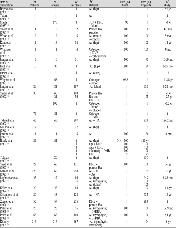
1980년부터 1999년까지 상악동 거상술에 대해 보고한 Medline 을 검색하였다. 문헌에서 보고한 이식재의 유형에 대한 개요는 Table 1에서 보여주고 있다. Medline 검색도구는 United States National Library of Medicine이였으며, 검색시 사용한 주요단어 (keyword)는 sinus augmentation이었다.
Table 1.Types of Graft : Sinus inlay A. Block
1. Nonvascularized Iliac
Calvarium Rib
Mandible : symphysis Maxilla : tuberosity Source unknown B. Particulate
1. Autogenous Iliac Tibia
Mandible : ramus and coronoid process 2. Alloplastic plus allogenic : HA + DFDB 3. Autogenous plus allogenic : iliac + DFDB 4. Autogenous plus alloplastic : iliac + HA,
Source unknown + HA
임프란트 식립을 위한 상악동점막 거상술: 문헌고찰
김수관∙강태호∙박정열
조선대학교 치과대학 구강악안면외과학교실, 구강생물학연구소
Abstract
MAXILLARY SINUS GRAFT FOR ENDOSSEOUS IMPLANT PLACEMENT : REVIEW OF THE LITERATURES
Su-Gwan Kim, Soo-Min Kim, In-Soon Park
Department of Oral & Maxillofacial Surgery, Oral Biology Research Institute, College of Dentistry, Chosun University
A review of the literature, provided by a MEDLINE search from 1980 through June 1999, was performed. This study was screened that 649 patients received 679 sinus lift grafts in which 2056 implants were placed. The types of grafts in sinus augmentation were auto- genous bone, corticocancellous block bone, allogenic bone, and a variety of alloplastic materials. Results of these grafts are presented.
The most frequent complications was the infection of maxillary sinus. Long-term follow-up is necessary to advance the sinus elevation and to support posterior maxillary restorations.
Key words: MEDLINE, Sinus augmentation, Long-term follow-up
김 수 관
501-717, 광주광역시 동구 서석동 588 조선대학교 치과대학 구강악안면외과학교실 Su-Gwan Kim
Dept. of OMFS, College of Dentistry, Chosun University 588, Seosuk-Dong, Dong-Ku, Kwongju-City, 501-717, Korea Tel. 82-62-220-3601 Fax. 82-62-224-9172
※ 본 연구는 보건복지부 보건의료기술연구개발사업의 지원에 의하여 이루어진 것임. (HMP-00-B-20507-0191)
Table 2.Longitudinal reports on sinus elevation
Boyne et al 11 ? ? Au (hip) ? ? ?-4 yr
(1980)27)
Tatum ? ? ? Au ? ? ?
(1986)28)
Misch ? 170 ? TCP + DMB 98 ? ?-4 yr
(1987)29) + blood
Smiler et al 4 5 12 Porous HA 100 100 4-6 mo
(1987)30) particles
Wood et al 2 2 5 Au (ramus, 100 100 6 mo
(1988)31) coronoid)
Kent et al 11 18 54 Au (hip) 100 100 1-4 yr
(1989)32)
Whittaker 1 1 4 Osteogen 100 100 8 mo
et al + DMB
(1989)33) + cortical bone
Jensen et al 5 10 51 Au (hip) 100 75 10-29 mo
(1990)34)
Hall et al 15 30 ? Au (hip) 100 90 1-26 mo
(1991)35)
Hirsch et al ? ? ? Au (chin) ? ? ?
(1991)36)
Wagner et al ? 63 ? Osteogen 96.8 ? ?-3.5 yr
(1991)37) + blood
Jensen et al 26 31 107 Au (chin) ? 93.5 6-32 mo
(1991)38)
Smiler et al 36 66 198 Porous HA ? ? ?-6 yr
(1992)39) ? 21 56 Bio-oss + ? 95 ?-2.5 yr
DMB 3:1
? 106 ? Osteogen ? ? ?-4.5 yr
+ blood + collagen
72 81 ? Osteogen ? ? ?
+ DMB
Tidwell et al 48 48 267 Au + HA ? 93.6 12-32 mo
(1992)40)
Loukota et al 7 ? 27 Au (hip) ? ? ?
(1992)41)
Jensen et al 1 1 5 Al 100 80 18 mo
(1992)42)
Block et al 32 51 ? Au (hip) 94.4 100 1-10 yr
(1993)43) ? hip + DMB 100 100
? chin + DMB 100 100
? tuberosity + DMB 100 100
? DMB 75 75
Tolman ? 20 ? Au (hip) ? 93.1 ?
(1993)44)
Small et al 27 45 111 DMB + 100 100 1-5 yr
(1993)45) porous HA
Lozada et al 120 69 298 Au + Al ? 85 1-5 yr
(1993)46) + Ap
Raghoebar et al 25 47 86 Au (hip) ? 94.2 6-36 mo
(1993)47) 6 Au (symphysis) ? 100
1 Au (tuber) ? 100
Keller et al 20 23 83 Au (hip) ? 92 1-6 yr
(1994)48)
Chiapasco et al 30 43 124 Au + HA ? 93.5 1-2 yr
(1994)49)
Zinner et al 50 57 215 DMB + ? 98.6 ?
(1996)50) porous HA
Peleg et al 20 20 55 Au (symphysis) 100 100 15-39 mo
(1998)51) + DFDBA
Peleg et al 63 63 160 Au (symphysis) 100 100 2-4 yr
(1999)52) + DFDBA
Khoury 216 216 467 Au (symphysis, ? 94 6 yr
(1999)53) retromolar)
Note, Au : Autogenous bone grafts, Al : Allogenic bone, Ap : Alloplastic materials
Osteogen : HA resorb, DMB : Demineralized bone, DFDBA : Demineralized freeze-dried bone allograft, tuber : Maxillary tuberosity, ? : unknown, yr : years, mo : months
Author Number Number Number Type Success Success Length
Date of of of of of Rate (%) Rate (%) of
publication Patients Sinuses Implants Material Graft Implants study
Ⅲ. 연구결과
총 84개의 문헌을 검색하였는데, 이 중 52개는 임상적인 면에, 9개는 기초적인 과학을 시행하였으며, 나머지 23개는 어느 쪽도 특별하게 관련되지 않았다.
총 649명의 환자에게 679개의 골이식술을, 2056개의 임프란트 를 식립하였다. 이들 임프란트 중 131개가 실패하여 93.6%의 성 공율을 보였으며, 추적기간은 0�10년까지 다양하였다. 비록 대 부분의 보고가 장기적인 추적검사는 시행하지 않았지만 Table 2 에 제시된 바와 같이 대부분은 우수한 성공율을 보였다.
Ⅳ. 총괄 및 고찰
무치악 상태의 상악 구치부는 치조제와 상악동 기저부의 양 방 향에서 생리적인 골흡수가 진행되므로 다른 부위와 비교하였을 때 골량이 절대적으로 부족한 경우가 많다. 또한 이 부위는 하악 골에 비교하여 골질이 현저하게 약하므로 임프란트가 식립될 수 있는 유효 골높이가 부족하면 통상적인 방법으로 임프란트를 성 공시키기 어려우므로 골이식술을 통해 임프란트의 식립이 가능 하도록 해야 한다54).
건전한 상악동 점막은 면역학적으로 항상성을 유지하며, 점액 등으로 덮히며, 항체도 분비되어 세균의 발육을 억제하고, 직모 운동에 의해 상악동을 청결하게 유지한다. 상악동 점막의 회복 기능도 빠르므로 상악동 골이식술 후 빠르게 치유된다24).
여러 종류의 골이식재와 이물성형재료(alloplastic material)들을 사용한 여러가지의 외과적 술식들이 추천되고 있다12-23). 상악동 점막 거상술은 상악 후방 구치부의 임프란트를 식립할 때 많이 사용하고 있는 술식으로, Boyne와 James27)가 처음 소개하였고, 그 이후 Tatum28), Wood와 Moore31)에 의해 변형되어 사용되고 있다.
많은 문헌들에서 이 술식을 지지하고,높은 성공율을 보고하고 있다55).
기존에는 상악동점막 거상술을 측방 접근법으로 시행하였다
43,56,57). 협측 피판은 상방으로 젖혀지고, 상악 측벽은 상악동 점막
쪽으로 회전시킨다. 이 때 수술전에 상악동에 질병이 있는 가를 알아내는 것이 중요하다. 상악동 기저부에 있는 중격(septa)의 존 재는 이러한 수술을 하는 동안 합병증을 유발할 수도 있다58).
술자가 상악 측벽에 window를 형성하고 hinge door를 거상하 는 동안 몇 개의 중격을 포함하고 있는 alveolar recess로부터 상악 동 점막을 올릴 때 점막을 손상시킬 위험성이 있다27,59). 상악동 점 막의 천공은 가능하며, 이 때 크기가 작다면 치료를 시행하지 않 거나 5-0 Vicryl (Ethicon, Norderstedt, Germany)로 봉합을 하며, 크 기가 크다면 천공부위를 폐쇄하기 위해 흡수성 재료인 콜라겐 막 등을 사용하여 폐쇄한다43,53).
더우기 중격은 상악동저의 시야를 방해할 수 있으며, 골이식재 들의 위치를 방해하여 상악동 기저부의 적절한 충전(filling)을 방 해한다. Orthopantomogram이 중격을 명확하게 탐지할 수 없으므 로 수술전 단층사진(computed tomogram)이 유용한 것처럼 보인 다59,60).
수술 전에 골의 높이는 골유착(osseointegration)을 유지한 다음 에도 임프란트의 성패에 영향를 미치는 중요한 인자이다. 현재 임프란트의 탈락은 골고경이 원래 작은 경우에 일어나기 쉬우며 낮은 골고경시에서는 상악동저 골이식을 시행해야 한다24). Misch29)는 측방 접근법을 통한 상악동점막 거상술의 적응증으로 필요로 하는 임프란트 부위에서 상악동 하방에 있는 이용가능한 골의 높이 (< 8mm)를 기준으로 하였다. 임프란트를 식립하기에 충분한 10�12mm의 골이 있다면 이식과 동시에 임프란트를 식 립할 수 있다. 10mm 이하의 골이 있다면 상악동점막 거상술과 이식을 시행한 후 6�9개월의 치유기간을 부여한 후 임프란트를
식립한다11,58).
상악동점막 거상술의 금기증에는 급성 상악동염, 낭종, 종양, 치근이 상악동내에 잔존하는 경우 등이 있다11,39). 또한 심한 흡연 자도 이 술식을 시행할 때 주의를 요한다24).
Jensen 등61)은 3mm 미만의 기존의 골을 가진 경우에 성공률이 매우 낮았으며, 7�9mm의 골이 있는 경우 이식을 시행하였을 때 결과가 가장 좋았다고 보고하였다. 2�4개의 15mm 길이의 implant body가 상악동내에 식립될 수 있으며, 이는 상악동의 크 기에 따라 식립된다. Wheeler 등62)은 상악동 골이식술후 가장 이 상적인 임프란트 길이는 13mm라고 추천하였다.
임프란트를 골이식과 동시에 식립해야 하는지 2차적으로 식립 해야 하는지는 명확하지 않으나, 대개는 치조골의 흡수 및 퇴축 의 정도와 임프란트의 primary stabilization에 따라 시행한다. 2차 적 이식에서는 식립 전에 견고한 기초가 되어 있고, 적절한 식립 부위를 선택하는 것도 가능하며, 확실한 임프란트의 고정도 얻 을 수 있고, 이식골의 생검도 가능하다. 또 즉시 식립하려고 하였 으나 상악동 점막이 찢어지거나, 골질이 좋지 않아 중지한 경우 에 또 한번 이식하는 경우에도 유용하다. 이상적인 위치에 식립 하고, 방법이 쉬우며, 보다 안전하다. 한편 즉시 이식은 1회의 수 술로 끝나고, 침습과 비용, 시간이 적게 걸린다. 2차적 이식과 즉 시 이식의 성공율은 통계적으로 유의차는 없고, 또 증례에 따라 어느 방법이 좋은가도 결론내릴 수 없다24).. Blomqvist 등63)은 1차 또는 2차 상악동 골이식술후 임프란트의 각도와 위치에 대한 비 교를 하였으며, 2차 수술시 임프란트의 적절한 위치를 제공하기 위한 보다 좋은 조건들을 제공하였다.
흔히 임프란트의 노출은 식립후 6�9개월 뒤에 시행한다12). 2차 수술시 provisional prosthesis의 장기간 사용을 추천한다. provi- sional prosthesis는 임상가에게 transition phase동안 계획된 최종 보철물의 설계를 변경할 수 있는 기회를 제공하고, 최종 보철물 의 template로 작용하며, composite alloplastic graft의 성숙을 위한 시간을 제공한다12,14).
임프란트의 설계에도 불구하고 잘 맞는 임프란트가 식립되고 보철물에 대한 lateral torque가 최소화되어야 한다50).
이식을 위한 기준(criteria)에는 상악동내에서 골을 잘 형성하 고, 골이식술과 동시에 임프란트를 위치시킨 후 안정성, 낮은 감 염의 위험성, 쉬운 이용 가능성, 낮은 항원성, 높은 신빙성(relia- bility) 등이 있다43,64).
선택되는 이식재는 초기에 임프란트를 안정시키고 골유착을
촉진시킬 수 있는 적절한 생존가능한 골(viable bone)을 제공해야 한다. 이식골의 활력은 임프란트를 길게 유지시키기 위해서 중 요하고, 이식골이 골유착을 유지하며, 약간의 손상에 대해 골유 착을 수복할 수 있을 정도로 충분한 골의 활성이 필요하다24). Momtaheni 등65)은 상악동내에 cortical cancellous strut를 사용하여 자가해면골 이식재(autogenous cancellous graft)를 고정하는 술식 을 제안하였다. 이 strut들은 해면골(cancellous bone) 상방에 위치 되었고 titanium screw를 사용하여 buttress와 anterior sinus에서 견 고하게 고정하였다.
많은 다른 이식재들이 상악동 골이식술에 사용되고 있다13,30,66-
74). 환자로부터 채취한 자가골이 가장 이상적이며,다른 이식재 들과 비교하였을 때 표준(standard) 역할을 한다. 자가골은 골결 손부에 최적의 이식재료로 동종골보다도 큰 가능성을 가지고 있 지만, 치유기간 중에 부하가 가해지면, 개조(remodeling)하고 있 는 이식골과의 골유착이 파괴된다24). 자가골은 더 빠른 골형성과 개조와 더불어 높은 acceptibility, 증가된 크기와 골밀도를 가진 다. 그러나 자가골은 제2의 외과적 술식이 필요하다는 단점이 있 다. 공여부는 장골능, 하악지, 상악결절, 하악 정중부 (Table 1) 등 에서 분말이나 조각, 절편 등의 다양한 형태로 사용할 수 있다
13,27,31,35,41,42,47,74-76). 대부분의 외과의사들은 2mm 미만의 잔존 치조제
가 존재하는 경우 이식재로 자가골을 추천하고 있다67).
동종골을 상악동내에 이식한 경우에는 형성되는 골은 제한되 며, 상악동저에 가까운 부위만 형성된다. 게다가 충분한 경도를 갖지 않으며, 반흔조직이 많고, 상악동저에서 떨어지면 골의 활 력은 적어진다24).
자가골과 demineralized bone을 1:1의 비로 혼합하여 사용하 므로써 이식재의 용적을 증가시키고 transplanted cell의 밀도를 증가시킨다. Synergistic response는 한 가지의 이식재를 사용하는 것보다 더 많은 골형성을 가져온다43,75).
이물성형재료들이 상악동저에 골형성을 위해 사용되고 있다.
이물성형재료는 2번째 수술부위가 없으며 흡수가 적고 이용하 기가 쉽다는 장점이 있으며, 단점으로는 압출(extrusion)과 감염 의 위험성이 있다43,67).
Wagner 등37)은 상악동거상을 위해 신선한 정맥혈과 흡수성 hydroxyapatite (Osteogen)를 사용하였다. 상악동점막 거상술과 골이식술을 시행한 63증례중 2증례에서만 실패하였다. 이 실패 는 provisional prostheses에 의해 임프란트에 premature loading를 가한 결과로 발생하였다.
Nishibori 등13)은 상악동점막 거상술후 demineralized freeze-dried bone (DFDB)을 이식한 군과 자가골(장골)을 이식한 군으로 나누 어 연구하였다. 자가골을 이식한 군에서는 임프란트 식립을 위 한 적절한 양과 질을 가진 골형성을 보인 반면 DFDB을 이식한 군에서는 임프란트 식립을 위해 필요한 양과 질이 불충분하였으 며 개조도 완전하게 되지 않았다. freeze-dried bone을 이식한 경 우에는 이식재의 성숙(maturation)을 위해 12개월 이상의 기간이 필요하다62).
Jensen 등61)은 lateral window에 Gore-tex barrier를 사용하여 상악 동점막 거상술후 이식과 임프란트의 성공에 기여하였다고 보고
하였다.
상악동점막 거상술에 대해 보고한 문헌들27-53)에서 이식재의 성 공율은 Block 등43)이 demineralized bone을 사용하여 75%의 성공 율을 보인 경우를 제외하고 대부분 95% 이상의 성공율을 보였으 며, 임프란트의 성공율은 Jensen 등34,42), Block 등43)이 demineralized bone을 사용한 경우, Lozada 등46)을 제외하고는 90% 이상의 성공 율을 보였다. 이와 같이 상악동점막 거상술은 퇴축된 상악 무치 악의 임프란트 식립을 위해 사용하였을 때 성공율이 높은 술식 이다.
상악동점막 거상술후 이병율(morbidity)은 수술의 형태, 이식이 필요한 치조제의 높이에 달려있다. 수술 후에 발생가능한 합병 증들에는 상악동의 감염, 골소실, hemosinus, 구강상악동루 (oroantral fistula), incisional breakdown, 수술 후 상악에 발생하는 낭종 등이 있다12,28,29,58,77-83). 상악동 감염의 임상 징후(sign)들에는 안 면부 동통, 종창, 압통, 절개선을 따라 배농, 화농성 비분비(dis- charge) 등이 있다.
상악동점막 거상술후 발생가능한 합병증에는 상악동 점막이 찢어지거나 이식재의 감염, 임프란트의 탈락 등이 있으며, 장기 간에 미치는 상악동의 합병증은 드물다. 이 중 가장 흔한 합병증 은 상악동 수술 후에 발생한 감염이며, 대부분의 환자들은 항생 제, 항히스타민제와 충혈제거제(decongestant) 치료, 적절한 영양 등으로 치료하였다. 항생제는 보통 수술 전 하루 전에 시작하여 하루에 3번씩 수술후 1주일동안 투여한다84).. 상악동에 염증이 발 생한 경우에 가장 적절한 항생제로는 amoxicillin (Amoxil), clin- damycin (Cleocin), metronidazole 등이 있다12,37,58,79,84).
문헌에 의하면 상악동 골이식술후 상악동염의 발생은 0�20%
에서 나타난다고 보고되고 있다40,78,85-87). Misch29)는 구강상악동루가 2증례에서 발생하였다고 보고하였으며, Small 등45)은 수술후 감 염이 27명의 환자들 중 2명에서 발생하였으며, 2명 모두 흡연자 들이었다.
상악동 골이식술을 시행받은 모든 환자의 수술후 지시사항으 로는 금연하도록 하고, 그들의 코를 불지 않도록 한다43).
생체역학적 요인과 더불어 생리적 요인도 상악동 골이식술의 성패에 관계된다. 흡연은 병태생리학적으로 보아 중요한 요인이 며, 상악동 골이식술에 유해하다. 흡연자는 비흡연자에 비해 구 치부에서 임프란트의 실패율이 높게 된다. 그러므로 흡연은 상 악동 골이식술을 결정할 때 중요한 생리적 위험인자로 생각된다
24). 흡연은 골이식의 치유도 저하한다. 흡연은 혈류를 저해하고, 혈소판의 응집력을 높인다. 흡연에 의해 생산된 화학물질들(시 안화수소와 일산화탄소)은 창상치유를 지연시키고, 니코틴은 세 포 증식을 저해한다. 또한 흡연은 골아세포의 기능을 저해하고, 골형성능을 약화시킨다. 흡연자에서는 골에 포함된 무기질이 적 고, 남성에서 10�20%, 여성에서 15�30% 낮다. 장기간동안 흡연 한 사람은 골에 포함된 무기질량이 보통 사람보다 2�6배 낮다24).
만일 흡연자가 환자로서 상악동 골이식술을 원한다면 금연을 할 계획을 세우고, 수술 전부터 흡연의 영향을 줄인다. 그리고 골 이식술을 시행받은 몇 주간은 금연하고, 이식골이 생착하기까지 는 금연을 계속하도록 교육시킨다24).
상악동 골이식술을 시행받은 환자는 치유기간동안 가급적 부 하가 가해지지 않도록 하는 것이 필요하다24).
Ⅴ. 결 론
상악동점막 거상술은 무치악의 퇴축된 상악의 임프란트 식립 을 위해 사용되고 있으며, 그 합병증은 비교적 적고 성공율은 높 은 것으로 나타났다. 많은 정보들을 제공한 문헌 고찰에서, 특히 자가골과 탈회 또는 비탈회 동결건조골이 함께 사용된 경우 더 욱 많은 정보가 필요한 것으로 생각되었다. 또한 상악동점막 거 상술의 술식을 진보시키고 상악 후방부 보철물을 지지할 수 있 도록 장기적이면서도 보다 자세한 추적연구가 필요하리라 사료 된다.
참 고 문 헌
1. Branemark PI, Albrektsson RA, Lekholm U, et al : An experimental and clinical study of osseointegrated penetrating the nasal cavity and maxillary sinus. J Oral Maxillofac Surg 42:497, 1984.
2. Barber HD, Betts NJ, Edwards ML : The status of implant training in oral and maxillofacial surgery residency programs. 52:1058, 1994.
3. Krekmanov L : A modified method of simultaneous bone grafting and placement of endosseous implants in the severely atrophic maxilla. Int J Oral Maxillofac Implants 10:682, 1995.
4. Smith DE, Zarb GA : Criteria for success of osseointegrated endosseous implants. J Prosthet Dent 62:567, 1989.
5. Saadoun AP, Le Gall MG : Implant site preparation with osteotomes:
principles and clinical application. Pract Periodontics Aesthet Dent 8:453, 1996.
6. Summers RB : The osteotome technique: Part 4-Future site develop- ment. Compend Contin Educ Dent 16:1080, 1995.
7. Summers RB : The osteotome technique: Part 3-Less invasive meth- ods of elevating the sinus floor. Compendium 15:698, 1994.
8. Summers RB : A new concept in maxillary implant surgery: the osteotome technique. Compendium 15:152, 1994.
9. Coatoam GW, Krieger JT : A four-year study examining the results of indirect sinus augmentation procedures. J Oral Implantol 23:117, 1997.
10. Coatoam GW : Indirect sinus augmentation procedures using one- stage anatomically shaped root-form implants. J Oral Implantol 23:25, 1997.
11. 여환호, 김수관 : 구강악안면 영역의 외과적 술식. 한서의학사, 1999.
12. Zinner ID, Small SA, Panno FV, et al : Provisional and definitive prostheses following sinus lift and augmentation procedures.
Implant Dent 3:24, 1994.
13. Nishibori M, Betts NJ, Salama H, et al : Short-term healing of auto- genous and allogeneic bone grafts after sinus augmentation: a report of 2 cases. J Periodontol 65:958, 1994.
14. Coatoam GW, Krieger JT : A four-year study examing the results of indirect sinus augmentation procedures. J Oral Implantol 23:117, 1997.
15. Neyt LF, De Clercq CA, Abeloos JV, et al : Reconstruction of the severely resorbed maxilla with a combination of sinus augmenta- tion, onlay bone grafting, and implants. J Oral Maxillofac Surg 55:1397, 1997.
16. Nevins M, Kirker-Head C, Nevins M, et al : Bone formation in the goat maxillary sinus induced by absorbable collagen sponge implants impregnated with recombinant human bone morpho- genetic protein-2. Int J Periodontics Restorative Dent 16:8, 1996.
17. Wallace SS, Froum SJ, Tarnow DP : Histologic evaluation of a sinus elevation procedure: a clinical report. Int J Periodontics Restorative Dent 16:46, 1996.
18. Rinaldi M, Mottola A : Pianificazione ed intervento di rialzo bilat- erale dei pavimenti sinusali nella chirurgia implantologica.
Presentazione di un caso. Minerva Stomatol 43:179, 1994.
19. Boskovic MM : Maxillary sinus floor elevation and subantral aug- mentation for dental implants. Impressions 12:16, 1990.
20. Feigel A, Makek M : The significance of sinus elevation for blade implantology: report of an autopsy case. J Oral Implantol 15:237, 1989.
21. Tong DC, Rioux K, Drangsholt M, et al : A review of survival rates for implants placed in grafted maxillary sinuses using meta-analy- sis. Int J Oral Maxillofac Implants 13:175, 1998.
22. Knabe C, Hoffmeister B : The use of implant-supported ceramomet- al titanium prostheses following sinus lift and augmentation proce- dures: a clinical report. Int J Oral Maxillofac Implants 13:102, 1998.
23. Bruschi GB, Scipioni A, Calesini G, et al : Localized management of sinus floor with simultaneous implant placement: a clinical report.
Int J Oral Maxillofac Implants 13:219, 1998.
24. 김현철 : Sinus lift의 consensus. 한국퀸테센스저널 1:26-48, 1999.
25. Block MS, Kent JN, Kallukaran FU, et al : Bone maintenance 5 to 10 years after sinus grafting. J Oral Maxillofac Surg 56:706, 1998.
26. Takahashi T, Fukuda M, Yamaguchi T, et al : Use of endosseous implants for dental reconstruction of patients with grafted alveolar clefts. J Oral Maxillofac Surg 55:576, 1997.
27. Boyne PJ, James RA, Linda L : Grafting of the maxillary sinus floor with autogenous marrow and bone. J Oral Surg 38:613, 1980.
28. Tatum H : Maxillary and sinus implant reconstructions. Dent Clinics North Am 30:207, 1986.
29. Misch CE : Maxillary sinus augmentation for endosteal implants:
organized alternative treatment plans. Int J Oral Implantol 4:49, 1987.
30. Smiler DG, Holmes RE : Sinus lift procedure using porous hydrox- yapatite: a prelimianry clinical report. J Oral Implantol 13:239, 1987.
31. Wood RM, Moore DL : Grafting of the maxillary sinus with intraoral- ly harvested autogenous bone prior to implant placement. Int J Oral Maxillofac Implants 3:209, 1988.
32. Kent JN, Block MS : Simultaneous maxillary sinus floor bone graft- ing and placement of hydroxyapatite-coated implants. J Oral Maxillofac Surg 47:238, 1989.
33. Whittaker JM, James RA, Lozada J, et al : Histological response and clinical evaluation of heterograft and allograft materials in the ele- vation of the maxillary sinus for the preparation of endosteal dental implant sites. Simultaneous sinus elevation and root form implanta- tion: an eight-month autopsy report. J Oral Implantol 15:141, 1989.
34. Jensen J, Simonsen K, Sindet-Pedersen S : Reconstruction of the severely atrophied maxilla with bone grafting and osseointegrated implants: a prelimianry report. J Oral Maxillofac Surg 48:27, 1990.
35. Hall HD, McKenna SJ : Bone graft of the maxillary sinus floor for Branemark implants: a prelimianry report. Oral Maxillofac Surg Clinics North America 3:869, 1991.
36. Hirsch JM, Ericsson I : Maxillary sinus augmentation using mandibu- lar bone grafts and simultaneously installation of implants. Clin Oral Implants Res 2:91, 1991.
37. Wagner JR : A 3½-year clinical evaluation of resorbable hydroxyap- atite osteogen(HA resorb) used for sinus lift augmentations in con- junction with the insertion of endosseous implants. J Oral Implantol 17:152, 1991.
38. Jensen J, Sindet-Pedersen S : Autogenous mandibular bone grafts and osseointegrated implants for reconstruction of the severely atrophied maxilla: a prelimianry report. J Oral Maxillofac Surg 49:1277, 1991.
39. Smiler DG, Johnson PW, Lozada JL, et al : Sinus lift grafts and endosseous implants. Dent Clinics North America 36:151, 1992.
40. Tidwell JK, Blijdorp PA, Stoelinga PJW, et al : Composite grafting of the maxillary sinus for placement of endosteal implants: a prelimi- nary report of 48 patients. Int J Oral Maxillofac Surg 21:204, 1992.
41. Loukota RA, Isaksson SG, Linner ELJ, et al : A technique for insert- ing endosseous implants in the atrophic maxilla in a single stage procedure. British J Oral Maxillofac Surg 30:46, 1992.
42. Jensen OT, Perkins S, Van De Water FW : Nasal fossa and maxillary sinus grafting of implants from a palatal approach: report of a case.
J Oral Maxillofac Surg 50:415, 1992.
43. Block MS, Kent JN : Maxillary sinus grafting for totally and partially edentulous patients. JADA 124:139, 1993.
44. Tolman DE : Advanced residual ridge resorption: surgical manage- ment. Int J Prosthodontics 6:118, 1993.
45. Small SA, Zinner ID, Panno FV, et al : Augmenting the maxillary sinus for implants: report of 27 patients. Int J Oral Maxillofac Implants 8:523, 1993.
46. Lozada JL, Emanuelli S, James RA, et al : Root-form implants placed in subantral grafted sites. CDA J 21:31, 1993.
47. Raghoebar GM, Brouwer TJ, Reintsema H, et al : Augmentation of the maxillary sinus floor with autogenous bone for the placement of endosseous implants: a preliminary report. J Oral Maxillofac Surg 51:1198, 1993.
48. Keller EE, Eckert SE, Tolman DE : Maxillary antral and nasal one- stage inlay composite bone graft: preliminary report on 30 recipient sites. J Oral Maxillofac Surg 52:438, 1994.
49. Chiapasco M, Ronchi P : Sinus lift and endosseous implants - pre- limianry surgical and prosthetic results. Eur J Prosthodont Rest Dent 3:15, 1994.
50. Zinner ID, Small SA : Sinus-lift graft: using the maxillary sinuses to support implants. JADA 127:51, 1996.
51. Peleg M, Mazor Z, Chaushu G, et al : Sinus floor augmentation with simultaneous implant placement in the severely atrophic maxilla. J Periodontol 69:1397-1403, 1998.
52. Peleg M, Mazor Z, Garg AK : Augmentation grafting of the maxillary sinus and simultaneous implant placement in patients with 3 to 5 mm of residual alveolar bone height. Int J Oral Maxillofac Implants 14:549-556, 1999.
53. Khoury F : Augmentation of the sinus floor with mandibular bone block and simultaneous implantation: a 6-year clinical investigation.
Int J Oral Maxillofac Implants 14:557-564, 1999.
54. 최장우 : 상악동저 거상술-골이식을 포함하는 개념. 치과임상
18:66-68, 1998.
55. Fugazzotto PA : Maxillary sinus grafting with and without simultane- ous implant placement: technical considerations and case reports.
Int J Periodont Rest Dent 14:545, 1994.
56. Zitzmann NU, Scharer P : Sinus elevation procedures in the resorbed posterior maxilla. Comparison of the crestal and lateral approaches. Oral Surg Oral Med Oral Pathol Oral Radiol Endod 85:8, 1998.
57. Babbush CA : Sinus lift revisited: an update on current implant-relat- ed procedures [interview]. Dent Implantol 9:1, 1998.
58. Misch CE : Contemporary implant dentistry. Mosby 1999.
59. Ulm CW, Solar P, Krennmair G, et al : Incidence and suggested sur- gical management of septa in sinus-lift procedures. Int J Oral Maxillofac Implants 10:462, 1995.
60. Betts NJ : Modification of the sinus lift procedure for septa in the maxillary antrum. J Oral Maxillofac Surg 52:332, 1994.
61. Jensen OT, Greer R : Immediate placement of osseointegrated implants into the maxillary sinus with mineralized cancellous allo- graft and Gore-tex: second stage surgical and histological findings.
Quintessence, Chicago. 321-332, 1991.
62. Wheeler SL, Holmes RE, Calhoun CJ : Six-year clinical and histologic study of sinus-lift grafts. Int J Oral Maxillofac Implants 11:26, 1996.
63. Blomqvist JE, Alberius P, Isaksson S : Sinus inlay bone augmenta- tion: comparison of implant positioning after one- or two-staged procedures. J Oral Maxillofac Surg 55:804, 1997.
64. Choung PH, Choung YH : Vascularized bone flap for access the maxillary sinus. J Oral Maxillofac Surg 55:832, 1997.
65. Momtaheni DM, Schweitzer K, Muenchinger F : Technique for stabi- lization of autogenous cancellous bone grafts in sinus lift proce-
dures. Oral Surg Oral Med Oral Pathol 78:14, 1994.
66. Moy PK, Lundgren S, Holmes RE : Maxillary sinus augmentation:
histomorphometric analysis of graft materials for maxillary sinus floor augmentation. J Oral Maxillofac Surg 51:857, 1993.
67. Wheeler SL : Sinus augmentation for dental implants: the use of alloplastic materials. J Oral Maxillofac Surg 55:1287, 1997.
68. Furusawa T, Mizunuma K : Osteoconductive properties and efficacy of resorbable bioactive glass as a bone-grafting material. Implant Dent 6:93, 1997.
69. Lorenzetti M, Mozzati M, Campanino PP, et al : Bone augmentation of the inferior floor of the maxillary sinus with autogenous bone or composite bone grafts: a histologic-histomorphometric preliminary report. Int J Oral Maxillofac Implants 13:69, 1998.
70. Wetzel AC, Stich H, Caffesse RG : Bone apposition onto oral implants in the sinus area filled with different grafting materials. A histological study in beagle dogs. Clin Oral Implants Res 6:155, 1995.
71. Suba Z, Szabo G, Haris A, et al : Tapasztalatok a HTR polymer klinikai alkalmazasaval. Sinus elevatio, human szovettani vizsgala- tok. Fogorv Sz, 84:75, 1991.
72. Hurzeler MB, Quinones CR, Kirsch A, et al : Maxillary sinus aug- mentation using different grafting materials and dental implants in monkeys. Part III. Evaluation of autogenous bone combined with porous hydroxyapatite. Clin Oral Implants Res 8:401, 1997.
73. Valentini P, Abensur D : Maxillary sinus floor elevation for implant placement with demineralized freeze-dried bone and bovine bone (Bio-Oss): a clinical study of 20 patients. Int J Periodontics Restorative Dent 17:232, 1997.
74. 이성재, 장현석, 이부규, 권종진, 임재석 : 임프란트 식립을 위한 상
악동 거상술의 임상적 연구. 대한악안면성형재건외과학회지 21:376-381, 1999.
75. Block MS, Kent JN : Sinus augmentation for dental implants: the use of autogenous bone. J Oral Maxillofac Surg 55:1281, 1997.
76. Dario LJ, English RJ : Chin bone harvesting for autogenous grafting in the maxillary sinus: a clinical report. Pract Periodontics Aesthet Dent 6:87, 1994.
77. Lazzara RJ : The sinus elevation procedure in endosseous implant therapy. Periodontology 3:178, 1996.
78. Chanavaz M : Maxillary sinusitis: anatomy, physiology, surgery, and bone grafting related to implantology: eleven years of surgical experience (1979-1990). J Oral Implantol 16:199, 1990.
79. Misch CE : Treatment planning for edentulous maxillary posterior region. In Contemporary Implant Dentistry. Mosby-Year Book, St.
Louis. 241-255, 1993.
80. Misch CM, Misch CE, Resnik RR, et al : Post-operative maxillary cyst associated with a maxillary sinus elevation procedure: a case report. J Oral Implantol 17:432, 1991.
81. DeFreitas J, Lucente FE : The Caldwell-Luc procedure: institutional review of 670 cases: 1975-1985. Laryngoscope 98:1297, 1988.
82. Golden AL, Foote J, Lally E, et al : Dentigerous cyst of the maxillary sinus causing elevation of the orbital floor. Report of a case. Oral Surg Oral Med Oral Pathol 52:133, 1981.
83. Moses JJ, Arredondo A : Sinus lift complications: avoiding problems and finding solutions [interview]. Dent Implantol 8:70, 1997.
84. Misch CE : The pharmacologic management of maxillary sinus ele- vation surgery. J Oral Implant 18:15, 1992.
85. Timmenga NM, Raghoebar GM, Boering G, et al : Maxillary sinus function after sinus lifts for the insertion of dental implants. J Oral Maxillofac Surg 55:936, 1997.
86. Quiney R, Brimble E, Hodge M : Maxillary sinusitis from dental osseointegrated implants. J Laryngol Otol 104:333, 1990.
87. Ueda M, Kaneda T : Maxillary sinusitis caused by dental implants:
report of two cases. J Oral Maxillofac Surg 50:285, 1992.