개 관절 윤활액 유래 중간엽 줄기세포의 특성과 분화능 분석
이정현, 이성림*
경상대학교 수의과대학 수의학과
Characterization and Differentiation of Synovial Fluid Derived Mesenchymal Stem Cells from Dog
Jeong-Hyeon Lee and Sung-Lim Lee
*College of Veterinary Medicine, Gyeongsang National University, Jinju 660-701, Republic of Korea
ABSTRACT
The synovial tissues are a valuable MSCs source for cartilage tissue engineering because these cells are easily obtainable by the intra-articular biopsy during diagnosis. In this study, we isolated and characterized the canine MSCs derived from synovial fluid of female and male donors.
Synovial fluid was flushed with saline solution from pre and post-puberty male (cM1-sMSC and cM2-sMSC) and female (cF1-sMSC and cF2-sMSC) dogs, and cells were isolated and cultured in advanced-DMEM (A-DMEM) supplemented with 10% FBS in a humidified 5% CO2 atmosphere at 38.5℃. The cells were evaluated for the ex- pression of the early transcriptional factors, such as Oct3/4, Nanog and Sox2 by RT-PCR. The cells were induced under conditions conductive for adipogenic, osteogenic, and chondrogenic lineages, then evaluated by specific staining (Oil red O, von Kossa, and Alcian Blue staining, respectively) and analyzed for lineage specific markers by RT-PCR.
All cell types were positive for alkaline phosphatase (AP) activity and early transcriptional factors (Oct3/4 and Sox2) were also positively detected. However, Nanog were not positively detected in all cells. Further, these MSCs were observed to differentiate into mesenchymal lineages, such as adipocytes (Oil red O staining), osteocytes (von Kossa staining), and chondrocytes (Alcian Blue staining) by cell specific staining. Lineage-specific genes (osteocyte;
osteonectin and Runx2, adipocytes; PRAR-γ2, FABP and LEP, and chondrocytes; collagen type-2 and Sox9) were also detected in all cells.
In this study, we successfully established synovial fluid derived mesenchymal stem cells from female and male dogs, and determined their basic biological properties and differentiation ability. These results suggested that synovial fluid is a valuable stem cell source for cartilage regeneration therapy, and it is easily accessible from osteoarthritic knee.
(Key words : dog, synovial fluid, mesenchymal stem cells, differentiation)
†
This study was supported by BioGreen 21, Rural Development Administration (grant No. 20110701-305-533-001-02-00) and National Research Foundation of Korea (grant No. 2011-0010252) of the Republic of Korea.
*
Correspondence : E-mail : [email protected]
서 론
최근 퇴행성 질환 및 사고, 질병 등으로 주요한 장기나 기 관의 기능이 상실된 경우, 줄기세포를 이용한 줄기세포 재생 치료기술을 접목한 근본적이고 영구적인 치료 가능성이 높아 지고 있다. 성체줄기세포를 이용한 세포재생 치료의 경우 재 생치료의 목적에 맞는 간단한 세포추출 방법의 개발과 효율 적인 분화능을 가진 줄기세포주를 구축이 필요하며, 이를 위 하여 사람뿐만 아니라 마우스, 랫트, 돼지, 소 등 다양한 종에 서 연구가 이루어지고 있다. 개의 경우, 이미 반세기 전에 골 수 유래 줄기세포를 성공적으로 이식(Thomas 등, 1999)한 대
표적인 줄기세포 재생 연구의 전임상 실험동물로써 다양한 줄기세포 연구에 활용이 가능하므로 다양한 in vitro와 in vivo 연구가 이루어져야 하지만 in vitro 연구는 다른 실험동물 종 에 비하여 미비한 실정이다.
중간엽 줄기세포는 골수(Friedenstein 등, 1970)에서 처음으 로 추출되고 구축된 후, 지방조직(Zuk 등, 2001)과 간, 신장, 폐, 비장, 뇌, 근육, 흉선, 췌장 등(Da Silva Meirelles 등, 2006) 다양한 장기로부터 중간엽 줄기세포를 추출할 수 있지만, 여 전히 골수유래 중간엽 줄기세포를 이용한 줄기세포치료 연구 가 활발하다. 줄기세포 치료 기술을 효율적으로 활용할 수 있 는 조직으로는 혈관의 분포가 거의 없고, 지지조직으로의 기
능을 하는 연골이 가장 적합한 것으로 알려져 있다. 그리고 중 간엽 줄기세포의 연골(Johnstone 등, 1998, Mackay 등, 2008) 과 뼈(Jaiswal 등, 1997)세포로 분화능을 이용함으로써 노령인 구의 증가에 따른 퇴행성 관절질환과 외상 등에 의한 손상 연 골조직의 재생 치료가 가능하다(Koga 등, 2007, 2008과 2009;
Horile 등 2009).
다양한 중간엽 줄기세포의 추출원 중에서 관절윤활막 유래 중간엽 줄기세포의 연골세포 분화능이 우수하다는 보고(Saka- guchi 2005; Yoshimura 2007; Koga 등, 2008)가 있다. 해부학 적 구조로써 관절막은 관절 내를 덮고 있는 얇은 막으로 관절 을 외과적 수술할 때 다량 추출이 가능하지만, 비외과적으로 추출이 어려워서 우수한 연골 분화능에도 불구하고 중간엽 줄기세포의 추출원으로 한계가 있다. 그러나 관절 윤활액은 관절강 내에 고여있는 액체로써 주사기로도 채취가 용이한 조 직으로 관절 윤활액을 세포 추출원으로 활용 가능할 경우, 관 절연골 손상 치료에 적용이 용이할 것이다. 연골세포 재생의 경우, 퇴행성으로 진행된 연골손상에 적용될 가능성이 높지만 노령의 개체로부터 채취된 중간엽 줄기세포의 증식능과 분화 능이 저조한 것으로 보고(Mueller 등, 2001; Stenderup 등, 2003) 되고 있어서 공여개체의 연령과 성별에 따른 연구가 필요하다.
따라서 본 연구에서는 줄기세포 자가 및 타인 이식 연구에 적용가능한 전임상 실험동물인 비글견을 이용하여 퇴행성 관 절염과 외상성 손상 등이 발생하였을 경우, 진단과 외과적 시술 을 하는 과정에서 쉽게 채취가능하고 반복적으로 채취가 가 능한 관절 윤활액 유래 중간엽 줄기세포를 구축하고자 하였다.
특히 퇴행성관절염의 경우 연령과 성별에 따른 발병차이가 인 정되므로 성성숙 전과 후의 비글견 암수에서 각각 추출된 관절 윤활액 유래 중간엽 줄기세포를 구축하고 특성을 비교하였다.
재료 및 방법
1. 관절 윤활액 유래 세포의 추출과 배양
본 연구는 6개월령 이전의 성성숙이 되지 않은 수컷(cM1- sMSC)과 암컷(cF1-sMSC) 비글견과 1년령 이상의 성성숙이 된 수컷(cM2-sMSC)과 암컷(cM2-sMSC) 비글견 총 4마리로 부터 세포를 구축하였으며, 경상대학교 동물실험 윤리기준에 따라 이 루어졌다. 공시동물의 대퇴경골관절(femoro-tibial joint)에 26 게이지 주사기를 이용하여 관절 윤활액을 채취하고, Dulbecco’s phosphate buffered saline(DPBS)를 이용하여 300×g로 10분간 원심분리하고, 10% FBS가 첨가된 advanced Dulbecco’s modified Eagle medium (ADMEM, GIBCO-BRL, Grand Island. USA) 에서 38.5℃, 5% CO2와 humidity 조건에서 배양하였다. 부착 배 양되지 않는 세포는 일차배양 2일 후 조심스럽게 제거하였으 며, 부착 배양된 confluent 세포는 0.25%(w/v) trypsin-EDTA으 로 처리 후 1 × 10⁴ cells/cm2로 계대배양하여 실험에 공시하였다.
2. 세포의 Alkaline Phosphatase (AP) Activity와 초기 전사 인자 발현 분석
배양된 관절 윤활액 유래 세포에서 중간엽 줄기세포의 특성 을 규명하기 위하여 AP activity를 BCIP/NBT(Promega, Madi- son, WI, USA)를 이용하여 염색하였으며, 초기 전사인자인 Oct-4, Sox-2, Nanog에 대한 발현을 확인하기 위하여 3-passage인 각 세포를 reverse transcription polymerase chain reaction(RT-PCR) 를 Maxime PCR Premix(iNtRON Biotechonology, Seoul, rea) 를 이용하여 시행하였다. Oct-4, Sox-2, Nanog 유전자의 pri- mer는 Table 1에 설명된 것과 같으며, GAPDH를 house-kee- ping gene으로 사용하였다. Total RNA는 RNeasy mini kit(Qia- gen, Hilden, Germany)로 추출하였으며 cDNA 합성은 Omni- scropt reverse transcription kit(Qiagen, Valencia, USA)를 이용 하여 37℃에서 60분간 시행하였다. PCR의 predenaturation은 94℃에서 10분간, denaturation은 94℃에서 1분간, annealing은 55~58℃에서 1분간, elongation은 72℃에서 1분간, extension 은 72℃에서 10분간 하는 과정을 30 cycle하였다. 그리고 1%
agarose gel과 1 ug/ml ethidium bromide에서 전기영동으로 분 리하여 확인하였다.
3. 분화 유도와 분화능 평가
구축된 관절 윤활액 유래 중간엽 줄기세포(cM1-sMSC, cF1- sMSC, cM2-sMSC 그리고 cM2-sMSC)를 골, 연골, 지방으로 분 화시키고, 각 세포유형으로 분화된 것으로 확인하기 위하여 세 포특이 염색과 RT-PCR로 표지자인 세포특이 유전자의 발현 을 분석하였다.
1) 골, 연골, 지방세포로 분화 유도
성성숙 전후의 암수 관절 윤활액 유래 중간엽 줄기세포는 3- passage까지 동일한 배양조건에서 계대배양하여 약 70% con- fluence에 도달하면 골, 연골 그리고 지방세포로 3주간 분화배 양액에서 배양하였다. 각 세포의 분화배양액은 골세포는 10%
FBS가 첨가된 ADMEM에 0.1 μM dexamethasone, 50 μM as- corbate-2-phosphate, 그리고 10 mM β-glycerol phosphate를 첨가하였으며, 지방세포는 10% FBS가 첨가된 ADMEM에 1 μM dexamethasone, 10 μM insulin, 200 μM indomethacin 그리 고 500 μM 3-isobutyl-1-methylxanthine(IBMX)를 첨가하였다.
연골세포 분화배양액은 chondrogenic medium(Hyclone, Logan, USA)에 10% FBS를 첨가하여 배양하였다.
2) 세포특이 염색과 유전자 발현 분석
3주간 분화 배양액에 배양한 세포의 각 세포로 분화를 확 인하기 위하여 세포학적 특성과 유전적 특성을 분석하였다. 골, 연골 그리고 지방 분화배양액에서 배양된 세포는 3.7% formal- dehyde로 실온에서 1시간 고정하고 각각 von Kossa (5% sil-
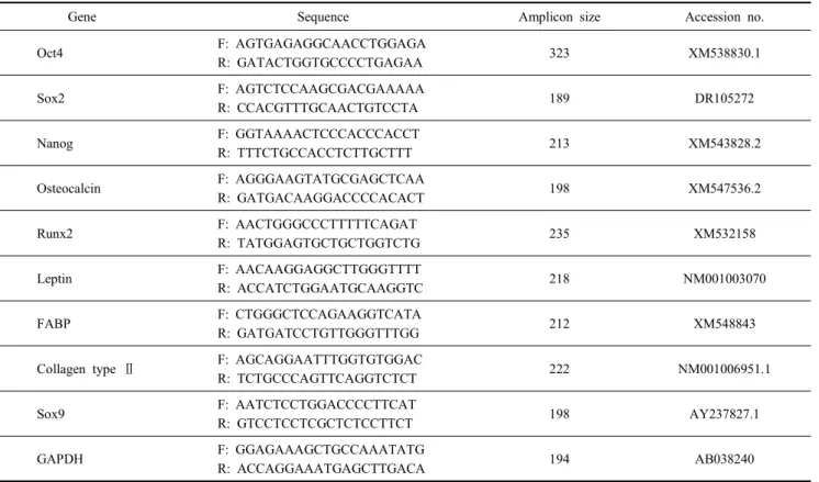
Table 1. Primers used for real-time PCR
Gene Sequence Amplicon size Accession no.
Oct4 F: AGTGAGAGGCAACCTGGAGA
R: GATACTGGTGCCCCTGAGAA 323 XM538830.1
Sox2 F: AGTCTCCAAGCGACGAAAAA
R: CCACGTTTGCAACTGTCCTA 189 DR105272
Nanog F: GGTAAAACTCCCACCCACCT
R: TTTCTGCCACCTCTTGCTTT 213 XM543828.2
Osteocalcin F: AGGGAAGTATGCGAGCTCAA
R: GATGACAAGGACCCCACACT 198 XM547536.2
Runx2 F: AACTGGGCCCTTTTTCAGAT
R: TATGGAGTGCTGCTGGTCTG 235 XM532158
Leptin F: AACAAGGAGGCTTGGGTTTT
R: ACCATCTGGAATGCAAGGTC 218 NM001003070
FABP F: CTGGGCTCCAGAAGGTCATA
R: GATGATCCTGTTGGGTTTGG 212 XM548843
Collagen type Ⅱ F: AGCAGGAATTTGGTGTGGAC
R: TCTGCCCAGTTCAGGTCTCT 222 NM001006951.1
Sox9 F: AATCTCCTGGACCCCTTCAT
R: GTCCTCCTCGCTCTCCTTCT 198 AY237827.1
GAPDH F: GGAGAAAGCTGCCAAATATG
R: ACCAGGAAATGAGCTTGACA 194 AB038240
ver nitrate) solution (Sigma-Aldrich, St. Louis, MO)과 1% Alcian blue 8GX solution (Sigma-Aldrich, St. Louis, MO)에서 30분 간 염색하여 골과 연골 기질염색을 확인하고 0.5% Oil red O solution (Sigma-Aldrich, St. Louis, MO)에서 1시간 염색하여 지방방울 염색을 확인하였다.
그리고 세포 특이 유전자를 RT-PCR를 이용하여 확인하였 으며, 각 유전자의 primer는 Table 1의 설명과 같다. 골 세포 분 화 유도된 세포는 Osteocalcin과 Runt-related transcription fac- tor 2(Runx2), 연골세포 분화 유도된 세포는 collagen type Ⅱ 와 sex determining region Y-box 9(Sox9) 그리고 지방 세포 분화 유도된 세포는 peroxisome proliferators activated recep- tor gamma 2(PPARγ2), fatty acid-binding protein(FABP) 그 리고 leptin(LEP)의 발현을 확인하였다.
결 과
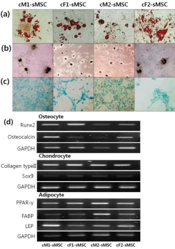
1. 성별과 연령이 다른 개의 관절 윤활액 유래 세포 구축 성별이 서로 다르고 성성숙 전과 이후인 임상적으로 건강하 다고 판정된 비글견의 경골관절에서 주사기를 이용하여 관절 윤활액을 약 300~500 ul 정도 채취할 수 있었다. 관절 윤활액 의 색상은 병변이 없는 상태로 투명한 유색을 띠고 있었으며, 가시적으로 보이는 부유물은 관찰되지 않았지만 300×g로 10
분간 원심분리하여 얻은 세포를 1차 배양하여 섬유아세포 유 사 형태의 세포와 함께 다양한 크기의 colony가 형성됨을 확인 할 수 있었다(Fig. 1a). 구축된 서로 다른 연령과 성별의 4가지 세포 (cM1-sMSC, cF1-sMSC, cM2-sMSC 그리고 cM2-sMSC) 를 3-passage까지 계대배양 후 AP activity를 확인하기 위하여 BCIP/NBT로 염색한 결과, 모든 세포에서 자주색으로 나타남 으로써 positive한 결과를 얻을 수 있었다(Fig. 1b). 줄기세포 의 초기 전사인자 발현을 확인하기 위하여 3-passage인 4가지 세포의 Oct-4, Sox-2, Nanog 발현을 RT-PCR로 확인한 결과, 성별과 연령이 다른 개체의 관절 윤활액 유래 세포는 Oct-4와 Sox-2는 발현을 확인할 수 있었으나, Nanog는 발현이 거의 되지 않았다(Fig. 1c).
2. 관절 윤활액 유래 세포의 분화능
성별과 연령이 서로 다른 개체의 관절 윤활액 유래 세포를 각각 골, 연골, 지방으로 분화 유도하기 위하여 분화배양액에 서 3주간 배양 후 세포의 형태 변화를 관찰하고 분화를 확인 하였다. 4종류의 세포를 지방세포 분화 유도 배양액에서 3주 간 배양 후 Oil red O로 염색한 결과, 붉은색으로 나타나는 지 방 방울들이 모여서 형성된 것을 확인할 수 있었으며 (Fig 2a), 골세포 분화 유도 배양액에서 3주간 배양 후 von Kossa 염색을 한 결과, 짙은 흑갈색으로 염색된 골기질의 구조를 관찰
Fig. 1. Primary colony formed synovial fluid MSCs derived from pre and post-puberty male (cM1-sMSC and cM2-sMSC) and female (cF1-sMSC and cF2-sMSC) female and male beagle dogs (a), specifically detected AP activity (b) and expression of the early transcription factors (Oct4 and Sox2) but Nanog was weakly expressed (c).
할 수 있었다 (Fig. 2b). 그리고 4종류의 세포를 연골세포 분화 유도 배양액에 3주간 배양 후 Alcian blue로 염색한 결과, 밝은 푸른색으로 염색된 연골기질을 확인할 수 있었다(Fig. 2c).
또한 골, 연골, 지방세포로 분화를 확인하기 위하여 표지자인 세포 특이 유전자의 발현을 RT-PCR로 검증하였다(Fig. 2d).
그 결과, 성별과 성성숙 정도가 다른 개체의 관절 윤활액 유래 중간엽 줄기세포를 골, 연골, 지방세포로 분화하였을 때 각 세 포 특이 유전자들이 모두 발현되었음을 확인할 수 있었다. 구체 적으로 골 분화 세포에서는 골 세포 표지자인 Osteocalcin과 Runx2, 연골세포 분화 유도된 세포는 연골세포 표지자인 colla- gen type II와 Sox9 그리고 지방세포 분화 유도된 세포는 지방 세포 표지자인 PPARγ2, FABP, LEP가 발현됨을 확인할 수 있었다.
고 찰
본 연구에서는 줄기세포의 연골 재생 연구와 실용화에 적합 한 세포원으로써 잠재성이 있는 관절 윤활액 유래 줄기세포 를 다양한 성별과 연령으로부터 구축하고, 분화능을 분석함으 로써 그 가능성을 제시하였다. 중간엽 줄기세포의 공여개체와 추출부위에 대한 다양한 연구가 이루어져 오고 있으며, 노화, 골다공증, 관절염과 같은 다양한 병리생리학적 요인에 의해서
Fig. 2. sMSCs are differentiated into adipogenic, osteogeniand and chondrogenic lineages. Adipogenic induction was assessed by oil red O staining for accumulation of the lipid droplets (a), osteogenic induction was assessed by von Kossa staining for identification of the mineralized matrix (b), and Alcian blue 8GX solution staining for synthesis of glycosaminogly- cans in chondrocytes (c). Markers for differentiation were detected by RT-PCR (d).
골수 유래 줄기세포의 증식능과 분화능이 감소될 수 있는 것 으로 보고(Rodriguez 등, 1999; Mueller 등, 2001; Stenderup 등, 2003)되어 있다. 그리고 중간엽 줄기세포를 추출하는 개체의 연령이 노령인 경우 젊은 개체보다 골수 유래 중간엽 줄기세포 가 유의적을 적게 존재한다는 보고(Stenderup 등, 2003)는 알 려져 있다. 그러나 퇴행성으로 진행되거나 여성에서 다발하는 것으로 알려진 퇴행성관절염이나 류마티스관절염과 같은 골관 절질환에 줄기세포 치료 기술을 효율적으로 적용하기 위하여 줄기세포 추출원의 성별과 연령에 따른 연구가 이루어져 한다.
최근 관절윤활막 유래 중간엽 줄기세포주를 구축하였다는 보고 (De Bari 등, 2001)가 있으며, 관절윤활막이 좋은 중간엽 줄기 세포원으로 보고되지만, 관절 윤활액으로 부터 연골 및 골 분화 유도능이 우수한 중간엽 줄기세포를 구축할 수 있다면 관절연
골 세포치료에 매우 유용한 맞춤식 세포 추출원이 될 것이다.
본 연구에서는 중간엽 줄기세포의 자가 및 타인이식 치료에 가장 적합한 동물 모델로 알려진 개(Bruder 등, 1998; Arinzeh 등, 2003; Vela 등, 2009)를 이용하여 관절을 비롯한 신체에 질병이 없는 임상적으로 건강하다고 판정 후, 경골관절에 주 사기를 주입하여 관절 윤활액을 채취하였다. 이렇게 채취된 관 절 윤활액은 약 300~500 ul 정도로 개체에 따른 차이가 있었는 데 서로 다른 연령의 개체로부터 채취하게 됨으로써 성성숙 전 의 개체의 경우 6개월령 미만으로 몸무게가 약 5 kg 미만이기 때문에, 관절강의 용적이 비교적 작아서 약 300 ul 정도 채취 가 가능했다. 그러나 성성숙이 지난 개체의 경우, 완전한 성체 로 몸무게가 15 kg 전후로 관절강의 용적이 비교적 크기 때문 에 약 500 ul 정도의 관절 윤활액의 채취가 가능하였다. 그리 고 분리한 세포를 초기 배양시 섬유아세포 유사세포들과 함 께 개 중간엽 줄기세포 특유의 선명한 돌출형의 colony들이 다양한 크기로 형성됨을 확인할 수 있었다(Fig. 1a).
이렇게 구축된 정상관절의 윤활액 유래 세포의 특성과 분 화능 분석은 동일한 조건에서 3-passage의 계대배양된 세포에 서 이루어졌다. 4종류의 세포에서 AP activity 염색에서 모두 positive가 확인되었으며, 줄기세포의 초기 전사인자인 Oct-4, Sox-2도 모두 발현됨을 확인할 수 있었다(Fig. 1b와 c). 그러나 4종류의 세포에서 모두 Nanog의 발현은 거의 미비하여 인정하 기 어려웠다. 반면, 골 및 연골, 지방세포로 분화를 유도하고 분 석한 결과, 모든 세포 종류에서 분화가 이루어짐을 확인함으 로써 Nanog의 발현이 분화 유도능에 미치는 직접적인 영향이 없는 것으로 보인다. 구축된 4종류의 세포를 골, 연골, 지방으 로 각각 분화 유도 후 해당 세포의 표지자인 세포 특이 유전자 의 발현을 확인한 결과 성별과 연령에 따라 발현 정도의 차이 는 존재하는 것으로 보이지만 연계성은 부족하였다(Fig 2d).
줄기세포를 추출한 개체의 연령이 세포의 분화능에 영향을 미친다는 보고들이 있지만 대부분 사람과 랫트 등 개 이외의 동물종에서 골수유래 중간엽줄기세포를 이용한 증식과 분화 율에 대한 연구가 이루어져 왔다(Osyczka 등, 2009; Stolzing 등, 2006; Zheng 등, 2007). 노령의 개체로부터 추출된 중간엽 줄기세포가 젊은 개체로부터 추출된 세포보다 인대나 골 세포 분화 형성이 저조하다고 보고(Mendes 등, 2002; Dressler 등, 2005)되고 있지만, 일반적으로 노령 개체 유래 줄기세포의 분 화능뿐만 아니라 줄기세포 증식능도 저조한 것을 알려져 있 다. 그러나 생체실험을 위한 동물모델의 경우, 사람과 노화의 과정이 다르기 때문에, 노령의 기준이 모호하고 사람과 달리 암컷의 경우 폐경이 존재하지 않기 때문에 생리적인 성성숙 기를 기준으로 미숙개체와 성숙개체로 구분할 필요가 있다.
본 연구에서는 이러한 성성숙 여부를 기준으로 구분하여 분 화능을 비교한 결과, 4종류의 세포에서 골세포로 분화 유도된 경우 Osteocalcin과 Runx2의 발현을 확인할 수 있었으며, 지방
세포로 분화 유도한 경우 PPARγ2, FABP 그리고 LEP의 발현 을 확인할 수 있었지만, 발현 정도에 다소 차이가 있는 것으 로 보이지만 연계성을 찾기는 어려웠다. 또한 연골세포로 분 화 유도한 경우 collagen type Ⅱ는 강하게 발현되었지만 Sox9 은 약한 발현을 나타내었다.
이러한 결과로 성별과 성성숙 상태가 다른 개의 관절 윤활 액에서 줄기세포의 구축이 가능하며, 세포주 특이적인 특성은 인정되지만 분화능의 차이를 인정할 만한 차이는 없는 것으 로 사료된다. 따라서 현재까지 가장 널리 연구되는 줄기세포 추출원인 골수의 경우 비교적 침습적이고 관절연골 치료의 경우 진단과 치료과정에 적용하기에 용이하지 않을 수 있는 한계를 극복할 수 있을 것이다. 일반적으로 관절염의 경우, 관절 윤활 액을 채취하여 검사하거나 관절경을 이용하여 치료 시술을 하 는 과정에서 관절 윤활액의 채취가 매우 용이하고, 반복적으로 채취가 가능한 조직이므로 자가 줄기세포 치료의 효율적인 추출원이 될 수 있을 것이다.
결 론
본 연구에서 성별과 성성숙 전후의 다양한 조건을 가진 공 여동물인 비글견의 관절 윤활액으로부터 중간엽 줄기세포를 구축할 수 있음을 확인하였다. 따라서 개에서 발생하는 다양 한 골 관절 연골질환에 관절윤활액에서 추출한 중간엽줄기세 포를 이용한 연골재생 치료에 효율적으로 이용가능 할 것으 로 사료되며, 더 나아가 사람의 연골재생 치료에도 응용가능 할 것이다. 그러나 줄기세포 공여개체의 성성숙 여부와 성별 차에 따른 영향을 명확히 분석하기 위하여 좀더 세부적으로 분화능을 분석하고, 성 호르몬의 첨가에 따른 영향을 규명할 필요가 있을 것으로 사료된다.
참고문헌
Arinzeh TL, Peter SJ, Archambault MP, van den Bos C, Gor- don S, Kraus K, Smith A and Kadiyala S. 2003. Alloge- neic mesenchymal stem cells regenerate bone in a critical- sized canine segmental defect. J. Bone Joint Surg. Am.
85-A:1927-1935.
Bruder SP, Kraus KH, Goldberg VM and Kadiyala S. 1998.
The effect of implants loaded with autologous mesenchy- mal stem cells on the healing of canine segmental bone defects. J. Bone Joint Surg. Am. 80:985-996.
Da Silva Meirelles L, Chagastelles PC and Nardi NB. 2006.
Mesenchymal stem cells reside in virtually all post-natal organs and tissues. J. Cell Sci. 119(Pt 11):2204-2213.
De Bari C, Dell'Accio F, Tylzanowski P and Luyten FP. 2001.
Multipotent mesenchymal stem cells from adult human sy- novial membrane. Arthritis & Rheumatism 44:1928-1942.
Dressler MR, Butler DL and Boivin GP. 2005. Effects of age on the repair ability of mesenchymal stem cells in rabbit tendon. J. Orthop. Res. 23:287-293.
Friedenstein AJ, Chailakhjan RK and Lalykina KS. 1970. The development of fibroblast colonies in monolayer cultures of guinea-pig bone marrow and spleen cells. Cell Tissue Ki- net. 3:393-403.
Horie M, Sekiya I, Muneta T, Ichinose S, Matsumoto K, Saito H, Murakami T and Kobayashi E. 2009. Intra-articular in- jected synovial stem cells differentiate into meniscal cells directly and promote meniscal regeneration without mobili- zation to distant organs in rat massive meniscal defect.
Stem Cells 27:878-887.
Jaiswal N, Haynesworth SE, Caplan AI and Bruder SP. 1997.
Osteogenic differentiation of purified, culture expanded hu- man mesenchymal stem cells in vitro. J. Cell Biochem.
64:295-312.
Johnstone B, Hering TM, Caplan AI, Goldberg VM and Yoo JU. 1998. In vitro chondrogenesis of bone marrow-derived mesenchymal progenitor cells. Exp. Cell Res. 238:265-272.
Koga H, Engebretsen L, Brinchmann JE, Muneta T and Sekiya I. 2009. Mesenchymal stem cell-based therapy for cartilage repair: a review. Knee. Surg. Sports. Traumatol. Arthrosc.
17:1289-1297.
Koga H, Muneta T, Ju YJ, Nagase T, Nimura A, Mochizuki T, Ichinose S, von der Mark K and Sekiya I. 2007. Synovial stem cells are regionally specified according to local micro- environments after implantation for cartilage regeneration.
Stem Cells 25:689-696.
Koga H, Muneta T, Nagase T, Nimura A, Ju YJ, Mochizuki T and Sekiya I. 2008. Comparison of mesenchymal tissues-de- rived stem cells for in vivo chondrogenesis: suitable condi- tions for cell therapy of cartilage defects in rabbit. Cell Tissue Res. 333:207-215.
Koga H, Shimaya M, Muneta T, Nimura A, Morito T, Hayashi M, Suzuki S, Ju YJ, Mochizuki T and Sekiya I. 2008. Lo- cal adherent technique for transplanting mesenchymal stem cells as a potential treatment of cartilage defect. Arthritis.
Res. Ther. 10:R84.
Mackay AM, Beck SC, Murphy JM, Barry FP, Chichester CO and Pittenger MF. 1998. Chondrogenic differentiation of cul- tured human mesenchymal stem cells from marrow. Tissue Eng. 4:415-428.
Mendes SC, Tibbe JM, Veenhof M, Bakker K, Both S, Platen- burg PP, Oner FC, de Bruijn JD and van Blitterswijk CA.
2002. Bone tissue-egineered implants using human bone marrow stromal cells: effects of culture conditions and donor age. Tissue Eng. 8:911-920.
Mueller SM and Glowacki J. 2001. Age-related decline in the osteogenic potential of human bone marrow cells cultured in three-dimensional collagen sponges. J. Cell Biochem.
82:583-590.
Osyczka AM, Damek-Poprawa M, Wojtowicz A and Akintoye SO. 2009. Age and skeletal sites affect BMP-2 responsive- ness of human bone marrow stromal cells. Connect. Tissue Res. 50:270-277.
Rodriguez JP, Garat S, Gajardo H, Pino AM and Seitz G.
1999. Abnormal osteogenesis in osteoporotic patients is reflected by altered mesenchymal stem cells dynamics. J.
Cell Biochem. 75:414-423.
Sakaguchi Y, Sekiya I, Yagishita K and Muneta T. 2005.
Comparison of human stem cells derived from various mesen- chymal tissues: superiority of synovium as a cell source.
Arthritis Rheum. 52:2521-2529.
Stenderup K, Justesen J, Clausen C and Kassem M. 2003. Aging is associated with decreased maximal life span and accele- rated senescence of bone marrow stromal cells. Bone 33:
919-926.
Stolzing A and Scutt A. 2006. Age-related impairment of me- senchymal progenitor cell function. Aging Cell 5:213-224.
Thomas ED and Storb R. 1999. The development of the scientific foundation of hematopoietic cell transplantation based on animal and human studies. In: Thomas, E. D.;
Blume, K. G.; Forman, S. J., eds. Hematopoietic Cell Trans- plantation, 2nd edition. Boston, MA:USA Blackwell Science, 1-11.
Vela DC, Silva GV, Assad JA, Sousa AL, Coulter S, Fernandes MR, Perin EC, Willerson JT and Buja LM. 2009. Histopa- thological study of healing after allogenic mesenchymal stem cell delivery in myocardial infarction in dogs. J. His- tochem. Cytochem. 57:167-176.
Volk SW, Wang Y and Hankenson KD. 2012. Effects of do- nor characteristics and ex vivo expansion on canine mesen- chymal stem cell properties: Implications for MSC-based therapies. Cell Transplant. In press.
Yoshimura H, Muneta T, Nimura A, Yokoyama A, Koga H and Sekiya I. 2007. Comparison of rat mesenchymal stem cells derived from bone marrow, synovium, periosteum,
adipose tissue, and muscle. Cell Tissue Res. 327:449-462.
Zheng H, Martin JA, Duwayri Y, Falcon G and Buckwalter JA. 2007. Impact of aging on rat bone marrow-derived stem cell chondrogenesis. J. Gerontol. A. Biol. Sci. Med. Sci.
62A:136-148.
Zuk PA, Zhu M, Mizuno H, Huang J, Futrell JW, Katz AJ,
Benhaim P, Lorenz HP and Hedrick MH. 2001. Multilineage cells from human adipose tissue: implications for cell-based therapies. Tissue Eng. 7:211-228.
(접수: 2012. 8. 19 / 심사: 2012. 8. 20 / 채택: 2012. 9. 5)