J O U R N A L O F
Veterinary Science
pISSN 1229-845X, eISSN 1976-555X J. Vet. Sci. (2013), 14(4), 495-497 http://dx.doi.org/10.4142/jvs.2013.14.4.495
Received: 14 Dec. 2011, Revised: 21 Aug. 2012, Accepted: 30 Aug. 2012
Case Report
*Corresponding author: Tel: +82-2-450-3664; Fax: +82-2-446-9876; E-mail: [email protected]
ⓒ 2013 The Korean Society of Veterinary Science.
This is an Open Access article distributed under the terms of the Creative Commons Attribution Non-Commercial License (http://creativecommons.org/licenses/by-nc/3.0) which permits unrestricted non-commercial use, distribution, and reproduction in any medium, provided the original work is properly cited.
Percutaneous transplantation of human umbilical cord-derived
mesenchymal stem cells in a dog suspected to have fibrocartilaginous embolic myelopathy
Wook-Hun Chung
1, Seon-Ah Park
1, Jae-Hoon Lee
1, Dai-Jung Chung
1, Wo-Jong Yang
1, Eun-Hee Kang
1, Chi-Bong Choi
1, Hwa-Seok Chang
1, Dae-Hyun Kim
1, Soo-Han Hwang
2, Hoon Han
2, Hwi-Yool Kim
1,*
1
Department of Veterinary Surgery, College of Veterinary Medicine, Konkuk University, Seoul 143-701, Korea
2
Seoul Cord Blood Bank, Histostem Co., Seongnam 462-807, Korea
The use of human umbilical cord blood-derived mesenchymal stem cells for cell transplantation therapy holds great promise for repairing spinal cord injury. Here we report the first clinical trial transplantation of human umbilical cord (hUCB)-derived mesenchymal stem cells (MSCs) into the spinal cord of a dog suspected to have fibrocartilaginous embolic myelopathy (FCEM) and that experienced a loss of deep pain sensation.
Locomotor functions improved following transplantation in a dog. Based on our findings, we suggest that transplantation of hUCB-derived MSCs will have beneficial therapeutic effects on FCEM patients lacking deep pain sensation.
Keywords: dog, fibrocartilaginous embolic myelopathy, human umbilical cord-derived mesenchymal stem cells, percutaneous transplantation, xenotransplantaion
Fibrocartilaginous embolic myelopathy (FCEM) is a common cause of ischemic myelopathy in dogs and is a syndrome of acute spinal cord infarction due to embolization of fibrocarctilageous materials identified as the nucleus pulposus of intervertebral discs [3,5,9,12]. The pathogenesis of FCEM is still unclear [5,9,11]. A definitive diagnosis of this disease is confirmed by histopathological evaluation; therefore, antemortem diagnosis of FCEM is based on patient history, clinical examination, Cerebrospinal fluid (CSF) analysis, Computed tomography (CT) data, and magnetic resonance imaging (MRI) findings [1,5,8,11,12]. MRI is especially helpful for making an antemoterm diagnosis of ischemic myelopathy and eliminating other causes of myelopathy including degenerative disc disease. This technique can also be used to visualize signal intensity changes indicative of ischemic
myelopathy [1,5,8,11,12].
The prognosis of FCEM depends on ischemia severity and extent of the affected area [5]. Previous reports described negative prognostic factors of FCEM including a loss of deep pain sensation, CSF changes, spinal cord swelling, and lack of improvement within the first two weeks of disease onset [3,5,7]. The absence of deep pain sensation after any spinal cord injury is indicative of a poor prognosis and significantly associated with bilateral damage to the gray and white matter [2,7]. Therefore, FCEM patients lacking deep pain sensation were given a guarded prognosis in previous reports [7]. Furthermore, methods for treating cases of FCEM without deep pain sensation have not been reported.
In a previous study using rat models of SCI, treatment with human umbilical cord blood-derived mesenchymal stem cells (hUCB-MSCs) led to significant improvement of locomotor function [4]. hUCB-MSCs were also found to be capable of differentiating into neural cells in vivo [10].
In fact, our previous studies have demonstrated that hUCB-MSCs promote regenerative potential in cases of SCI [10].
We encountered a dog (7-year-old female cocker spaniel) that was referred to the Konkuk University Veterinary Teaching Hospital (Korea) with an acute onset of hindlimb paralysis in the absence of any history of trauma.
Neurological examination revealed that the dog suffered
from hindlimb paralysis. No postural reaction was seen in
the bilateral hindlimb including a lack of paw positioning,
hopping, and extensor postural thrust. Additionally, no
voluntary urination was observed. Spinal reflexes were
normal. Deep pain sensation in the dog was absented at the
496 Wook-Hun Chung et al.
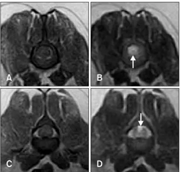
Fig. 1. T1-weighted (TR 530, TE 26) and T2-weighted (TR 3500, TE 90) MRI images of the dog 12 h (A∼D) after the onset of clinical signs. The transverse T1-weighted image showed isointensity in the parenchyma of L2 (A) and L3 (C). Arrows:
transverse T2-weighted image showing hyperintensity in the parenchyma of L2 (B) and L3 (D).
Fig. 2. T1-weighted (TR 550.0, TE 12.6) and T2-weighted (TR 4400, TE 96) MRI images of the dog 12 weeks after transplantation. The transverse T1-weighted image showed isointensity in the parenchyma of L3 (A). In the transverse T2-weighted image of L3, an area of hyperintensity was visible in the dorsal aspect of the spinal cord (B).
first neurological examination.
Methylprednisolone succinate (Koreamypharm, Korea) was administered intravenously (30 mg/kg) to reduce secondary spinal cord damage. MRI (0.3T; Esaote, Italy) was performed 24 h after clinical onset of symptoms (Fig. 1).
There was no evidence of disc extrusion. An intramedullary lesion involving the gray matter alone, appearing as an area of hypointensity from L2 and L3 in the transverse image, was found on the T2-weighted image. CSF analysis was performed as well and did not produce any remarkable findings. We tentatively diagnosed the dog with FCEM based on the clinical symptoms, MRI findings, and CSF analysis according to previous studies [1,5,8,12]. Dorsal laminectomy was performed on the dog from L1 to L3 to promote decompression. The spinal cord was significantly swollen. However, no evidence of disc material extrusion or hemorrhage was found during surgery.
Deep pain sensation and motor function did not improve until 1 week after surgery. We notified the owners of the dog about the possibility of an adverse prognosis given the absence of deep pain sensation and significant spinal cord swelling. Participation of the dog in this clinical trial involving hUCB-derived MSC transplantation was offered and owner gave consent. Before transplantation, sensory evoked potential (SEP) analysis was performed (Sierra Wave, 2006; Cadwell Laboratories, USA). Based on the results, we confirmed that the dog did not show SEPs.
Transplantation of hUCB-derived MSCs was performed 7 days after decompression surgery. The hUCB-derived MSCs were provided for pure research purposes by the Seoul Cord Bank (Histostem, Korea). For the transplantation procedure, 1 × 10
6hUCB-derived MSCs in a total 0.3 mL volume of saline was injected directly into three separate spinal cord segments (L1∼L2, L2∼L3, and L3∼L4) using a spinal needle. Immunosuppressants were not administered to the dog. A modified Tarlove scale [13]
was used to evaluate locomotor function. This scale grades hindlimb movement on a scale of 0 (no movement) to 5 (normal gait). Deep pain sensation was assessed using a hemostat to pinch the digit.
The owner of the dog reported that manual bladder expression was not needed 3 weeks after transplantation and the dog voluntarily urinated at a given location at 4 weeks during follow-up. The dog exhibited gradual improvements in hindlimb locomotor functions starting at 4 weeks post-transplantation. Modified Tarlov scores increased from 0 to 4 by 10 weeks post-transplantation. One year after transplantation, modified Tarlov scores were maintained. However, gait performance further improved although deep pain sensation was not restored after transplantation. MRI (3T; Oxford Medinus, Korea) studies were performed 3 months (Fig. 2) after transplantation.
Hyperintense signals were still observed in the dorsal portion of the spinal cord even 3 months after the initial MRI examination.
The presence of FCEM is confirmed by
neurohistopathologic examination showing occlusion of
the spinal vasculature to nucleus pulposus that causes
ischemic necrosis of dependent regions in the spinal cord
parenchyma [5,7]. However, neurohistopathological
examination could not be performed in the present study
because the dog was still alive. Therefore, we diagnosed
FCEM according to clinical symptoms, MRI results based
on previous studies [1,5,8,12], and observations made
during surgery. The criteria of MRI findings for diagnosing
FCEM include the presence of a focal, relatively sharply
hUCB-MSC for treating a dog with FCEM 497