Human Adipose Tissue-Derived Mesenchymal Stem
Cells 배양 시 효율적인 Extracellular Matrix의 증명
The Extracellular Matrix Affected Proliferation and Cell Adhesion of Human Adipose Tissue Derived Mesenchymal Stem Cells in vitro Purpose: Human mesenchymal stem cells (hMSCs) have the potency for self-renewal and
differentiation into various kinds of cells. The hMSCs are obtained from the various tissues, including adipose tissue, bone marrow and cord blood. The extracellular matrix (ECM) is an important factor that affects cell adherence, growth, migration, apoptosis and differentiation both
in vitro and vivo. The adipose-derived mesenchymal stem cells (AD-MSCs) have CD29 (integrin)
on the cell surface, which is the receptor for fibronectin. The aim of this study is to validate the efficacy of ECM, and especially fibronectin, for cell expansion.
Methods: The AD-MSCs were obtained from the abdominal fat of humans. These cells were
seeded onto culture plates coated with fibronectin-Human (FN) and plates without ECM (control). The cells were incubated for 3 passages and the cellular morphology was simultaneously observed with microscopy. CCK-8 assay was performed to compare the proliferation ability in each condition at the same passage. Immunocytochemistry staining for integrin-beta1 was performed to observe the cell to cell interaction.
Results: The hAD-MSCs in the FN-coated and non-coated plates exhibited cytoplasm staining
for integrin-beta1. In all the cultures, extended fibroblastic-shaped cells that turned into rhomboid cells were most frequently observed. The cell growth rates for the non coated culture plate were lower than those for the FN coated plates. After 72 hour culture under the different coated concentrations of FN and the non coated condition (control), the control group had a lower growth rate. In the culture with a FN coated plate, a significant change was observed as compared with that of the control group. We observed an increase in cell proliferation, with a maximum of 140%, on the FN coated plate by performing CCK-8 assay. In comparison, integrin β1 on the cells was more expressed in the FN-coated plates than that in the non-coated plates.
Conclusion: The cell morphology can be changed faster in the FN coated culture plates than
that in the non coated culture plates. Because proliferation and adhesion with FN can enhance the expansion, the culture within a FN coated plate is needed to encourage hAD-MSCs to proliferate in vitro. 민선옥1, 이상우1,2, 최새별3, 김경식1,4 연세대학교 의과대학 1외과학교실, 2연세 대학교 대학원 나노과학 기술 협동과정, 3 고려대학교 의과대학 외과학교실, 4연세 대학교 의과대학 세브란스병원 세포치료 센터
Seon Ok Min, B.S.1, Sang Woo Lee, B.S.1,2, Sae Byeol Choi, M.D., Ph.D.3, Kyung Sik Kim, M.D., Ph.D.1,4
1Department of Surgery, Yonsei
University College of Medicine,
2Graduate Program of Nano Science
and Technology Graduate School of Yonsei University, 3Department of
Surgery, Korea University College of Medicine, 4Cell Therapy Center,
Severance Hospital, Yonsei University College of Medicine 책임저자 김 경 식 서울시 서대문구 성산로 250번지 연세대학교 의과대학 외과학교실 우편번호 120-752 Tel: 02-2228-2125, 2100 Fax: 02-313-8289 E-mail: kskim88@yuhs.ac
Key Words : Adipose tissue-derived mesenchymal stem cells (hAD-MSCs), Extracellular matrix, Fibronectin
중심단어 : 지방유래간엽줄기세포, 세포 외 기질, Fibronectin
Received: 2009. 9. 2 Accepted: 2009. 12. 9
서
론
인체 지방 유래 간엽줄기 세포(Human adipose
tissue-deri-ved Mesenchymal stem cells, hADSCs)는 다능성의 줄기세포 (multi-potent stem cells)로서 생체 내(in vivo) 및 생체 외(in
vitro)에서 뼈, 지방, 연골, 간세포 등의 다른 세포로 분화가
가능하다.1 이런 특성을 이용하여 조직 공학적으로 활용하거
나 다른 조직의 세포로 분화를 시켜 이식을 함으로써 질병 치 료에 각광 받고 있다.1 하지만 지방유래 간엽 줄기 세포 역시
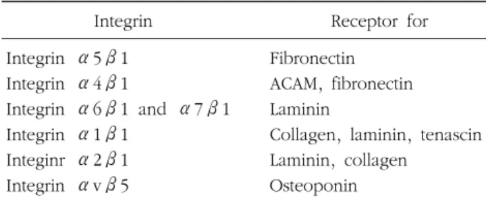
Table 1. Integrin and receptors
Integrin Receptor for Integrin α5β1 Fibronectin
Integrin α4β1 ACAM, fibronectin Integrin α6β1 and α7β1 Laminin
Integrin α1β1 Collagen, laminin, tenascin Integinr α2β1 Laminin, collagen
Integrin αvβ5 Osteoponin 인체에서 대량으로 세포를 얻기가 쉽지 않고, 수명도 제한적 이기 때문에 단 시간 내에 보다 많은 세포를 얻는 것이 중요하 다. 그러므로 간엽줄기세포를 임상적으로 적용하기 전에 효율 적인 증식 조건을 연구하는 것이 우선 해결해야 할 과제이다.2 이로 인해 간엽줄기세포의 배양 방법이 중요시 되고 있는 데, 배양 시 가장 기본적인 방법인 배양 용기에 대한 세포기질 의 도포는 체외에서 세포의 초기 퍼짐이나 형태, 증식 및 분화 에 영향을 미치는 것으로 생각된다.3 특히 이에 관여하는 In-tegrin family는 세포와 세포 또는 세포와 세포외기질 결합 모 두에 관여하는 세포 수용체이다.4 적혈구를 제외한 모든 세포 는 한 종류의 인테그린(integrin)을 갖고 있고, 세포외기질에 관한 정보는 세포 표면 수용체를 통해서 세포 내로 이송하게 된다. 인테그린은 알파 아단위(α-subunit)와 베타 아단위(β- subunit)로 구성되어 있는데 각각의 아단위(subunit)에 따라 서 리간드(ligand)가 조금씩 달라진다(Table 1).4 세포외 기질을 구성하는 피브로넥틴(Fibronectin), 라미닌 (Laminin)의 짧은 단백질 서열에 부착하는데, 인테그린은 세 포외 기질의 아르지닌-글라이신-아스파라진(arg-gly-asp, RGD) 서열 부위에 부착한다.5 이러한 세포외기질 단백질의 수용체 로 대표적인 것이 콜라겐과 피브로넥틴이 있다. 콜라겐은 조 직의 구조 단백질로, 여러 형태가 존재하는데, 대부분 다량체 형 원섬유이며, 주로 인테그린과 직접 상호작용한다. 피브로 넥틴은 회전타원체(spheroid)의 형태로 거의 모든 조직에 존 재하는 단백질로서, 결합조직의 주된 부착단백질로 조직에서 는 거의 모든 세포가 피브로넥틴과 상호작용한다. 대부분의 조직 유래 세포들은 점착하는 위치에 의존하며, 생존과 성장 을 위해서는 세포간질 표면에 붙어야 한다. 세포의 퍼짐, 접착 및 이동은 각각 세포외기질과의 상호작용과 연관되어 있다.6 지방 유래 간엽 줄기 세포 역시 간엽 줄기세포로서, 점착성 세포이므로 이러한 세포외 기질과의 영향을 받을 것으로 생각 된다. 기존의 간엽 줄기세포의 배양 도포 방법으로 콜라젠과 피브로넥틴 등을 많이 이용하여 도포하고 있지만 간엽 줄기세 포의 배양 시 좀 더 효율적인 도포 방법과 그 영향에 대한 연구는 많이 시행되고 있지 않다. 그러므로 지방 간엽줄기세 포를 다른 종류의 세포로 분화시키기 위한 초기 배양 시 세포 외기질의 효능성을 알아보고자 하였다.
방 법
1. 지방 유래 간엽줄기세포의 분리 건강한 성인의 복부로부터 지방을 채취한 후 혈액을 제거하 기 위해 3번 정도 PBS로 세정한다. 지방 조직에 붙어 있는 혈관, 림프구 등을 제거한 후 collagenase (Wako, Japan)가 녹아 있는 PBS에 지방조직을 넣은 후 37oC에서 shaking, 1 시간 정도 둔다. 1시간 후 효소 불활성을 위해서 10% FBS 들어간 DMEM을 첨가하고, 4oC, 1,200 G에서 10분간 원심분 리 한다. 상층의 지방 덩어리와 상층액을 버리고 pellet에 RBC lysis buffer 10 mL을 넣고 상온에서 5분 방치 한 후 4oC, 350 G에서 5분 원심분리 후 3 mL 배지를 넣고 세포 수 측정한 다. 6 well culture plate (corning Incorporated, USA)에 5,000 cells/cm2 분주 후 37oC 5% CO2에서 배양한다. 분리된 세포의증식을 위해 다음과 같은 배지 60% DMEM-LG (GibcoBRL, Grand Island, NY, USA), 40% MCDB-201 (Sigma), 1X in-sulin transferrin-selenium (ITS) (GibcoBRL, Grand Island, NY, USA), 10-9M dexamethasone (Sigma), 10-4M ascorbic acid 2-phosphate (Sigma), 10 ng/mL rhEGF (Daewoong Pharmaceuticals, Korea), antibiotic/antimycotic fibronectin (GibcoBRL, Grand Island, NY, USA) 및 10% FBS (Wel-GENE, Daegu, Korea)를 사용하여 1주에 두 번 증식배지를 교환하였다. 초대배양(Primary culture)후 집락(confluence) 이 80%정도 되었을 때 1X Trypsin-EDTA (Invitrogen, USA)사 용하여 계대 배양 해준다. 첫 번째 계대 배양 시 각각의 도말 배양용기(coated plate)에 분주한다.
2. 세포 배양 도포(Cell culture plate coating)
세포와 세포외기질과의 결합에 관여하는 integrin family와 이와 결합하는 수용체인 피브로넥틴(BD, England)을 사용시
Fig. 1. The surface of cell culture plate by SEM (×20,000). (A) Non-coated. (B) FN Non-coated. MSCs culture의 효율성을 증명하기 위해 fibronectin (5μg/ cm2)을 도포한 후, 3차례 계대 배양한 지방 간엽 줄기세포를 분주 했으며, 이에 대조군으로 도포 하지 않은 것을 사용하 였다. 피브로넥틴 도포한 배양용기와 도포 하지 않은 배양 용기의 표면 상태를 비교하기 위하여 주사 현미경(Scanning electron microscope, SEM)을 이용하여 배양 용기의 표면을 촬영하였다.
3. 증식 분석
72시간 배양한 3번째 계대 배양된 간엽 줄기세포를 72시간 배양한 뒤 96 well plate (NUNCLON, DENMARK)에 5,000 cells/well 분주하여 37oC, 5% CO2 조건에서 72시간 배양한
다. 72시간 후 cell counting kit-8 (CCK-8) (Dojindo Labora-tories, JAPAN)을 각 well 당 10μL씩 분주한다. 1∼4시간 정 도 보육(incubation)한 뒤 450 nm microplate reader로 측정 한다. 4. CD29-integrin beta 1에 대한 면역 세포 화학 염색 각 도말 조건에서 지방유래 간엽 줄기세포의 증식의 차이를 확인하기 위해서 integrin-ß1을 면역세포화학법을 이용하여 검출하였다.
Chamber slide (Nunc Lab Tek. USA)에 각각 도포 후 간엽 줄기세포 배양한다. 25% 집락(confluence) 되었을 시 4% para-formaldehyde를 이용해 30분 동안 37oC에서 고정한다. 고정
후 0.2% Triton X-100이 포함된 PBS로 37oC, 30분 동안 처리 후 1% Bovine serum albumin에서 1시간 반응시킨다. 그 후 1:250으로 희석된 primary antibody integrin β1 (rabbit anti human integrin β1), (Santa Cruz Biotechnology, Cali-fornia, USA)로 4oC overnight 처리 후 PBS로 수세한다. 다음 1:250으로 희석 된 secondary antibody integrin β1 (goat anti-rabbit IgG conjugated Texas Red), (Santa Cruz Bio-technology, California, USA)으로 45분 처리한다. 핵을 염색 하기 위한 DAPI 염색 후, permount를 사용해 mounting 후 cover slide로 덮은 후 형광 현미경으로 확인한다.
결 과
1. 세포 외 기질-도포(ECM-coated): 피브로넥틴 도포된 배양 용기의 주사현미경 소견 도말하지 않은 배양 용기의 주사 현미경 사진에서 볼 수 있는 L-lysine의 물질들이 피브로넥틴에 의해 덮히므로 편평 한 것을 관찰할 수 있었다(Fig. 1A, B). 1) 지방유래 간엽줄기세포의 형태학적 변화 피브로넥틴 도포 배양 용기의 지방유래 간엽줄기세포(hAD-SCs)의 변화가 빠르게 일어났으며, 퍼짐(spreading) 역시 육안 으로 더 빠른 것을 관찰할 수 있었으며, 그 모양 또한 방추모 양에서 지방유래 간엽줄기세포의 형태인 장사방형(rhomboid) 의 모양으로 대부분 변했으나, 비도포배양용기에서는 대부분 이 방추형의 형태인 것을 볼 수 있었다(Fig. 2A, B).Fig. 2. The morpholgy of mesenchymal stem cells by inverted microscope. (A, C) Non-coated plate (A: ×40) (C: ×100) & (B, D) FN-coated plate (B: ×40) (D: ×100).
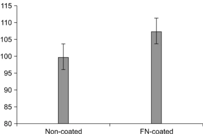
Fig. 3. Cell proliferation assay on non-coated and FN-coated plates.
2) 세포 증식
세포외기질으로서 피브로넥틴-도말의 증식을 비교하기 위 하여 지방유래 간엽줄기세포 #3의 배양 후 3일 째 CCK-8 assay를 통해 실험하였으며, 흡광도는 450 nm에서 96-well micro test spectrophotometer를 이용해 측정하였다. 피브로
넥틴의 농도는 5μg/cm2으로 도포하였다. 세포 증식은 피브
로넥틴 도포군이 비도포군보다 수치가 더 높은 것을 볼 수 있 었다(Fig. 3).
3) 인테그린 베타-1의 활성
면역세포형광분석 결과를 hADSC #3 배양 후 3일 촬영한 사 진이다. 피브로넥틴(5μg/cm2) 도포되었다(Fig. 4A∼C). non- coating 지방유래 간엽줄기세포사진이다(Fig. 4D∼F). Blue color는 핵이 염색된 것이며(Fig. 4A, D), Red color는 세포
Fig. 4. The nuclei and the integrin beta-1 expression of mesenchymal stem cells 3 days by immunocytocehmistry assay. (A∼C) Non-coated plate (×200) & (D∼F) FN-coated plate (×200).
표면 표지자인 인테그린 베타-1의 발현을 나타낸 것이다(Fig. 4B, E). 두 개의 color가 혼합된 사진은 핵과 인테그린 베타-1 의 발현을 merge한 그림이다(Fig. 4C, F). 피브로넥틴 도포을 한 실험군의 지방유래 간엽 줄기세포에서 인테그린 베타-1의 발현(Red color)이 비도포군에서의 발현 정도에 비해 훨씬 많 이 발현 한 것을 관찰 할 수 있었다(Fig. 4B, C, E, F).
고 찰
줄기 세포를 이용한 질병 치료는 보다 근본적인 치료의 가 능성이 있으며, 그 중에서도 성체 간엽 줄기세포는 배아 줄기 세포에서 문제 되고 있는 윤리적인 문제가 그다지 크지 않고 여러 장기에서 얻기가 용이하다는 장점이 있다.7 간엽 줄기세 포는 자가 증식을 하며,8 다른 장기의 세포로의 분화가 가능하 다는 특징을 갖고 있다.9,10 이런 특징을 이용하여 조직공학이 나 유전자 치료를 하기도 한다.11 이런 치료 방법들은 자신의 간엽줄기세포를 이용한 자가 이식을 통해 면역거부반응을 줄 이거나 거의 일어나지 않게 할 수 있을 가능성을 갖고 있다.12 우리는 복부에서 지방조직을 떼어내 간엽줄기세포를 얻었는 데,7,13 이유는 지방으로부터 얻은 간엽 줄기세포는 다른 장기 로부터 얻은 것에 비해 가장 긴 세포 주기를 갖고 있고 증식을 비교하였을 때 가장 좋은 결과를 보여주기 때문이다.14 그러나 간엽 줄기세포를 생체 외(in vitro)에서 배양하여 다른 세포로 의 분화 등의 실험을 시행하기 위해서는 최적의 배양 조건이 필요하다. 이런 조건은 간엽줄기세포의 모양과 간엽줄기세포 가 분화하는 세포의 전형적 단백질 발현에 큰 영향을 끼친 다.15 기존의 연구에서는 피브로넥틴으로 도포된 배양용기의 간 엽줄기세포와 콜라겐, 그리고 도포되지 않은 배양용기에서의 간엽줄기세포를 비교한 결과 피브로넥틴으로 도포된 배양용 기에서 간엽줄기세포가 가장 잘 부착된다는 것 이외에는 세포 증식의 비교에서는 거의 차이가 없었다고 하였다.2 하지만 우 리는 앞의 결과에서 언급된 바와 같이 피브로넥틴으로 도포 된 배양용기에서의 간엽줄기세포가 도포되지 않은 용기의 세 포에 비해 성장이나 증식에서 더 효과적인 결과를 얻을 수 있 어서 세포 외 기질과 간엽줄기세포간의 상호작용을 통해 부착 과 증식에 영향을 받는다는 것을 알 수 있었다. 비도포군의 배양용기에서 자란 간엽줄기세포는 증식 능력이 도포된 배양 용기의 세포에 비해 낮은 수준을 보였으며, 세포 모양 또한 느리게 변화되는 것을 볼 수 있었다. 세포 외 기질과 세포의 결합에 관여하는 세포의 표면에 있 는 인테그린 베타-1와 같은 수용체의 발현 정도를 비교하여보았는데 피브로넥틴도포된 배양용기의 세포에서 인테그린 베타-1 수용체가 더 많이 발현된 것을 볼 수 있었다. 이것은 세포 외 기질과 이와 결합하는 세포 표면의 수용체인 인테그 린을 통한 결합이 활성화되면서 세포수가 빠른 시간에 증식되 며, 이로 인해 세포와 세포간 상호작용이 활발해 짐을 의미한 다. 간엽줄기세포는 세포 팽창 중에는 다분화성이 줄어든다고 하며, 반대로 분화 중에는 세포의 팽창이 줄어든다는 연구도 발표된 바 있다.16-18 간엽줄기세포의 빠른 성장과 증식은 짧은 시간 내에 효과적으로 번식 또는 분화하여 임상적용을 할 수 있다는 강점이 있다.12 그러므로 이러한 세포 외 기질(ECM)을 이용하면 아마 생체 외에서 간엽줄기세포 배양 시 세포 증식 을 빠르게 증가시키는 역할을 할 것이다. 세포 부착에 관여하는 세포 표면 수용체로 알려져 있는 Integrin family의 한 종류인 인테그린 베타-1은 콜라젠, 피브 로넥틴, 라미닌 등 세포외 기질 단백질에 수용체로서 작용한 다. 이것은 세포와 세포외 기질 간의 부착을 중개하는 물질로 서, 세포 신호 기작이나 세포의 모양, 이동, 그리고 세포 주기 등을 조절한다고 알려져 있다. 그 구조는 α와 β unit으로 구성되어 있으며, 두 아단위의 결합으로 작용한다.19 α-sub-unit은 18개가 존재하며 β-subα-sub-unit은 8개가 존재한다. 이 중 에서 세포 외 기질과의 결합에 관여하는 대표적인 것은 인테 그린 베타-1이다. 이러한 Integrin family와 세포 외 기질이 부 착을 이루어 세포의 신호 기작 등에 관여하여 세포 죽음, 이 동, 부착, 분화 등을 조절하게 되는 것이다.19 본 연구 결과에서 이 수용체의 발현 정도를 비교하여 지방 유래 간엽줄기세포와 피브로넥틴과의 부착 정도를 알아볼 수 있었다. 지방유래 간엽줄기세포에서 피브로넥틴 도포는 세포 성장과 증식을 좀 더 활성화 시키는 역할을 하는 것으로 생각 된다. 특히 피브로넥틴 도포에서 인간슈반세포의 생체외 배양 시 눈에 띄게 증식의 증가를 볼 수 있었다는 연구도 발표 된 바 있다.20 세포 외 기질 단백질의 종류로서 피브로넥틴 이외에도 콜라 젠이나 피브릴린(fibrilin), 비트로넥틴(vitrornectin) 등이 존재 하는데 이러한 단백질 역시 간엽줄기세포의 증식이나, 신호 기작, 분화에 관여한다.2 실제로 collagen type I은 osteogenic 분화를 개시하고 좀 더 빠른 속도로 일어난다고 알려져 있 다.21 그러므로 좀 더 효율적인 지방유래 간엽세포의 증식, 분 화, 이동을 유도하기 위해서 다양한 세포 외 기질을 이용한 연구가 필요할 것으로 사료된다.
감사의 글
본 연구는 보건복지가족부 보건의료기술연구개발사업의 지 원에 의하여 이루어진 것임(A084120).참 고 문 헌
1. Pittenger MF, Mackay AM, Beck SC, et al. Multilineage potential of adult human mesenchymal stem cells. Science 1999;284:143-147.
2. Song GB, Ju Y, Soyama H. Growth and proliferation of bone marrow mesenchymal stem cells affected by type I collagen, fibronectin and bFGF. Materials Science & Engineering CB 2008;28:1467-1471.
3. Hutchings H, Ortega N, Plouët J. Extracellular matrix-bound vascular endothelial growth factor promotes endothelial cell adhesion, migration, and survival through integrin ligation. FASEB J 2003;17:1520-1522.
4. Goessler UR, Bugert P, Bieback K, et al. Integrin expression in stem cells from bone marrow and adipose tissue during chondrogenic differentiation. Int J Mol Med 2008;21:271-279. 5. Hynes RO. Integrins: Bidirectional, allosteric signaling
ma-chines. Cell 2002;110:673-687.
6. Flaim CJ, Chien S, Bhatia SN. An extracellular matrix micro-array for probing cellular differentiation. Nat Methods 2005; 2:119-125.
7. Taléns-Visconti R, Bonora A, Jover R, et al. Human mesen-chymal stem cells from adipose tissue: Differentiation into-hepatic lineage. Toxicology in vitro 2007;21:324-329. 8. Kolf CM, Cho E, Tuan RS. Mesenchymal stromal cells.
Biology of adult mesenchymal stem cells: regulation of niche, self- renewal and differentiation. Arthritis Res Ther 2007;9: 204.
9. Gimble JM, Katz AJ, Bunnell BA. Adipose-derived stem cells for regenerative medicine. Circ Res 2007;100:1249-1260. 10. Prockop DJ. Marrow stromal cells as stem cells for continual
renewal of nonhematopoietic tissues and as potential vectors for gene therapy. J Cell Biochem Suppl 1998;30-31:284-285. 11. Etheridge SL, Spencer GJ, Heath DJ, Genever PG. Expression
profiling and functional analysis of Wnt signaling mechanisms in mesenchymal stem cells. Stem Cells 2004;22:849-860. 12. Qian L, Saltzman WM. Improving the expansion and
neu-ronal differentiation of mesenchymal stem cells through cul-ture surface modification. Biomaterials 2004;25:1331-1337. 13. Choi FJ, Kwon JY, Kim Ho, Kim SH, Choi YJ, Cho JA. The
fat, cord blood, placenta tissues. Korean J HBP Surgery 2006; 10:1-6.
14. Kern S, Eichler H, Stoeve J, Klüter H, Bieback K. Compara-tive analysis of mesenchymal stem cells from bone marrow, umbilical cord blood, or adipose tissue. Stem Cells 2006; 24:1294-1301.
15. Deng W, Obrocka M, Fischer I, Prockop DJ. in vitro differen-tiation of human marrow stromal cells into early progenitors of neural cells by conditions that increase intracellular cyclic AMP. Biochem Biophys Res Commun 2001;282:148-152. 16. Banfi A, Muraglia A, Dozin B, Mastrogiacomo M, Cancedda
R, Quarto R. Proliferation kinetics and differentiation potential of ex vivo expanded human bone marrow stromal cells: im-plications for their use in cell therapy. Exp Hematol 2000; 28:707-715.
17. Digirolamo CM, Stokes D, Colter D, Phinney DG, Class R,
Prockop DJ. Propagation and senescence of human marrow stromal cells in culture: a simple colony-forming assay iden-tifies samples with the greatest potential to propagate and differentiate. Br J Haematol 1999;107:275-281.
18. Conget PA, Minguell JJ. Phenotypical and functional proper-ties of human bone marrow mesenchymal progenitor cells. J Cell Physiol 1999;181:67-73.
19. Hynes RO. Integrins: versatility, modulation, and signaling in cell adhesion. Cell 1992;69:11-25.
20. Harnett EM, Alderman J, Wood T. The surface energy of various biomaterials coated with adhesion molecules used in cell culture. Colloids Surf B-Biointerfaces 2007;55:90-97. 21. Salasznyk RM, Williams WA, Boskey A, Batorsky A, Plopper
GE. Adhesion to vitronectin and collagen I promotes osteo-genic differentiation of human mesenchymal stem cells. J Biomed Biotechnol 2004;2004:24-34.


