Anti-inflammatory Activity of the Methanol Extract from the Stem of Coriandrum Sativum in RAW 264.7 Cells
Ji Yun Jung
#, Chung A Park
*College of Korean Medicine, Daegu Haany University, Gyeongsan, Republic of Korea
ABSTRACT
Objectives : Coriandrum sativum is a medicinal herb that is used to enhance organoleptic quality and food flavor and as source of natural antioxidants. This research investigated the anti-inflammatory activity of Coriandrum sativum stem methanol extract (CSSE) using RAW 264.7 cells.
Methods : Production of tumor necrosis factor-α (TNF-α ), interleukin (IL)-1β , IL-6, and nitric oxide (NO) in the culture supernatant, protein expression of inducible nitric oxide synthase (iNOS), cyclooxygenase-2 (COX-2), and nuclear factor-kappa B (NF-κ B) in the extract were assayed.
Results : Treatment with CSSE (100 ㎍/㎖) resulted in inhibited levels of protein expression of lipopolysaccharide- (LPS-) induced iNOS, COX-2, and NF-κB as well as production of TNF-α , IL-1β, IL-6, and NO induced by LPS.
Conclusions : These results demonstrate that CSSE exhibits anti-inflammatory activities via decreasing production of pro-inflammatory mediators through suppression of the pathways of NF-κ B in LPS-induced RAW 264.7 cells.
Thus, CSSE may have therapeutic potential for a variety of inflammation-mediated diseases.
1)
Key words : Coriandrum sativum stem, Anti-inflammation, Nuclear factor-kappa B, pro-inflammatory mediators
Ⅰ. Introduction
Coriandrum sativum is a medicinal herb of the Apiaceae family that is used to enhance organoleptic quality and aromatic flavor and as source of natural antioxidants. In addition, this plant is extensively cultivated in Central Europe, Russia, India, and Morocco to be used in food industry
1-3). Different parts of the plant are traditionally used to alleviate giddiness, bronchitis, gout, gastric complaints, and spasms
4). Previous reports on the C. sativum indicate their diverse medicinal activities, such as antioxidant, anticancer, antidiabetic, hypocholesterolemic, antibacterial, hepatoprotective, and anxiolytic properties
5,6). The phenolic compounds, catechin, apigenin and p-coumaric acid, and alkanals and aliphatic alkenals were found in C. sativum aerial parts (stem and leaf)
7,8).
Inflammation, indicated by the important signs (heat, redness, pain, and swelling) is a host response to
invading pathogens. This response may cause progressive damage, contributing to the pathogenesis of various disorders (arthritis and asthma) in the long term
9). Several pro-inflammatory mediators are involved in inflammatory response. Among them, COX-2, iNOS and cytokines (TNF-α , IL-1β and IL-6) play important roles in the inflammatory processes
9,10). COX-2 and iNOS which contribute to production of PGE
2and NO are proteins whose expression is regulated by activation of NF-κB
11). NF-κB acts as a transcription factor involved in transactivation of a variety of genes associated with regulation of immune and inflammatory responses, cellular proliferation, and tumorigenesis
12). Moreover, involvement of inflammatory cytokines (TNF-α , IL-1β and IL-6) in promotion of inflammatory responses has been recognized
13,14).
According to several studies there is a dispute about C. sativum activities between antioxidant and anti- inflammatory effects
15,16). Furthermore, the molecular
*Corresponding author : Chung A Park. Department of Internal Medicine, Daegu Oriental hospital of Daegu Haany University, 136, Sincheondong-ro, Suseong-gu, Daegu 706-828, Republic of Korea.
·Tel : +82-53-770-2201 ·Fax : 82-53-770-2189 ·E-mail : [email protected]
#First author : Ji Yun Jung. College of Korean Medicine, Daegu Haany University.
·E-mail : [email protected]
·Received : 06 August 2018 ·Revised : 17 August 2018 ·Accepted : 25 September 2018
mechanisms underlying anti-inflammatory activities of Coriandrum sativum stem methanol extract (CSSE) has not been elucidated. Therefore, we investigated the effects of CSSE on NF-κB-signaling pathway and NF- κ B-regulated induction of inflammatory cytokines, COX-2 and iNOS in RAW 264.7 cells. These data provide a basis for the molecular mechanism for understanding the effects of CSSE on inflammation.
Ⅱ. Materials and methods
1. Chemicals and reagents
Anti-COX-2, anti-NF-κ B p65, anti-p-Iκ Bα, and anti-lamin A/C antibodies were obtained from Santa Cruz Biotechnology Inc. (Santa Cruz, CA, USA) and peroxidase-conjugated secondary antibody was purchased from Santa Cruz Biotechnology Inc. Anti-iNOS and Anti-β-actin antibodies were purchased from Calbiochem (San Diego, Calif.). In addition, the TNF-α , IL-1β and IL-6 ELISA kits were obtained from Pierce Endogen (Rockford, IL, USA). 3-(4,5-dimethylthiazol-2-yl)-2,5- diphenyl tetrazolium bromide (MTT), sulfanilamide, dimethyl sulfoxide, lipopolysaccharide (LPS) and all other chemicals were purchased from Sigma Aldrich Chemical Co. (St. Louis, MO, USA).
2. Preparation of CSSE
The dried stems of Coriandrum sativum were purchased from Seomun market (Daegu, Republic of Korea). The stems of Coriandrum sativum (100 g) were extracted with 2 L of methanol for 3 h, and filtered through a filter paper (Hyundai Micro No. 20). The supernatant was filtered through a 0.2 µm filter (Nalgene, New York, USA), and the filtrate was then lyophilized. The yield of lyophilized CSSE was 19.8%. The lyophilized powder of CSSE was dissolved in dimethyl sulfoxide prior to use.
3. Cell culture
RAW 264.7 cells (American Type Culture Collection) were maintained in Dulbecco’ s modified Eagle’ s medium (DMEM; Hyclone, Thermo Scientific Inc., Bremen, Germany) supplemented with 10% heat-inactivated fetal bovine serum (FBS; Sigma, St. Louis, MO, USA), 100 U/㎖ of penicillin, and 100 ㎍/㎖ of streptomycin (Gibco/BRL, Grand Island, NY) at 37 ℃ in a 5% CO
2incubator.
4. MTT assay for cell viability
For determination of cytotoxic concentrations of CSSE, RAW 264.7 cells were plated in a 96-well plate at a density of 5 × 10
4cells per well. Cells were serum- starved for 16 h, followed by pretreatment with 30 and 100 ㎍/㎖ of CSSE for 1 h, followed by stimulation with 1 ㎍/㎖ of LPS. Cells were then incubated for the next 18 h at 37 ℃, in a 5% CO
2incubator. Following incubation of the cells, viable cells were stained with MTT (0.5 ㎎/㎖, 4 h). Media were then removed and formazan crystals produced in the wells were dissolved by addition of 200 ㎕ dimethyl sulfoxide. Absorbance was measured at 570 ㎚ using an ELISA microplate reader (Tecan). Cell viability was defined relative to untreated control cells (that is, viability (% of control) = 100 × (absorbance of treated sample)/(absorbance of control)).
5. Measurement of NO production
Following pre-incubation of RAW 264.7 cells (5 × 10
5cells/㎖) for 24 h, cells were pretreated with 30 and 100 ㎍/㎖ of CSSE for 1 h, followed by stimulation with 1 ㎍/㎖ of LPS. Cells were then incubated for 18 h at 37 ℃, in a 5% CO
2incubator, followed by collection of culture supernatants. Nitric oxide was measured by reaction with 100 ㎕ of Griess reagent (1% sulfanilamide and 0.1% N-[1-naphthy]-ethylenediamine dihydrochloride in 5% phosphoric acid; Roche) to 100 ㎕ of culture supernatant for 15 min at room temperature in the dark. Absorbance was determined at 540 ㎚ using an ELISA microplate reader (Tecan, USA). A standard curve was generated in the same fashion using NaNO
2.
6. TNF-α , IL-1β , and IL-6 assays
RAW 264.7 cells (5 × 10
5cells/ml) were pre-incubated for 24 h. Cells were then pretreated with 30 and 100
㎍/㎖ of CSSE for 1 h, followed by stimulation with 1 ㎍/㎖
of LPS. Following collection of culture supernatants at 18 h after LPS stimulation, enzyme-linked immunosorbent assay (ELISA) was performed according to the manufacturer’ s protocol for quantification of levels of TNF-α , IL-1β and IL-6.
7. Western blot analysis
Control and CSSE-treated RAW 264.7 cells were
collected by centrifugation and washed once with
phosphate-buffered saline (PBS). Washed cell pellets
were resuspended in extraction lysis buffer [50 mM HEPES
(pH 7.0), 250 mM NaCl, 5 mM EDTA, 0.1% Nonidet
P-40, 1 mM PMSF, 0.5 mM dithiothreitol, 5 mM NaF,
and 0.5 mM sodium orthovanadate] containing 5 ㎍/㎖
each of leupeptin and aprotinin and incubated for 20 min at 4 ℃. Microcentrifugation was performed for removal of cell debris, followed by rapid freezing of supernatants.
Bio-Rad protein assay reagent was used for determination of protein concentrations, according to the manufacturer's instructions. The cellular proteins from treated or untreated cell extracts were separated on 8% SDS- polyacrylamide gels, followed by electroblotting onto nitrocellulose membranes, followed by incubation overnight with blocking solution (5% skim milk) at 4 ℃, and then with primary antibody for 2 h. Blots were then washed three times with Tween 20/Tris-buffered saline (TTBS), incubated with a 1:1000 dilution of horseradish peroxidase-conjugated secondary antibody for 1 h at room temperature, and rewashed three times with TTBS. ECL Western detection reagents (Amersham Bioscience, Piscataway, NJ) were used for development of blots.
8. Preparation of nuclear extracts
Dishes were washed with ice-cold PBS, scraped, and transferred to microtubes. Cells were allowed to swell by addition of lysis buffer [10 mM HEPES (pH 7.9), 10 mM KCl, 1.5 mM MgCl
2, 1 mM dithiothreitol, 0.2%
Nonidet P-40, and protease inhibitor cocktail (Roche Diagnostics, Indianapolis, IN, USA)]. Samples were incubated for 10 min on ice and centrifuged for 5 min at 4 ℃. Pellets containing crude nuclei were resuspended in 50 ㎕ of extraction buffer containing 20 mM HEPES (pH 7.9), 1.5 mM MgCl
2, 1 mM dithiothreitol, 420 mM NaCl, 20% glycerol, and protease inhibitor cocktail, followed by incubation for 30 min on a shaker at 4 ℃.
Samples were centrifuged at 16,000 × g for 10 min in order to obtain supernatant containing nuclear extracts.
9. Statistical analysis
Data were expressed as mean ± S.D. Multiple comparison tests were performed for different dose groups. The Levene test was used for examination of variance homogeneity. If results of the Levene test indicated no significant deviations from variance homogeneity, the obtained data were analyzed using a one way ANOVA test followed by least-significant differences (LSD) multicomparison test, for determination of which pairs of the group comparison was significantly different, and independent t -test. SPSS for Windows (Release 14.0K, SPSS Inc., Chicago, IL, USA) was used in performance of statistical analyses. Differences were considered significant at p < 0.05.
Ⅲ. Results
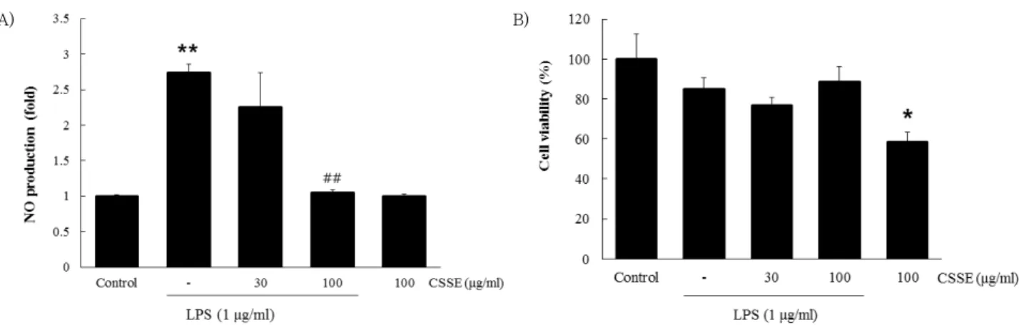
1. Effects of CSSE on LPS-induced production of NO and cell viability
We examined the effects of CSSE on LPS-inducible production of NO. Different dosages of CSSE (30 and 100 ㎍/㎖) were used for evaluation of the inhibitory effects of CSSE on LPS-induced production of NO in RAW 264.7 cells. Compared to control, treatment with LPS resulted in significantly increased production ( p <
0.01) of NO. However, treatment with CSSE (100 ㎍/㎖) resulted in significantly reduced production ( p < 0.01) of LPS-induced NO (Fig. 1A). Therefore, CSSE exhibited an inhibitory activity for induction of NO in macrophages.
In addition, the MTT assay was used for examination of possible cytotoxic effects of CSSE in RAW 264.7 cells.
As shown in Fig. 1B, no significant change in cell viability was observed according to the different concentrations of CSSE treatment.
A) B)
Fig. 1. Inhibitory effects of CSSE on LPS-induced production of NO (A) in RAW 264.7 cells. The cells (5 × 10⁵cells/㎖) were treated with various concentrations (30 and 100 ㎍/㎖) of CSSE for 1 h, followed by continuous incubation with LPS (1 ㎍/㎖) for the next 18 h.
Concentrations of NO in culture medium were monitored as described in the methods section. The MTT assay was used for measurement of viability of cells exposed to CSSE (B). Data represent the mean ± S.D. from three separate experiments. *p < 0.05, **p < 0.01 significant compared with vehicle-treated control; ##p < 0.01 significant compared with LPS alone.
2. Effects of CSSE on LPS-induced production of pro-inflammatory cytokines (TNF-α , IL-1β , and IL-6)
We evaluated the effects of CSSE on LPS-inducible production of TNF-α, IL-1β , and IL-6 by enzyme immunoassay.
Compared to control, treatment with LPS resulted in significantly increased production ( p < 0.01) of TNF-α , IL-1β , and IL-6 in culture supernatants of RAW 264.7 cells. However, at the concentrations of 30 and 100 ㎍/㎖, treatment with CSSE resulted in significantly ( p < 0.01) inhibited LPS-induced production of TNF-α , IL-1β , and IL-6 (Fig. 2 A, B, and C). Therefore, these results indicate that CSSE is a potent inhibitor of induction of TNF-α , IL-1β, and IL-6.
A) B)
C)
Fig. 2. Inhibitory effects of CSSE on LPS-induced production of pro-inflammatory cytokines (TNF-α (A), IL-1β (B), and IL-6 (C)) in RAW 264.7 cells. The cells (5 × 10⁵cells/㎖) were treated with various concentrations (30 and 100 ㎍/㎖) of CSSE for 1 h, followed by continuous incubation with LPS (1 ㎍/㎖) for the next 18 h. Concentrations of pro-inflammatory cytokines in culture medium were monitored as described in the methods section. Data represent the mean ± S.D. from three separate experiments. **p < 0.01 significant compared with vehicle- treated control; ##p < 0.01 significant compared with LPS alone.
3. Effects of CSSE on LPS-induced protein expression of iNOS and COX-2
As shown in Fig. 3, outstandingly up-regulated levels of iNOS and COX-2 protein were observed in response to LPS. Treatment with CSSE (100 ㎕/㎖) resulted in significant inhibition of levels of LPS-stimulated iNOS and COX-2 protein (Fig. 3A) ( p < 0.01 for iNOS (B); p < 0.05 for COX-2 (C), Fig. 3). Accordingly, these data show that CSSE is an inhibitor of induction of iNOS and COX-2 protein.
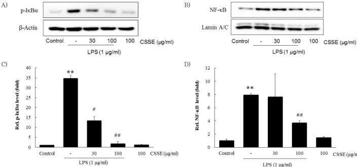
4. Effects of CSSE on LPS-induced activation of NF-κ B
To examine if the reduction of nuclear translocation of NF-κ B (p65) by CSSE was due to the inhibition of p-Iκ Bα , Western blot analysis was conducted to evaluate levels of p-IκBα and nuclear NF-κ B (p65). Treatment with LPS significantly increased the level of p-Iκ Bα ( p < 0.01) and CSSE (100 ㎍/㎖) significantly blocked the LPS-stimulated increase ( p < 0.01, Fig. 4A and C). In addition, NF-κ B (p65) was accumulated in the nucleus after treatment with LPS and the LPS-stimulated NF-κB accumulation was significantly inhibited by CSSE (100 ㎍/㎖) ( p < 0.01, Fig.
4B and D).
A)
B) C)
Fig. 3. Inhibitory effects of CSSE on LPS-induced expressions of iNOS and COX-2 (A). RAW 264.7 cells (5 × 10⁵cells/㎖) were treated with various concentrations (30 and 100 ㎍/㎖) of CSSE for 1 h, followed by continuous incubation with LPS (1 ㎍/㎖) for the next 18 h. Control cells were incubated with vehicle alone. Western blot analysis was performed for determination of protein levels of iNOS and COX-2. β- Actin was used as a loading control. The blots shown are representative of three blots yielding similar results. The densities of iNOS (B) and COX-2 (C) were measured via densitometry. Data represent the mean ± S.D. from three separate experiments. *p < 0.05, **p < 0.01 significant compared with vehicle-treated control; #p < 0.05, ##p < 0.01 indicate significant differences from the LPS-induced group.
A) B)
C) D)
Fig. 4. Inhibitory effects of CSSE on the LPS-induced activation of p-IκBα (A and C) and nuclear NF-κB (p65) (B and D). RAW 264.7 cells at a concentration of 5 × 10⁵cells/㎖ were treated with CSSE (30 and 100 ㎍/㎖) for 1 h, then with LPS (1 ㎍/㎖) for 15 min. Control cells were treated with vehicle only. Western blot analysis was performed to determine the protein levels of p-IκBα and NF-κB (p65). The densities of p-IκBα and NF-κB (p65) were measured by densitometry. Data are given as the mean ± S.D. of three replicates for each sample.
**p < 0.01 significant compared with vehicle-treated control. #p < 0.05 and ##p < 0.01 significant compared with LPS only group.