책임저자: 함대현, 서울시 동대문구 회기동
130-702, 경희대학교 한의과대학 침구경락과학연구 센터
Tel: 02-961-0366, E-mail: [email protected] 접수: 2011년 2월 15일, 심사: 2011년 3월 10일 게재승인: 2011년 3월 12일
본 연구는 보건복지부 보건의료기술진흥사업의 지원에 의하여 수행되었음(과제번호: A091037).
오징어 유래 포스파티딜세린(phosphatidylserine)의 in vitro 항염 효과
*경희대학교 침구경락과학연구센터, †경희대학교 한의과대학 기초한의과학과, ‡(주)두산글로넷,
§한국기초과학지원연구원 춘천지원, ¶원광대학교 인체과학연구소
염 미 정 *ㆍ박 현 정 *ㆍ심 현 수 *ㆍ심 인 섭 *,†ㆍ한 정 준‡ㆍ정 국 훈‡ㆍ허 송 욱§ㆍ정 동 명¶ㆍ함 대 현 *,†
In vitro Anti-inflammatory Activity of Squid Lecithin-transphosphatidylated Phosphatidylserine in Raw 264.7 Cells
Mijung Yeom*, Hyun-Jung Park*, Hyun-Soo Shim*, Insop Shim*,†, Jeong-Jun Han‡, Guk Hoon Chung‡, Song Her§, Dong-Myong Jeong¶, Dae-Hyun Hahm*,†
*Acupuncture and Meridian Science Research Center, Kyung Hee University, Seoul, †The Graduate School of Basic Science of Oriental Medicine, College of Oriental Medicine, Kyung Hee University, Seoul, ‡Glonet BU, Doosan Co., Youngin-si, Gyeonggi-do, §Korea Basic Science Institute Chuncheon Branch, Gangwon-do, ¶Institute of Somatic Science, Wonkwang University, Chonbuk, Korea
In the present study, the effects of squid-derived phosphatidylserine (PS) have been evaluated on lipopolysaccharide (LPS)- induced release of tumor necrosis factor-alpha (TNF-α) and interleukin-1 beta (IL-1β) in the macrophage Raw 264.7 cells. The cytotoxicity of PS in Raw 264.7 macrophages was measured by MTT-based cytotoxicity assay. The mRNA and protein levels of tumor necrosis factor (TNF)-α and interleukin (IL)-1β were measured by quantitative reverse-transcriptase polymerase chain reaction (RT-PCR) and by enzyme-linked immunosorbent assay (ELISA), respectively. Cytotoxic effects of PS in Raw 264.7 macrophages were not observed at concentration up to 100 μg/ml. Squid PS inhibited the mRNA and protein expression of TNF-α but not IL-1β. Taken together, these results indicated that squid-originated PS exhibited a significant activity of anti-inflammation in vitro and might be a useful candidate for treating various inflammatory diseases. (Korean J Str Res 2011;19:89∼95)
Key Words: Squid, Phosphatidylserine, Inflammation, Raw 264.7, Lipopolysaccharide (LPS)
서 론
인체에 가해지는 외적, 정신적 스트레스를 막론하고 모 든 스트레스는 hypothalamic pituitary adrenocotical axis (HPA 축)을 활성화하여 혈중 부신피질 자극호르몬(ACTH)과 cortisol (설치류의 경우 corticosterone)의 농도를 증가시킨다 (Young & Akil, 1985).
병원균의 대표적인 내독소인 lipopolysaccharide (LPS)의 주
입은 대식세포나 단핵구를 활성화를 통해 다양한 염증성 cytokine을 분비시켜서, 열, 시상하부, 부신피질 등의 활성 을 유도하여 감염이나 염증을 유발한다(Bret-Dibat et al., 1997). 또한 LPS의 주입은 혈중 내 corticosterone의 농도를 증가시키는데, 이를 염증 스트레스라 한다(Johnson et al., 1996).
염증형성 과정에서 대식세포는 tumor necrosis factor-α (TNF-α), interleukin-1β (IL-1β) 등의 염증성 cytokine, prostagrandin, free radicals 등 다양한 매개물질을 생산하여 초기 염증 반응에서 감염 방어에 중요한 역할을 한다 (Higuchi et al., 1990; Weller, 1997; Seo et al., 2002). 그러나 대 식세포의 과다한 활성은 이러한 염증 인자의 과잉 생산을 통해 오히려 염증 반응을 심화시키고, 인체에 유해한 방향 으로 진행될 수 있다. 따라서 이러한 염증의 제어가 만성 적인 염증 질환의 예방 및 치료에 유의하게 작용할 수 있 을 것이라 사료된다.
Phosphatidylserine (PS)는 인산지방질의 하나로, 생물의 세 포막을 구성하는 중요한 성분이며, 그 중에서도 뇌의 신경 세포막에 많이 포함되어 있다. 정상적으로 PS는 세포막의 속판(inner leaflet)에 존재하지만, 세포사멸신호를 받은 세포 의 세포막 외부(out leaflet)로 노출된다(Fadeel, 2003; Lauber et al., 2004). 대식세포가 세포표면의 수용체를 통해 세포막 외부로 노출된 PS를 인식하여 식균작용에 의해 apoptotic cell을 제거한다(Fadok et al., 2000). 이런 이유로 PS는 염증 완화의 중요한 부분인 apoptotic cell의 제거의 시작에 중요 한 것으로 알려져 있다. PS 리포좀은 케리지난으로 유도된 생쥐의 발바닥 부종을 억제했는데, 이때 PS 리포좀에 의한 proliferator-activated receptors (PPARs)의 활성화가 관여하는 것으로 나타났다(Ramos et al., 2007). 이런 항염 효과는 PS 리포좀이 apoptotic cell의 세포막을 모방에 따른 것으로 사 료된다. PS의 항스트레스 효과 또한 보고되고 있다. 예를 들어, PS의 장기 복용은 스트레스로 유도된 HPA 축의 활 성화에 대응하였고(Monteleone et al., 1992), 스트레스 호르 몬의 증가나 면역 체계의 이상 등을 억제하였다(Kelly, 1999).
전통적으로, 의약품이나 식이보충제에서 사용되는 PS는 소의 뇌에서 분리하였다. 그러나 최근 광우병에 대한 우려 로 그 사용이 금지되고 있다. 따라서, 대두 유래 PS가 개발 되어 대체 사용되고 있고, 화학적 구조 등에 차이가 있음 에도 불구하고, 소의 뇌 유래 PS와 그 기능이 유사하다고 일부 알려져 있으나, 실제로 그 효과에 대하여 논란이 많 다. 이러한 이유로 소의 뇌 유래 PS를 대체한 동물성 PS의
개발에 많은 관심이 집중되고 있다. 대표적인 예로 오징어 나 새우와 같은 해산물 유래 PS가 개발되고 있으며, 그 효 능이 발표되고 있다.
따라서 본 연구의 목적은 LPS로 활성화된 Raw 264.7 세 포에서 TNF-α 및 IL-1β와 같은 pro-inflammatory cytokine 의 생성에 미치는 오징어 유래 PS의 영향을 조사함을 통해 PS의 염증 스트레스에 적용 가능성을 확인하는 것이다.
재료 및 방법
1. 세포 배양
Murine macrophage Raw 264.7 세포는 한국세포주은행 (KCLB)에서 분양받았으며, 10% fetal bovine serum (FBS) 및 penicillin (100μg/ml), streptomycin (100 U/ml)이 포함된 DMEM 배지에서 37oC, 5% CO2 incubator에서 배양하였다.
2. 오징어 유래 PS
본 실험에서 사용된 오징어 추출 phosphatidyserine은 (주) 두산글로넷에서 제공하였고 오징어에서 추출된 lecithin을 원료로 효소를 이용하는 transphosphatidylation 반응을 통해 제조하였다. 그 제조된 오징어 PS (92%)의 조성은 다음과 같다: 1% myristic, 23.3% palmitic, 0.8% palmitoleic, 5.5%
stearic, 2.7% oleic, 0.2% linoleic, 4% eicosanoic, 13.5% EPA, 40.7% DHA, 8.3% others.
3. 염증 반응 유도 및 PS 처리
Raw 264.7 대식세포를 2×10⁵ cells/well을 6 well에 plating 하고 16시간 후 lipopolysaccharide (LPS, 1μg/ml)를 처리하였 다. 염증 반응에 대한 PS의 효과를 알아보기 위해, LPS 처 리와 동시에 1, 10, 100μg/ml의 농도로 첨가하여 24시간 동안 배양하였다. 오징어 유래 phosphatidylserine (PS)는 두산 으로부터 제공받았다.
4. 세포 독성 시험
세포 독성은 EZ-Cytox Enhanced cell viability assay kit (DaeilLab Service, Korea)를 이용하여 실시하였다. 96 well plate에 2×10⁵ cells/well로 세포를 준비하여, 여러 농도의 시 료 용액을 처치하였다. 24시간 및 48시간 후에 kit reagent를 넣고 2시간 동안 incubation 후 wave length 450 nm에서 흡광 도를 측정하였다.
Table 1. Primers for RT-PCR analysis.
Gene PCR primers
TNF-α Sense: 5'-GCA GAA GAG GCA CTC CCC CA- 3' Antisense: 5'-GAT CCA TGC CGT TGG CCA GG- 3' IL-1β Sense: 5'-GGC TGT GGA GAA GCT GTG GC- 3'
Antisense: 5'-GGG TGG GTG TGC CGT CTT TC- 3' GAPDH Sense: 5'-ACA CAT TGG GGG TAG GAA CA- 3' Antisense: 5'-AAC TTT GGC ATT GTG GAA GG- 3'
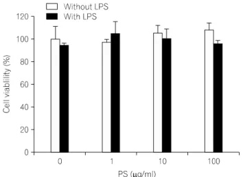
Fig. 1. Effects of squid-derived phosphatidylserine (PS) on cell viability Raw 264.7 macrophages. After treatment with 1, 10, and 100μg/ml of PS for 24 h, cell viability was determined by MTT assay.
5. RNA 분리 및 RT-PCR
LPS 및 다양한 농도의 PS (1, 10, or 100μg/ml)를 24시간 처리한 후, total RNA를 TRIzol reagent (InVitrogen, USA)를 이용하여 제조사의 지침에 따라 Raw 264.7 세포로부터 분 리하였다. RNA의 농도는 spectrophotometer를 이용하여 260 nm에서 측정하였고, RNA의 순도는 260 nm와 280 nm에서 측정한 흡광도의 비율을 이용하여 평가하였다. 역전사 반 응은 Moloney murine leukemia virus reverse transcriptase (M- MLV RTase; TaKaRa Inc., Japen)와 random hexamer를 이용하 여 이루어졌는데, 1μg의 total RNA로부터 cDNA를 합성하 였다. Pro-inflammatory cytokine인 IL-1β와 TNF-α의 mRNA 발현 정도를 확인하기 위해 각각의 염증매개물질에 대한 특이적인 primer (Table 1)를 사용하여 PCR 반응을 통해 증 폭하였다. PCR 반응 수행한 후, ethidium bromide를 사용하 여 1.5% argarose gel 상에서 band를 확인, 분석하였다.
6. 세포 배양액 내의 cytokines 측정
세포 배양액 내의 cytokines의 양을 측정하기 위해 Enzyme- Linked Immunosorbent Assay (ELISA)를 수행하였다. 세포에 1 μg/ml의 LPS 및 다양한 농도의 PS를 24시간 처리한 후 세 포배양액을 수거하여 cytokines 측정에 이용하였다. 배양액 을 적절한 농도로 희석한 후 TNF-α와 IL-1β 각각의 ELISA kit (BD biosciences, USA)를 사용하여, 제조사의 지침 에 따라 측정하였다. 각 실험군 당 well 수는 3으로 하였다.
7. 통계학적 분석
실험치의 값은 mean±SE로 나타냈으며, 통계적 분석은 one-way ANOVA analysis로 그 유의성을 분석하였다(*p
<0.05, **p<0.01, ***p<0.001).
결 과
1. PS가 세포 독성에 미치는 영향
마우스 대식세포 Raw 264.7에 대한 PS의 세포 독성을 알 아보기 위하여 MTT-based cytotoxicity assay를 실행하였다.
PS를 1, 10, 100μg/ml의 농도 별로 24시간 처리한 결과, 세 포의 생존율에는 거의 영향을 주지 않았다. 또한 LPS에 의 해 유도된 염증 조건 하에서도 100μg/ml 이하의 농도까지 의 PS는 세포의 생존율에는 거의 영향을 주지 않았다 (Fig.
1). 이러한 결과로 PS는 100μg/ml 이하의 농도까지는 Raw 264.7 대식세포에서 염증 조건 하에서조차 독성이 없음을 확인하였다.
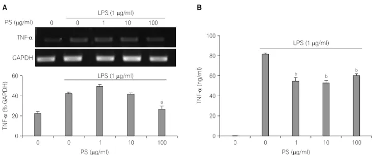
2. PS가 TNF-α의 mRNA 발현과 생성에 미치는 영향 LPS 처리에 의해 과형성 되는 TNF-α는 선천적 면역 체 계에서 중요한 역할을 한다(Lee AK et al., 2003). LPS 처리는 Raw 264.7 대식세포에서 TNF-α의 mRNA와 단백질의 발 현을 현저히 증가시켰다. LPS 처리에 의해 증가된 TNF-α mRNA의 발현은 PS에 의해 농도의존적 감소를 보였다(Fig.
2A). 특히 LPS 처리에 의해 증가된 TNF-α 단백질의 생성 은 1μg/ml의 PS에서부터 통계적으로 유의하게 강력히 억 제되었다(p<0.05; Fig. 2B).
3. PS가 IL-1β의 mRNA 발현과 생성에 미치는 영향 IL-1β는 TNF-α, IL-6 등과 함께 pro-inflammatory cytokine
Fig. 2. Effects of squid-derived phosphatidylserine (PS) on LPS-induced TNF-α mRNA expression and production in Raw 264.7 macrophages. Raw 264.7 cells were treated with/ without various concentrations of PS and 1μg/mL LPS in serum-free DMEM for 24 h. ap<0.01, bp<0.001 vs. LPS-treated group without PS.
Fig. 3. Effects of squid-derived phosphatidylserine (PS) on LPS-induced IL-1β mRNA expression and production in Raw 264.7 macrophages. Raw 264.7 cells were treated with/without various concentrations of PS and 1μg/mL LPS in serum-free DMEM for 24 h.
으로서 여러 면역학적 작용들과 연관되어 있다(Delgado et al., 2003). LPS 처리는 Raw 264.7 대식세포에서 IL-1β의 mRNA와 단백질의 발현을 현저히 증가시켰다. PS는 LPS에 의해 증가된 IL-1β의 mRNA 발현 및 분비를 억제하는 경 향을 나타내었다(Fig. 3).
고 찰
포스파티딜세린(phosphatidylserine, PS)는 세포막의 속판 (inner leaflet)에 존재하는 주요 인산지방질의 하나이다. PS 는 세포막의 구성성분으로 뿐만 아니라, 신호전달, 분비소 포(secretory vesicle) 방출, 세포간의 밀접한 정보교환(cell-to- cell communication), 세포 성장 조절과 같은 신경세포의 세
포막 기능에 있어 중요한 역할을 하는 것으로 알려져 있다 (Vance & Steenbergen, 2005). 흥미로운 것은 PS의 복용이 운 동수행력(sport performance) 향상에 도움이 된다는 것이다.
실제로 PS의 복용은 골프와 같은 운동능력을 향상시키고 (Jager et al., 2007), 격렬한 운동에 따른 증가되는 코티졸의 분비를 억제한다(Kingsley, 2006).
최근 많은 연구들이 우울증과 같은 스트레스성 정신 질 환의 발병 원인으로 염증 반응을 손꼽고 있다(Smith, 1991;
Maes et al., 2009; Miller et al., 2009). 실제로 염증면역체계의 활성화는 신경내분비물질 및 신경전달물질의 발현 변화를 초래하는데, 이는 외적 또는 정신적 스트레스에 의한 변화 와 매우 유사하다. 이런 이유로 두뇌가 염증 면역반응 활 성화를 마치 스트레스 자극과 같이 받아들인다는 가설이 제안되기도 한다(Anisman, 2009). 또한 대표적인 병원균 내 독소인 LPS는 대식세포나 단핵구를 활성화를 통해 다양한 염증성 cytokine을 분비시켜 염증 반응은 물론 혈중 내 GC 의 농도를 증가시키는데, 이를 염증 스트레스라 한다 (Johnson et al., 1996).
따라서 본 연구진은 PS에 의한 코티졸 분비의 감소 보고 를 바탕으로 PS가 스트레스에 대해 효과적일 것이고 더 나 아가 스트레와 밀접한 관계가 있는 염증 반응에 효과가 있 을 것이라 추정하였으며, 이에 PS의 항염 효과를 Raw 264.7 macrophage에서 조사하고자 하였다.
전통적으로, 의약품이나 식이보충제에서 사용되는 PS는 소의 뇌에서 분리하였다. 그러나 최근 광우병에 대한 우려 로 그 사용이 금지되고 있다(Prusiner, 1991). 따라서, 대두 유래 PS가 개발되어 대체 사용되고 있다. 비록 화학적 구 조 등에 차이가 있으나, 소의 뇌 유래 PS와 그 기능이 유사 하다는 발표가 일부 있으나(Sakai et al., 1996; Kato-Kataoka et al., 2010), 그 효과에 대해서 논란이 많다. 이러한 이유로 많은 연구진들이 소의 뇌 유래 PS와 구조가 유사한 새로운 동물 유래 PS 개발에 박차를 가하고 있으며, 난황 또는 새 우나 오징어와 같은 해산물이 그 원천으로 주목 받고 있 다. 본 연구에서 사용된 오징어 유래 PS는 소의 뇌 유래 PS 와 구조적으로 매우 유사하며, 특히 Docosahexaenoic acid (DHA) 함량이 높다(Chen et al., 1989).
대식세포(macrophage)는 숙주의 면역방어 기전에 중요한 역할을 수행하며, 가장 중요한 염증성 cytokine의 근원이 된 다. 대표적인 세포 내 독소인 LPS는 대식세포를 자극하여 다양한 염증성 cytokine을 분비하고, 이러한 염증성 cytokine 들은 면역세포를 활성화시켜 세균의 침입을 효과적으로
방어하도록 도와준다. 그러나 LPS의 자극을 받은 대식세포 에 의한 다량의 TNF-α와 같은 염증성 cytokine의 분비는 정상 세포를 파괴할 수 있으므로 LPS에 의해 자극 받은 대 식세포의 면역작용을 억제할 수 있는 약물의 발굴은 항염 증 치료제 개발에 있어 중요하다.
염증성 cytokine 중 TNF-α는 LPS 반응의 주요 매개체로, 선천적 면역반응(innate immune response)에서 특히 급성 및 만성 염증반응을 자극하는데 중요한 역할을 한다(Lee et al., 2003). 또 다른 주요 염증성 cytokine인 IL-1β 또한 여러 면 역학적 작용들과 연관되어 있다. 예를 들어 T cell의 활성 화, B cell의 성숙화, NK cell의 활성화를 유도하며(Casey et al., 1993), prostaglandin, leukotriene, platelet-activating factor, nitric oxide 등의 합성을 증가시켜 염증반응을 나타낸다 (Takabayashi et al., 2004). PS의 처리는 LPS 자극에 의한 대식 세포의 TNF-α와 IL-1β의 생산을 억제시켰다. 이러한 항 염증 효과를 보이는 PS의 농도는 100μg/ml 이하의 농도로 세포 독성을 나타내지 않았다. 이는 PS의 항염증 효과는 세포 생존율의 감소에 의한 것이 아니라 물질의 고유한 특 성임을 의미한다.
이와 같은 PS의 항염증성 효과는 염증성 스트레스를 포 함한 만성 염증성 질환의 예방 및 치료에 대한 효과적인 응용 가능성을 의미한다.
참 고 문 헌
Anisman H (2009) Cascading effects of stressors and inflammatory immune system activation: implications for major depressive disorder. J. Psychiatry Neurosci. 34:4-20.
Bret-Dibat JL, Creminon C, Couraud JY et al. (1997) Systemic capsaicin pretreatment fails to block the decrease in food-motivated behavior induced by lipopolysaccharide and interleukin-1beta. Brain Res. Bull. 42:443-449.
Casey LC, Balk RA, Bone RC (1993) Plasma cytokine and endotoxin levels correlate with survival in patients with the sepsis syndrome. Ann. Intern. Med. 119:771-778.
Chen S, Benfenati E, Fanelli R et al. (1989) Molecular species analysis of phospholipids by negative ion fast atom bombardment mass spectrometry: application of surface precipitation technique.
Biomed. Environ. Mass. Spectrom. 18:1051-1056.
Delgado AV, Mcmanus AT, Chambers JP (2003) Production of tumor necrosis factor-alpha, interleukin 1-beta, interleukin 2, and interleukin 6 by rat leukocyte subpopulations after exposure to substance P. Neuropeptides 37:355-361.
Fadeel B (2003) Programmed cell clearance. Cell Mol. Life Sci. 60:
2575-2585.
Fadok VA, Bratton DL, Rose DM et al. (2000) A receptor for phosphatidylserine-specific clearance of apoptotic cells. Nature 405:85-90.
Higuchi M, Higashi N, Taki H et al. (1990) Cytolytic mechanisms of activated macrophages. Tumor necrosis factor and L-argi- nine-dependent mechanisms act synergistically as the major cytolytic mechanisms of activated macrophages. J. Immunol. 144:
1425-1431.
Jager R, Purpura M, Geiss KR et al. (2007) The effect of phosphatidylserine on golf performance. J. Int. Soc. Sports Nutr.
4:23.
Johnson RW, Propes MJ, Shavit Y (1996) Corticosterone modulates behavioral and metabolic effects of lipopolysaccharide. Am. J.
Physiol. 270:R192-R1928.
Kato-Kataoka A, Sakai M, Ebina R et al. (2010) Soybean-derived phosphatidylserine improves memory function of the elderly Japanese subjects with memory complaints. J. Clin. Biochem.
Nutr. 47:246-255.
Kelly GS (1999) Nutritional and botanical interventions to assist with the adaptation to stress. Altern. Med. Rev. 4:249-265.
Kingsley M (2006) Effects of phosphatidylserine supplementation on exercising humans. Sports Med. 36:657-669.
Lauber K, Blumenthal SG, Waibel M et al. (2004) Clearance of apoptotic cells: getting rid of the corpses. Mol. Cell 14: 277-287.
Lee AK, Sung SH, Kim YC et al. (2003) Inhibition of lipopolysa- ccharide-inducible nitric oxide synthase, TNF-alpha and COX-2 expression by sauchinone effects on I-kappaBalpha phos- phorylation, C/EBP and AP-1 activation. Br. J. Pharmacol. 139:
11-20.
Maes M, Yirmyia R, Noraberg J et al. (2009) The inflammatory &
neurodegenerative (I&ND) hypothesis of depression: leads for future research and new drug developments in depression. Metab.
Brain Dis. 24:27-53.
Miller AH, Maletic V, Raison CL (2009) Inflammation and its discontents: the role of cytokines in the pathophysiology of major depression. Biol. Psychiatry. 65:732-741.
Monteleone P, Maj M, Beinat L et al. (1992) Blunting by chronic phosphatidylserine administration of the stress-induced activation of the hypothalamo-pituitary-adrenal axis in healthy men. Eur. J.
Clin. Pharmacol. 42:385-388.
Prusiner SB (1991) Molecular biology of prion diseases. Science 252:
1515-1522.
Ramos GC, Fernandes D, Charao CT et al. (2007) Apoptotic mimicry: phosphatidylserine liposomes reduce inflammation through activation of peroxisome proliferator-activated receptors (PPARs) in vivo. Br. J. Pharmacol. 151:844-850.
Sakai M, Yamatoya H, Kudo S (1996) Pharmacological effects of phosphatidylserine enzymatically synthesized from soybean lecithin on brain functions in rodents. J. Nutr. Sci. Vitaminol.
(Tokyo) 42:47-54.
Seo SJ, Choi HG, Chung HJ et al. (2002) Time course of expression of mRNA of inducible nitric oxide synthase and generation of nitric oxide by ultraviolet B in keratinocyte cell lines. Br. J.
Dermatol. 147:655-662.
Smith RS (1991) The macrophage theory of depression. Med.
Hypotheses. 35:298-306.
Takabayashi T, Shimizu S, Clark BD et al. (2004) Interleukin-1 upregulates anaphylatoxin receptors on mononuclear cells. Surgery 135:544-554.
Vance JE, Steenbergen R (2005) Metabolism and functions of phosphatidylserine. Prog. Lipid Re. 44:207-234.
Weller R (1997) Nitric oxide--a newly discovered chemical transmit- ter in human skin. Br. J. Dermatol. 137:665-672.
Young EA, Akil H (1985) Corticotropin-releasing factor stimulation of adrenocorticotropin and beta-endorphin release: effects of acute and chronic stress. Endocrinology 117:23-30.
= 국문초록 =
본 연구에서는 Raw264.7 대식 세포주에서 lipopolysaccharide (LPS)로 유도된 tumor necrosis factor-alpha (TNF-α)와 interleukin-1 beta (IL-1β)의 분비에 대한 오징어 유래 포스파티딜세린(phosphatidylserine)의 효과를 확인하였다.
Raw264.7 대식 세포주에서 PS의 세포독성 효과는 MTT 분석으로 조사하였으며, TNF-α와 IL-1β의 mRNA 및 단백질 의 발현 정도는 각각 RT-PCR 및 ELISA 분석법으로 확인하였다. PS는 100μg/ml 농도까지는 세포 독성을 나타내지 않았으며, 세포 독성을 보이지 않는 농도에서 LPS로 유도된 IL-1β와 특히 TNF-α의 mRNA 및 단백질의 발현을 저해 하였다. 이러한 실험 결과는 염증성 질환을 위한 유용한 물질로써 PS의 개발 가능성을 시사한다고 사료된다.
중심단어: 포스파티딜세린, 염증, Raw264.7 대식 세포, lipopolysaccharide (LPS)