http://dx.doi.org/10.11620/IJOB.2017.42.4.163 pISSN 1226-7155, eISSN 2287-6618
Osteoporosis is a metabolic bone disease that is characterized by low bone mass resulting from an increase in bone resorption relative to bone formation. The most current therapies for osteoporosis have focused on inhibiting bone resorption by osteoclasts. The purpose of this study is to develop new anabolic agents for treatment of osteoporosis that have fewer risks compared to conventional therapies.
We searched the natural products that were derived from the traditional Asian medicines which have been used for treatment of bone related diseases. Icaritin is a flavonoid glycoside derived from the herb Epimedium which has beneficial effects on bone formation. To determine the effect of icaritin on bone formation, we examined the effect of icaritin on MC3T3-E1 cell proliferation and differentiation.
For determining the effects of icaritin on proliferation, we performed the MTT assay using MC3T3-E1 cells. To evaluate whether icaritin could promote the osteogenic differentiation of MC3T3-E1 cells, alkaline phosphatase (ALP) activity and mRNA expressions of Runx2, osteocalcin (OCN), RANKL, and osteoprotegerin (OPG) were determined. Icaritin
increased MC3T3-E1 cell proliferation. Icaritin increased the ALP activity of MC3T3-E1 cells on 72 hour culture in osteogenic media. mRNA expression of Runx2 was increased after 24 hour culture with icaritin. mRNA expression of osteocalcin was increased after 72 hour culture with icaritin.
In addition, icaritin increased the mRNA expressions of OPG and RANKL. However, icaritin increased the mRNA expression of OPG much more than that of RANKL, and then, it increased the OPG/RANKL ratio. These results suggest that icaritin promotes osteogenic differentiation of osteoblasts and decreases osteoclast formation regulated by osteoblasts.
Key words: Icaritin, Herb Epimedium, MC3T3-E1 cells, osteogenic differentiation
Introduction
Osteoporosis is a metabolic bone disease that is characterized low bone mass resulted from an increase in bone resorption relative to bone formation. The strategy for treatments of osteoporosis is to promote bone formation by osteoblasts or inhibit bone resorption. The most current therapies for osteoporosis have focused on inhibiting bone resorption by osteoclasts like bisphosphonates [1]. These drugs also reduce metabolic turn-over of bone, reducing both bone resorption and bone formation, therefore they do not rebuild the damaged trabecular bone. Although the conventional drugs have therapeutic benefits, they also have disadvantages such as breast cancer in estrogen replacement and osteonecrosis of jaw in
Icaritin, a Flavonoid Derived from the Herb Epimedium, Promotes Osteogenic Differentiation of MC3T3-E1 Cells
Dan-Bi Park1, Hee Su Lee2 and Seong-Hee Ko1,*
1Department of Pharmacology, 2Department of Oral anatomy, College of Dentistry and Research Institute of Oral Science, Gangneung-Wonju National University, Ganuneung, Korea
(received August 31, 2017; revised September 15, 2017; accepted September 16, 2017)
*Correspondence to: Seong-Hee Ko, Department of Pharmacology, College of Dentistry and Research Institute of Oral Science, Gangneung-Wonju National University, Ganuneung, Korea
Tel: +82-33-640-2453, Fax: +82-33-642-6410 E-mail: [email protected]
ORCID : 0000-0003-2714-9780
This is an Open-Access article distributed under the terms of the Creative Commons Attribution Non-Commercial License (http://creati- vecommons.org/licenses/by-nc/3.0) which permits unrestricted non- commercial use, distribution, and reproduction in any medium, pro- vided the original work is properly cited.
163
bisphosphonates therapy [2]. The purpose of this study is to develop the new anabolic agents for treatment of osteoporosis that have fewer risks compared to conventional therapies. We searched the natural products that derived from traditional Asian medicines which have been used for treatment of bone injuries and bone related diseases.
Herb Epimedii have been prescribed for improving cardiovascular and cerebrovascular functions as well as treatment of osteoporosis in traditional Asian medicine [3].
Epimedii contain a number of flavonoid glycosides or ligands including icariin I, II, icariside I, II, icaritin, epimedoside A–E, epimedin A–C, ikarisoside A–E [3]. Flavonoids including icariin, epimedin B, and epimedin C are the main active components from Epimedium plant having antiosteoporotic effects [4]. Among these, icariin is a major component that have a lot of pharmacologic activities such as anti-osteoporosis, neuroprotection, cardiovascular protection, anti-tumor, immunoprotection [5]. Icaritin is a common serum metabolite of seven flavonoid derived from herb Epimedium (epimedoside A, hexandraside F, epimedin A,B,C, icariin, baohuoside-I) [6].
Therefore, to determined the effect of icaritin on bone formation, we examined the effect of icaritin on MC3T3-E1 cell proliferation and differentiation.
Materials and Methods
Reagent
Icaritin was obtained from Dr. JW Lee (KIST Gangneung Institute of Natural Products, Gangneung-shi, Korea). Modified Eagle medium (α-MEM) and fetal bovine serum (FBS) were purchased from HyClone Laboratories (Logan, UT, USA). The easy-BLUE™ was ordered from iNtRON Biotechnology (Kyungki-Do, Korea) and the AccuPower RT-PreMix and AccuPower®2X GreenStar™ qPCR MasterMix were purchased from Bioneer (Daejeon, Korea). PCR primers were synthesized by Macrogen (Seoul, Korea). Thiazolyl blue tetrazolium bromide and alkaline phosphatase kit were purchased from Sigma-Aldrich (St. Louis, MO, USA). SensoLyte pNPP alkaline phosphatase assay kit was purchased from AnaSpec (Fremont, CA, USA).
Cell culture and cell proliferation assay
For an in vitro model for osteoblast, we used the MC3T3-E1 cell line. MC3T3-E1 cells were cultured in α-MEM with 10%
fetal bovine serum and penicillin 100 U/ml and streptomycin 100 μg/ml at 37℃ in a humidified atmosphere of 5% CO2.
The MTT (3-(4,5-dimethylthiazol-2-yl)-2,5-diphenyl-2H- tetrazolium bromide) assay was used as a measurement of cell proliferation. For MTT assay, MC3T3-E1 cells seeded in 24-well plates 1x 10⁵ cell per well. Cells were treated with 1, 10 100 nM icaritin or DMSO (control). After 24 hours, the culture solution containing 5 mg/ml thiazolyl blue tetrazolium bromide was added and incubated for 4 hours. The cells were washed with PBS. Formazan was dissolved using dimethylsulfoxide (DMSO), and detected using a microplate spectrophotometer (BioTek, St. Winooski, VT, USA) at 540 nm.
ALP activity assay and ALP staining
MC3T3-E1 cells were cultured for 72 hours in osteogenic differentiation media (α-MEM with 10% FBS with 50 μg/ml ascorbic acid, 10 mM β-glycerophosphate) at 1x 10⁵ cell per well in 24 well plate with 1, 10, 100 nM icaritin or DMSO (control). ALP activity assay was determined using SensoLyte pNPP alkaline phosphatase assay kit. The cells were lysed by scraping with a scraper in the assay buffer containing triton X-100 and incubated at 4℃ for 10 minutes.
After centrifugation for 10 minutes, only supernatant was used for ALP activity assay. ALP activity was detected using a microplate spectrophotometer (BioTek, St. Winooski, VT, USA) at 405nm.
ALP staining was determined using alkaline phosphatase kit.
The cell were fixed with citrate-acetone-formaldehyde fixative solution. After washed, alkaline-dye mixture was added and incubated for 15 minutes in a protected from light. For the nuclei staining, the cells were stained with hematoxylin.
Real-time quantitative RT-PCR
MC3T3-E1 cells were cultured in osteogenic differentiation media treated with 1, 10 100 nM icaritin or DMSO (control).
Total RNA was extracted using easy-blue reagent according to the manufactured protocol after 24 hours culture for runt-related transcription factor 2 (Runx2) and 72 hours culture for osteocalcin (OCN), receptor activator of nuclear factor κB ligand (RANKL) and osteoprotegerin (OPG) mRNA expression determination. The 2.5 μg total RNA was reverse-transcribed using the AccuPower RT PreMix under the following conditions:
42℃ 60min, 94℃ 5min. Quantitative real time-PCR for Runx2, osteocalcin, RANKL, OPG and glyceraldehyde-3-phosphate dehydrogenase (GAPDH) was carried out using mixture of
AccuPower®2X GreenStar ™ qPCR MasterMix and primer (table 1.) by CFX96 ™ system (Bio-Rad, Hercules, Califonia).
The cycling conditions were 3 min polymerase activation at 95 ℃ followed by 40 cycles of 95 ℃ for 10 sec, 58 ℃ for 30 sec. The sequences of the PCR primers used for real-time PCR are shown in Table 1. The target genes were normalized on the basis of its GAPDH.
Statistical analysis
Results were presented as the mean ± SD of these experiments and statistically analyzed by Student’s t-test between the means of the control and test groups. Statistical significance was set at p<0.05.
Results
Icaritin increased MC3T3-E1 cell proliferation in 1, 10 nM.
However, 100 nM icaritin did not change cell proliferation compared to control (Fig 1). To investigate the effects of icaritin on differentiation of MC3T3-E1 cells, we determined the ALP activity and ALP positive cells by ALP activity assay and ALP
Fig. 1. The effect of icaritin on proliferation of MC3T3-E1 cells.
For MTT assay, MC3T3-E1 cells seeded in 24-well plates 1x 10⁵ cell per well. Cells were treated with 1, 10 100 nM icaritin or DMSO (control). After 24 hours, 5 mg/ml thiazolyl blue tetrazolium bromide was added and incubated for 4 hours. The formazan was detected using a microplate reader. Results were presented with mean ± S.D. (N=4). *p<0.05, compared to control.
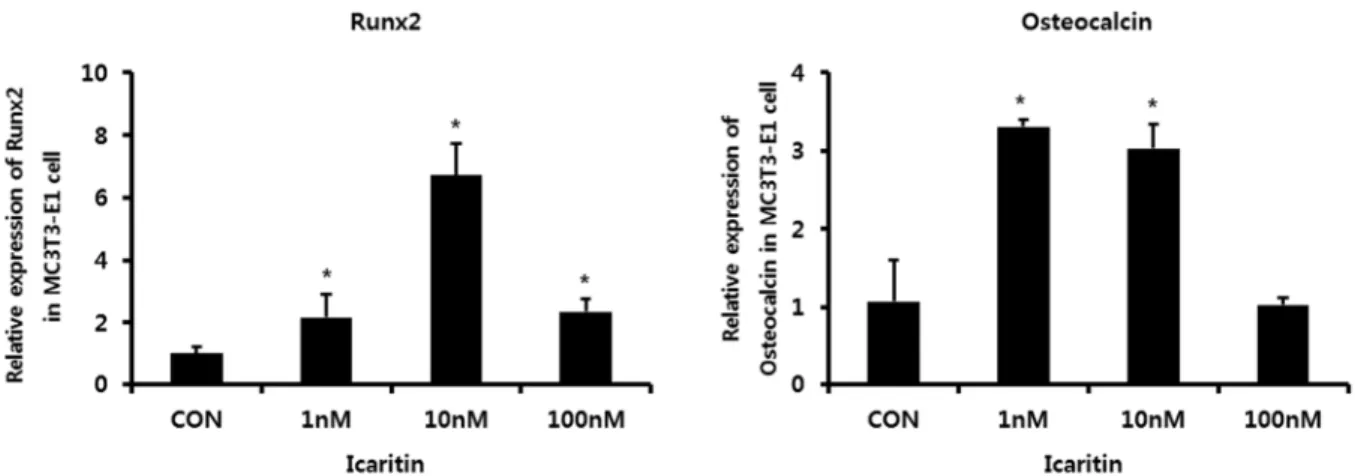
Fig. 2. The effect of icaritin on ALP activity of MC3T3-E1 cells.
MC3T3-E1 cells were cultured for 72 hours in osteogenic differentiation media (α-MEM with 10% FBS with 50 μg/ml ascorbic acid, 10 mM β-glycerophosphate) at 1x 10⁵ cell per well in 24 well plate with 0, 1, 10, 100 nM icaritin. (A) ALP-positive cells was determined by ALP staining (B) ALP activity assay was determined using SensoLyte pNPP Alkaline Phosphatase Assay Kit. Results were presented with mean ± S.D. (N=3). *p<0.05, compared to control.
staining. Icaritin increased ALP activity of MC3T3-E1 cells by 1.7, 2.0, 1.8-fold in 1, 10 100 nM as compared to control (Fig 2). Icaritin also increased the number of ALP positive cells (Fig 2). The extent of increase is the bigger in 10 nM icaritin than 1, 100 nM concentration. mRNA expression of Runx2 after 24 hours culture with icaritin was increased by 2.1, 6.7, 2.3–fold in 1, 10 100 nM icaritin respectively compared to control (Fig 3). mRNA expression of osteocalcin after 72 hours culture with icaritin was increased by 3.3, 3.0 -fold in 1, 10 nM respectively (Fig 3). In addition, icaritin increased mRNA expression of OPG by 22.9, 269.2, 64.9-fold in 1, 10 100 nM respectively (Fig 4). Icaritin also increased mRNA expression of RANKL by 5.1, 7.5, 7.7-fold in 1, 10 100 nM respectively (Fig 4).
However, icaritin increased mRNA expression of OPG much more than RANKL. Therefore, icaritin increased OPG/RANKL mRNA ratio (Fig 4).
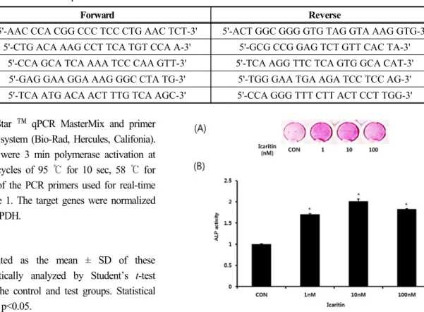
Genes Forward Reverse
Runx2 5'-AAC CCA CGG CCC TCC CTG AAC TCT-3' 5'-ACT GGC GGG GTG TAG GTA AAG GTG-3' Osteocalcin 5'-CTG ACA AAG CCT TCA TGT CCA A-3' 5'-GCG CCG GAG TCT GTT CAC TA-3'
RANKL 5'-CCA GCA TCA AAA TCC CAA GTT-3' 5'-TCA AGG TTC TCA GTG GCA CAT-3'
OPG 5'-GAG GAA GGA AAG GGC CTA TG-3' 5'-TGG GAA TGA AGA TCC TCC AG-3'
GAPDH 5'-TCA ATG ACA ACT TTG TCA AGC-3' 5'-CCA GGG TTT CTT ACT CCT TGG-3' Table 1. Primer Sequences for Real-time quantitative RT-PCR
Discussion
The strategies of osteoporosis therapy is to gain positive bone balance that is to increase bone formation or to decrease
bone resorption. Currently available therapeutic drugs are bisphosphonates which reduced bone resorption, and some anabolic agents such as parathyroid hormone (PTH) 1-84 and teriparpatide. The anabolic effect of PTH, when it is administered intermittently by subcutaneous, has been Fig. 3. The effects of icaritin on the mRNA Expression for (A) Runx2 and (B) osteocalcin. MC3T3-E1 cells were cultured in osteogenic differentiation media without or with icaritin. Cells were collected after 24 hours culture for Runx2 and 72 hours culture for osteocalcin mRNA expression determination. Results were presented with mean ± S.D. of duplicates. *p<0.05, compared to control.
Fig. 4. The effects of icaritin on the mRNA expression for RANKL and OPG. MC3T3-E1 cells were cultured in osteogenic differentiation media without or with icaritin. Cells were collected after 72 hours culture for mRNA expression determination (A). The ratio of OPG/RANKL mRNA was presented (B). Results were presented with mean ± S.D. of duplicates. *p<0.05, compared to control.
reported [7]. However, the side effects of PTH therapy, such as hypercalcemia and bone cancer and the anabolic window of 2 years which reduce the therapeutic effect of PTH after 2 year treatment, limited the PTH use in osteoporosis treatment.
Therefore, new anabolic agents such as anti-sclerostin antibodies are in development [8]. Hormone replacement therapy (HRT) also had been used because estrogen could prevent postmenopausal osteoporosis [9]. However, it has been reported that HRT has potential complications such as breast cancer, cardiovascular disease [10].
To search for new anabolic agent, we examined the effect of natural products which have been used in Asian traditional medicine for osteoporosis treatment. Natural products which have been used for long time are more suitable to long-term therapy without side effects. Herb Epimedium is one of agents that have been used for bone health in China, Japan and Korea.
Icariin is a active component of Epimedii and is metabolized to icaritin. We determined the effects of icaritin on bone formation in vitro. Icaritin increased proliferation of MC3T3-E1 cells (Fig 1). it is similar with results that icariin stimulates proliferation of mice primary osteoblasts, UMR-106 cells, human osteoblastic cell-line, MG-63 and mouse preosteoblastic cell, MC3T3-E1 cells [11,12,13]. In our study, icaritin at concentration 10 nM had the largest effect on cell proliferation, and had no effect at 100 nM concentration. Icariin also promoted osteogenic differentiation of various cell types [14]
Icariin increased mRNA expression or protein synthesis of osteogenic markers such as ALP, collagen type 1, Runx2.
Icaritin in our study also increased ALP activity, at 1-100 nM concentration in the osteogenic media that contained ascorbic acid and β–glycerophosphate. And icaritin increased the number of ALP-positive cells (Fig 2). To determine the effect of icaritin on osteoblast differentiation, we examined the mRNA expression of osteogenic marker after icaritin treatment for 24 or 72 hours by real-time RT-PCR (Fig 3). Icaritin increased the mRNA expression of Runx2 which is pivotal transcriptional factor in osteoblast differentiation after 24 hours treatment but not 72 hours (data not shown). Ostecalcin was increased after 72 hours icaritin treatment. Therefore, we suggest that icaritin promote the osteogenic gene expression, and may induce osteoblastic differentiation and bone formation. Icariin has been reported to increase the bone density in ovariectomized rat which has estrogen deficiency. And it was reported that icariin stimulated the osteoblast function through estrogen receptor [15]. Icaritin also has a similarity of chemical structure with
estrogen. Recently, icaritin has been found to induce osteoblastic cell differentiation through estrogen receptor [16].
Icariin has been reported to increase bone formation parameters and to decrease bone resorption parameters in vivo, and these effects were diminished in OPG-deficient mice [17].
These reports may mean that icariin inhibit osteoclast formation through OPG. Icariin decreased osteoclast differentiation in osteoblast - bone marrow cell coculture system [18].
Osteoclasts, which are multinucleated cells formed by fusion of mononulear precusors cells are responsible for the bone resorption. RANKL binding to its receptor RANK leads to differentiation of osteoclasts. OPG, decoy receptor of RANKL, prevent osteoclast differentiation. Osteoblasts secrete the RANKL and OPG, and RANKL/ OPG ratio is an important factor in bone mass [19, 20]. We examined the effects of icaritin in expression of mRNA of RANKL and OPG. Icaritin increased both mRNA. However, the extent of increase of OPG mRNA expression is much greater than RANKL. Icaritin increased OPG mRNA expression by 269 fold compared to control, and in case of RANKL, increased by 7.5 fold in 10 nM. Icaritin may inhibit the osteoclast formation by modulation of OPG/RANKL expression ratio. In conclusion, these results suggest that icaritin promote osteogenic differentiation of osteoblasts, and decrease osteoclast formation regulated by osteoblasts. These suggest that icaritin is a promising candidate for the anabolic agent of osteopororis treatment.
Conflict of interest
The authors declare that they have no conflicting interest.
References
1. Milat F, Ebeling PR. Osteoporosis treatment: a missed opportunity. Med J Aust 2016;205:185-190
2. McClung M, Harris ST, Miller PD, Bauer DC, Davison KS, Dian L. Bisphosphonate therapy for osteoporosis: benefits, risks, and drug holiday. Am J Med 2013;126:13-20. doi:
10.1016/j.amjmed.2012.06.023.
3. Ma H, He X, Yang Y, Li M, Hao D, Jia Z. The genus Epimedium: an ethnopharmacological and phytochemical review. J Ethnopharmacol. 2011 ;134:519-541. doi:10.1016/
j.jep.2011.01.001
4. Jia M, Nie Y, Cao DP, Xue YY, Wang JS, Zhao L, Rahman K, Zhang QY, Qin LP. Potential Antiosteoporotic Agents from Plants: A Comprehensive Review. Evid Based Complement
Alternat Med. 2012; 2012:1-28. doi:10.1155/2012/364604 5. Li C, Li Q, Mei Q, Lu T. Pharmacological effects and
pharmacokinetic properties of icariin, the major bioactive component in Herba Epimedii. Life Sci 2015;126:57-68.
http://dx.doi.org/10.1016/j.lfs.2015.01.006
6. Zhang G, Wang XL, Sheng H, Xie XH, He YX, Yao XS, Li ZR, Lee KM, He W, Leung KS, Qin L. Constitutional flavonoids derived from Epimedium dose-dependently reduce incidence of steroid-associated osteonecrosis not via direct action by themselves on potential cellular targets. PLoS One. 2009; 29:4(7):1-11. doi:10.1371/journal.pone.0006419 7. Poole KE, Reeve J. Parathyroid hormone - a bone anabolic
and catabolic agent. Curr Opin Pharmacol. 2005;5(6):
612-617. doi: 10.1016/j.coph.2005.07.004
8. Ponnapakkam T, Katikaneni R, Sakon J, Stratford R, Gensure RC. Treating osteoporosis by targeting parathyroid hormone to bone. Drug Discov Today. 2014; 19(3):204-208. doi:
10.1016/j.drudis.2013.07.015.
9. Anderson GL, Limacher M, Assaf AR, Bassford T, Beresford SA, Black H, Bonds D, Brunner R, Brzyski R,Caan B, Chlebowski R, Curb D, Gass M, Hays J, Heiss G, Hendrix S, Howard BV, Hsia J, Hubbell A, Jackson R, Johnson KC, Judd H, Kotchen JM, Kuller L, LaCroix AZ, Lane D, Langer RD, LasserN, Lewis CE, Manson J, Margolis K, Ockene J, O’SullivanMJ, Phillips L, Prentice RL, Ritenbaugh C, Robbins J, Rossouw JE, Sarto G, Stefanick ML, Van Horn L, Wactawski-Wende J, llace R, Wassertheil-Smoller S. Effects of conjugated equine estrogen in postmenopausal women with hysterectomy: The Women’s Health Initiative randomized controlled trial. JAMA 2004;291:1701-1712.
10. Beral V, Million women study collaborators. Breast cancer and hormone- replacement therapy in the million women study. Lancet 2003;362:419-427.
11. Hsieh TP, Sheu SY, Sun JS, Chen MH, Liu MH. Icariin isolated from Epimedium pubescens regulates osteoblasts anabolism through BMP-2, SMAD4, and Cbfa1 expression.
Phytomedicine. 2010;17(6):414-423. doi: 10.1016/j.phymed.
2009.08.007.
12. Mok SK, Chen WF, Lai WP, Leung PC, Wang XL, Yao XS, Wong MS. Icariin protects against bone loss induced by oestrogen deficiency and activates oestrogen receptor- dependent osteoblastic functions in UMR 106 cells. Br J Pharmacol. 2010 ;159(4):939-949. doi: 10.1111/j.1476-5381.
2009.00593.x.
13. Xia L, Li Y, Zhou Z, Dai Y, Liu H, Liu H. Icariin delivery porous PHBV scaffolds for promoting osteoblast expansion in vitro. Mater. Sci. Eng C. 2013;33 :3545-3552. doi:
10.1016/j.msec.2013.04.050.
14. An Ji, Yang H, Zhang Q, Liu C, Zhao J, Zhang L, Chen B.
Natural products for treatment of osteoporosis: The effects and mechanisms on promoting osteoblast-mediated bone formation. Life Sci. 2016;147:46-58. doi: 10.1016/j.lfs.
2016.01.024.
15. Xiao H-H, Fung C-Y, MokS-K, Wong K-C, Ho M-X, Wang X-L, Yao X-S, Wong M-S. Flavonoids from Herba epimedii selectively activate estrogen receptor alpha (ERα) and stimulate ER-dependent osteoblastic functions in UMR-106 cells. J Steroid Biochem & Mole Biol. 2014;143:141-151.
doi: 10.1016/j.jsbmb.2014.02.019.
16. Wu Z, Ou L, Wang C, Yang Li, Wang P, Liu H, Xiong Y, Sun K, Zhang R, Zhu X. Icaritin induces MC3T3-E1 subclone14 cell differentiation through estrogen receptor-mediated ERK1/2 and p38 signaling activation. Biomedicine &
Pharmacotherapy 2017;94:1-9. doi: 10.1016/j.biopha.2017.
07.071.
17. Zheng D, Peng S, Yang SH, Shao ZW, Yang C, Feng Y, Wu W, Zhen WX. The beneficial effect of icariin on bone is diminished in osteoprotegerin-deficient mice. Bone 2012;51:85-92. doi: 10.1016/j.bone.2012.04.006.
18. Hsieh TP, Sheu SY, Sun JS, Chen MH Icariin inhibits osteoclast differentiation and bone resorption by suppression of MAPKs/NF-κB regulated HIF-1α and PGE(2) synthesis.
Phytomedicine 2011;18(2-3):176-185. doi: 10.1016/j.phymed.
2010.04.003.
19. Yasuda H, Shima N, Nakagawa N, Yamaguchi K, Kinosaki M, Mochizuki S, Tomoyasu A, Yano K, Goto M, Murakami A, Tsuda E, Morinaga T, Higashio K, Udagawa N, Takahashi N, Suda T. Osteoclast differentiation factor is a ligand for osteoprotegerin/osteoclastogenesis-inhibitory factor and is identical to TRANCE/RANKL. Proc Natl Acad Sci U S A.
1998;95(7):3597-3602
20. Aubin JE, Bonnelye E. Osteoprotegerin and its ligand: A new paradigm for regulation of osteoclastogenesis and bone resorption. Osteoporos Int. 2000;11(11):905-913