Gene Expression of Supernumerary Dental Pulp Related to the Subculture Speed:
A Pilot Study
Yookyung Lee, Jongsoo Kim, Jisun Shin, Jongbin Kim
Department of Pediatric Dentistry, School of Dentistry, Dankook University
The purpose of this study was to investigate the odontoblast gene expression related to the subculture speed of supernumerary dental pulp stem cells (sDPSCs). The stem cell is undifferentiated cells which has the ability to differentiate into various cells. Specific stimulation or environment induces cell differentiation, and these differentiation leads to bone or muscle formation.
20 sDPSCs were obtained from 20 children under aseptic condition. During the culture through the 10th passage, the third passage cells which showed short subculture period and 10th passage cells which showed long subculture period were earned. Each cell was divided into differentiated group and non-differentiated group. Quantitative real-time polychain reaction (q-RT-PCR) was performed for each group. The genes related to odontoblast differentiation, Alkaline Phosphatase (ALP), Osteocalcin (OCN), Osteonectin (ONT), Dentin sialophosphoprotein (DSPP) and Dentin matrix acidic phosphoprotein 1 (DMP-1), were measured.
Differentiated cells showed more gene expression levels. Undifferentiated cells showed higher gene expression level in 10th passages but differentiated cells showed higher gene expression level in 3rd passages. Cells that showed faster subculture period showed relatively lower gene expression level except for OCN and DSPP.
Key words : Supernumerary Dental Pulp Stem Cells, Odontoblasts Abstract
Corresponding author : Jongbin Kim
Department of Pediatric Dentistry, School of Dentistry, Dankook University, 119 Dandaero, Dongnam-gu, Cheonan, 31116, Korea Tel: +82-41-550-1921 / E-mail: [email protected]
Received October 13, 2018 / Revised November 28, 2018 / Accepted November 28, 2018
Ⅰ. 서 론
과잉치는 정상 치열에서 과잉으로 존재하는 치아로 인접 영구 치의 맹출 지연 및 장애, 총생, 정중이개, 치성낭종 형성 등의 문 제를 유발할 가능성이 있어 합병증 발생 시 즉시 발치하는 것이 추천된다[1-4].
줄기세포는 여러 종류의 세포로 분화가 가능한 세포로 이에 대한 많은 연구들이 진행되어왔다. 그 중 치수 유래 줄기세포는 미분화 중간엽세포로 상아모세포 등으로 분화가 가능하다. 치수
조직의 세포밀집대에는 미분화 상태인 세포들이 존재하는데 이 로 인해 치수에서 추출한 세포들은 상아모세포, 골모세포, 연골 모세포, 뉴런, 간세포 등으로 분화가 가능하다. 이런 성질로 인해 인간의 치수 유래 줄기세포가 많은 인간의 질병과 관련된 재생 의학에서 사용될 수 있을 것이라 여겨지고 있다[5-13].
이전의 연구들을 통해 줄기세포는 계대 배양을 거침에 따라 계대 배양 속도 등 일부 특성이 변화한다는 사실이 밝혀졌으며, 제3대구치 유래 치수줄기세포는 9계대까지 상아모세포로의 분 화능을 보이나 그 이후 현저하게 감소하며 골모세포로의 분화능
만이 존재한다고 하였다[10]. 하지만 과잉치 유래 치수 줄기세포 의 배양 중 세포 간 계대 배양 속도에 차이가 존재하며 이에 대 한 연구는 드물다. 이 연구는 과잉치 치수 유래 줄기세포의 계 대 배양 속도에 따른 유전자의 발현양을 비교적 초기 계대인 3 계대와 후기 계대인 10계대 세포를 실시간 중합효소 연쇄반응 (Quantative real-time polychain reaction, q-RT-PCR)법을 이용 하여 알아보았다.
Ⅱ. 연구 대상 및 방법
1. 과잉치 치수 세포의 채취
사용된 과잉치는 단국대학교 치과대학 소아치과에서 전신질 환 및 의과 병력이 없는 만 5 - 9세 사이의 환아를 대상으로 발 치한 20개의 과잉치였다. 실험을 위한 과잉치의 발치는 단국대 학교 치과대학 기관윤리위원회의 승인(H-1506/006/001)을 받 아 서명 동의 하에 이루어졌다. 과잉치의 발치는 단국대학교 치 과병원 소아치과의 숙련된 술자에 의해 시행되었으며, 발치 즉 시 항생제인 100 U/mL of penicillin, 100 μg/mL streptomycin (Gibco BRL, Grand Island, NY, USA)과 fetal bovine serum 20%
(FBS; Gibco, Life Technologies Corporation, Carlsbad, Calif., USA)를 함유한 α-minimum essential medium (α-MEM; Gibco BRL, Grand Island, NY, USA)에 보관하여 실험실로 옮겼다.
2. 세포의 분리 및 배양
세포 추출을 위해 멸균된 공간에서 멸균된 고속 절삭기구로 과잉치에서 발수를 위해 Clean bench 내에서 멸균된 round bur 를 이용하여 과잉치의 백악법랑경계(Cementoenamel junction, CEJ)하방을 멸균된 Phosphate-buffered saline (PBS) 주수 하 에 치수가 노출되지 않을 정도로 절삭 후 치관부와 치근부 를 분리하였다. 노출된 치수는 멸균된 파일을 이용하여 발수 하였으며 1 mm3 이하로 작게 잘랐다. 이후 3 mg/mL의 Type I collagenase (Sigma-Aldrich Co., St. Louis, M.O., USA)와 4 mg/
mL의 Dispase (Sigma-Aldrich Co., St. Louis, M.O., USA)가 들어 있는 conical tube에 넣고 37℃에 1시간 동안 반응이 일어나도 록 하여 세포를 분리하였다. 이 후 20% fetal bovine serum (FBS;
Gibco, Life Technologies Corporation, Carlsbad, Calif., USA), 100 U/mL penicillin, 100 μg/mL streptomycin (Gibco BRL), 2mM L-glutamine (Gibco BRL, Grand Island, NY, USA) 그리고 10 mM L-ascorbic acid (Sigma, St. Louis, MO, USA)를 첨가한 α -MEM 배지에 넣은 후 37℃, 5% CO2의 습윤 항온기에서 배양하
였다. 이후 배지의 70 - 80% confluency를 보일 때 Trypsin-EDTA (CORNING Inc., N.Y., USA)로 배양 접시에서 세포를 분리시킨 후 1/4씩 나누어 계대 배양하였다. 이 과정에서 20개의 과잉치 중 계대 배양의 속도가 현저히 차이나는 세포 두 개를 선별하여 실 시간 중합효소 연쇄반응의 실험 대상으로 하였다.
3. 경조직 형성 세포 분화 유도
과잉치 치수유래줄기세포의 골모세포로의 분화유도능의 평가 를 위해 각 세포의 3계대와 10계대의 세포를 관찰하였다. 각 세 포들은 10% FBS와 5 mM β-glycerophosphate (Sigma-Aldrich Co., St. Louis, M.O., USA), 100 nM Dexamethasone (Sigma- Aldrich Co., St. Louis, M.O., USA) 그리고 100 μM Ascorbic acid (Sigma-Aldrich Co., St. Louis, M.O., USA)를 10 mL α-MEM (Gibco, LifeTechnologies, Grand Island, N.Y., USA)에 혼합하여 제작한 골분화 배지에서 배양하여 분화를 유도하였다. 배양액은 매 3일마다 교환해주었으며 8일간 분화를 유도하였다.
4. 실시간 중합효소 연쇄반응(Real-time polymerase chain reaction)
각 세포 별, 계대 별, 분화 여부 별 세포에서 Easy-spin total RNA extraction Kit (iNtRON Biotechnology, Gyeonggi-do, Korea) 를 사용하여 Total RNA의 추출을 시행하였으며, Nanodrop ND- 2000® (Thermo Scientific, Waltham, MA, USA)분광광도계를 이 용하여 RNA의 정량을 시행하였다. 이후 qPCR RT Master Mix kit (TOYOBO Co., Osaka, Japan)을 이용하여 RNA의 역전사를 통 해 cDNA (complementary DNA)를 합성하였다.
유전자의 증폭에 필요한 polymerase와 dNTP (Nucleoside triphosphate)가 포함되어 있는 SYBR® green master mix (Bio- Rad Labratories, Hercules, CA, USA)를 사용하였으며, Primer로 는 alkaline phosphatase (ALP), osteocalcin (OCN), osteonectin (ONT), dentin matrix acidic phosphoprotein 1 (DMP-1), dentin sialophosphoprotein (DSPP), 그리고 각 샘플의 유전자의 상 대적 발현량의 비교를 위해 대부분의 세포에서 고르게 발현하 며 외부조건에 잘 변하지 않고 일정 정도의 발현값을 보이는 housekeeping gene인 GAPDH (glyceraldehyde 3-phosphate dehydrogenase)를 control로 사용하였다.
유전자의 증폭 및 형광 검출은 StepOnePlus™ Real-time PCR (AB Applied Biosystems by life Technology, Waltham, M.A., USA)을 사용하였으며, 증폭 과정은 초기 변성(denaturation)을 95℃에서 20초, 소둔(annealing)은 각 primer의 적정 온도에서 1
분, 총 40 cycle 진행하고, 이후 65℃에서 95℃까지 해리(disso- ciation) 과정을 통해 융해 곡선(melting curve) 분석도 함께 시행 하였다. 사용한 primer의 sequence와 소둔 온도는 Table 1에 표 기하였다.
Ⅲ. 연구 성적
1. 과잉치 치수 유래 줄기세포의 배양
총 20개의 발치한 과잉치에서 유래한 치수줄기세포를 10계대 까지 배양하였으며 계대 배양 속도는 평균 2.3 - 3.8일이었다. 계 대 배양 속도가 가장 느린 세포와 가장 빠른 세포는 각 7세 남아
와 7세 여아의 발치한 과잉치에서 유래된 치수 유래 줄기세포였다.
2. qRT-PCR 분석
qRT-PCR을 통해 OCN, ONT, ALP, DMP-1, DSPP, 그리고 housekeeping Gene으로 GAPDH의 Ct (threshold cycle)값을 얻 었다. 이를 이용하여 △Ct (target gene - housekeeping gene)값 을 얻고 이를 이용하여 △△Ct 값을 계산하여 각 유전자의 상대 적 발현량을 얻었다[14].
각 군의 △Ct 값은 Table 2에서와 같이 나왔다. 모든 군의 △Ct 값은 대부분 ONT에서 가장 작은 값, DSPP에서 가장 높은 값을 보였다.
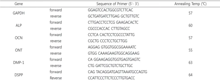
Table 1. Real Time quantitative RT-PCR primer sequences used in this study
Gene Sequence of Primer (5’- 3’) Annealing Temp (℃) GAPDH forward GGAGTCCACTGGCGTCTTCAC
reverse GCTGATGATCTTGAG GCTGTTGTC 57
ALP forward CTTGACCTCCTCG GAAGACACTC reverse CGCCCACCAC CTTGTAGCC 60
OCN forward CCTCA CACTCCTCGCCCTATTG reverse CGCTG CCCTCCTGCTTGG 57
ONT forward AGGAG GTGGTGGCGGAAAATC reverse GTGG CAAAGAAGTGGCAGGAAG 55
DMP-1 forward CA GGAAGAGGTGGTGAGTGAGTC reverse CTG GATTCGCTGTCTGCTTGC 63
DSPP forward CAG TACAGGATGAGTTAAATGCCAGTG Reverse CCATTCCCTTCTCCCTTGTGACC 64
GAPDH = Glyceraldehyde 3-phosphate dehydrogenase, ALP = Alkaline Phosphatase, OCN = Osteocalcin, ONT = Osteonectin, DMP-1 = Dentin matrix acidic phosphoprotein 1, DSPP = Dentin sialophosphoprotein
Table 2. The mean value of the difference between target gene and housekeeping gene of supernumerary dental pulp stem cells
Sample ONT OCN ALP DMP-1 DSPP
Fast
3-UD 3.89 12.92 14.63 14.71 15.44
3-D 3.15 12.68 8.39 14.41 14.91
10-UD 2.61 12.27 13.44 16.43 16.54
10-D 4.08 13.04 9.92 14.32 15.26
Slow
3-UD 2.44 13.25 9.71 19.63 17.67
3-D 0.21 10.68 7.70 11.04 12.31
10-UD 0.60 12.77 12.27 15.83 17.60
10-D -0.97 11.80 8.24 14.03 14.41
Fast = Fast growing supernumerary dental pulp stem cell, Slow = Slow growing supernumerary dental stem cell, UD = Undifferentied cell, D = Differenti-
ated cell, ALP = Alkaline Phosphatase, OCN = Osteocalcin, ONT = Osteonectin, DMP-1 = Dentin matrix acidic phosphoprotein 1, DSPP = Dentin sialophos-
phoprotein
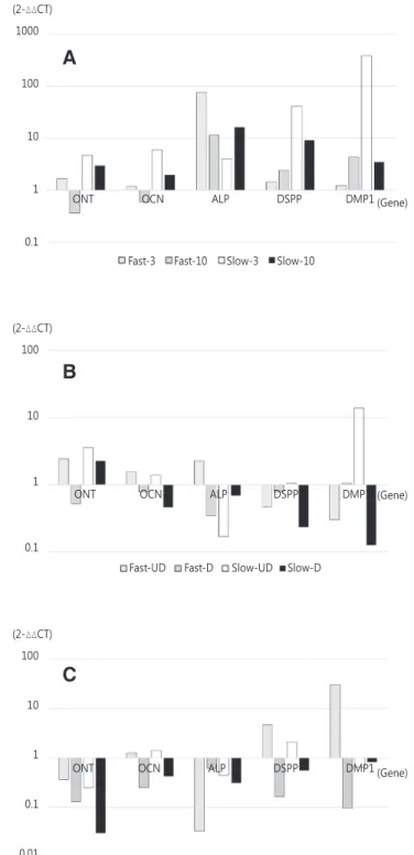
Fig. 1은 각 유전자의 △△Ct 값을 계산하여 얻은 유전자의 상 대적 발현량이다. Fig. 1A는 미분화된 세포에 대한 분화된 세포 의 유전자 발현도의 상대값을 나타낸 그래프이다. 계대 배양 속 도가 빠른 10계대의 세포의 ONT와 OCN을 제외한 모든 군에서 유전자 발현도가 분화된 세포에서 더 높은 값을 보였다. Fig. 1B 는 3계대 세포에 대한 10계대 세포의 유전자 발현도의 상대값 을 나타낸 그래프이다. 계대에 따른 유전자 발현도의 연관성은 뚜렷하게 나타나지 않지만 10계대에서 ONT의 발현도가 전반적 으로 높으며 ALP, DSPP, DMP1은 전반적으로 발현도가 낮은 것 으로 나타났다. Fig. 1C는 느리게 계대 배양된 세포에 대한 빠르 게 계대 배양된 세포의 유전자 발현도의 상대값을 나타낸 그래 프이다. 3계대 미분화 세포의 OCN, DSPP, DMP1의 발현도, 10계 대 미분화 세포의 OCN, DSPP의 발현도를 제외하고 각 군의 유 전자 상대적 발현도가 전반적으로 빠르게 계대 배양된 세포에서 낮게 나타났다.
IV. 총괄 및 고찰
이 연구의 목적은 과잉치 치수 유래 줄기세포의 계대 배양 속 도에 차이가 있는 두 세포 간 상아모세포 연관 유전자의 발현을 비교하는 것이다. 치수 유래 줄기세포의 특이한 표지자로는 조 상아모세포의 분화 표지자인 DSPP, DMP-1이 있다. DSPP는 조 상아모세포로부터 분비되는 특이한 단백질이며, DMP-1은 조 상아모세포로의 분화를 조절하는데 중요한 역할을 하는 단백질 이다[15,16]. 이 밖에 석회화 물질의 형성에 중요한 역할을 하는 ALP, OCN, ONT 등이 있다[5,17,18].
DSPP와 DMP-1은 뼈의 형성과 관련된 SIBLING (Small Integrin-Binding Ligand, N-linked Glycoprotein) 중 하나이다.
이들은 치아의 광화에 관여하는 비아교성 단백질로 상아모세 포에서 생성된다. DSPP는 BMP-1에 의해 치아의 구성성분인 dentin sialoprotein (DSP), dentin glycoprotein (DGP), dentin phosphoprotein (DPP)로 분해되며 DSPP에 관련된 유전자에 변 이가 생긴 환자는 제 3형 상아질 형성부전 등 치아 형성에 관련 된 질환을 가지게 된다[19-21]. 반면 DMP-1은 골모세포와 상아 모세포의 성숙과정에서 인산화되어 방출된다. DMP-1은 주로 골 의 광화와 혈중 칼륨의 농도를 조절하므로 결핍 시 저칼륨혈증, 골연화증 등의 질환이 발생된다[21]. ONT는 경조직의 광화를 시 작하는데 역할을 하는 골 특이성 단백질이다[22]. OCN은 골모 세포 또는 상아모세포에서 합성되며 골을 형성하고 신체의 물질 대사를 조절하는 역할을 하며 골의 광화와 혈액 내 칼슘이온의 농도 조절에도 관여하여 골 형성 관련 표지자로 사용되고 있다 [23]. ALP는 조직의 광화와 관련된 효소로 섬유아세포와 조상아 세포에서 생성되며 ALP수치의 증가는 조상아세포로의 초기 분
Fig. 1. A: Relative gene expression of Differentiated Super- numerary dental pulp stem cells, B: Relative gene expres- sion of 10
thgeneration Supernumerary dental pulp stem cells, C: Relative gene expression of Fast grown Supernu- merary dental pulp stem cells.
Fast-UD Fast-D Slow-UD Slow-D Fast-3 Fast-10 Slow-3 Slow-10 (2-△△CT)
1000
100
10
1
0.1
(Gene)
(Gene)
(Gene) (2-△△CT)
100
10
1
0.1
(2-△△CT) 100
10
1
0.1
0.01
A
B
C
ONT OCN ALP DSPP DMP1 ONT OCN ALP DSPP DMP1 ONT OCN ALP DSPP DMP1
3-UD 3-D 10-UD 10-D
화 단계를 의미하며 세포의 염증 상태를 의미하기도 한다[24].
이전의 연구에서 인간의 중간엽 줄기세포가 계대 배양을 거 침에 따라 변화하는 성장 역학, 골 형성능에 대해 연구한 논문에 서 계대 수가 증가함에 따라 세포의 분열 활성이 감소되는 것을 발견하였다[25]. 또한 인간의 중간엽세포의 계대 배양에 따른 골 형성능에 관한 논문에서 5계대 이후부터 세포 자체의 증식력과 osteoblast로의 분화능이 감소한다고 했다[26].
위 두 연구의 결과를 통해 세포의 분열 활성도라고 볼 수 있는 계대 배양 속도와 유전자 발현도 간 연관성을 예상할 수 있었다.
이 실험에서도 세포를 배양하는 동안 초기 5계대까지 평균 계대 배양 속도는 각 2.0일과 3.0일 이었던 반면 5계대에서 10계대까 지의 평균 계대 배양 속도는 각 2.8일과 4.2일로 계대 배양 속도 가 느려진 것을 확인할 수 있었다. 또한 10계대가 3계대에 비해 전반적으로 더 낮은 유전자 발현도를 보인 것을 확인 할 수 있었 으며, 빠르게 계대 배양된 세포에서 전반적으로 낮은 상대적 유 전자 발현을 보였다. 이는 빠르게 계대 배양된 세포가 상대적으 로 분화가 적게 일어난 것으로 유추할 수 있다.
줄기세포는 분열능과 분화능을 가지는 세포이다. 빠르게 분열 이 되는 세포는 분열능이 좋은 세포이며 아직 분화가 진행되지 않은 세포로 생각할 수 있다. 실제로 영구치 유래 치수줄기세포 에 비해 과잉치 유래 치수줄기세포는 세포의 성장속도가 빠르며 더 많은 계대 배양이 가능하다. 이런 사실과 이 연구의 결과를 통해 세포의 계대 배양 속도가 빠른 세포가 분화가 더 적게 진행 된 세포임을 알 수 있다.
과잉치는 인접 영구치의 맹출 장애 또는 지연 같은 합병증으 로 인해 제3대구치에 비해 비교적 이른 나이에 발치를 시행한 다. 젊은 사람에서 얻은 골수 줄기세포와 나이가 많은 사람에게 서 얻은 줄기세포의 골 형성능과 증식을 평가한 연구에서 젊은 사람에게서 얻은 골수 줄기세포가 더 증식 능력이 높으며, 골 형 성능이 더 높다는 것이 밝혀졌다[27]. 또한 유치 치수유래 줄기 세포가 영구치 유래 치수 줄기세포에 비해 증식 속도가 더 빠르 다는 사실이 밝혀졌다[28]. 이처럼 어린 나이에 얻어지는 줄기세 포는 세포의 분열 활성이 더 높다. 이런 특성은 줄기세포의 세포 치료에의 활용도가 더 뛰어나게 만든다.
과잉치 유래 치수줄기 세포는 기존에 가장 많이 사용하던 제 3대구치 유래 치수줄기세포에 비해 빠른 계대 배양 속도를 가진 다. 이는 이 실험 결과를 토대로 유추해보았을 때 과잉치 유래 치수줄기 세포가 제3대구치 유래 치수줄기세포에 비해 더 높은 세포 분열 활성을 가진다는 결론을 내릴 수 있다. 따라서 과잉치 유래 치수 줄기세포는 추후 더 많은 검증을 거친다면 좋은 중간 엽 세포의 공여부가 될 수 있을 것으로 보인다.
이 연구는 계대 배양 속도가 빠른 세포와 느린 세포 각 하나의
세포만 가지고 실험을 진행했다는 데 그 한계가 있다. 이 후 더 많은 세포를 이용한 실험을 통한 확인이 필요할 것으로 판단된 다.
Ⅴ. 결 론
이 연구의 결과들을 볼 때 과잉치 유래 치수줄기세포는 계대 배양됨에 따라 그 속도가 느려지며 분화가 일어남을 알 수 있으 며, 세포 분열 속도가 빠른 세포가 분화가 더 적게 일어난 것을 확인할 수 있었다.
이 연구는 적은 표본을 가지고 연구가 이루어져 있으므로 추 가적으로 더 많은 세포를 이용한 실험을 통한 검증이 필요할 것 으로 사료된다.
References
1. Meighani G, Pakdaman A : Diagnosis and management of supernumerary (mesiodens): a review of the literature. J Dent (Tehran) , 7:41-49, 2010.
2. Van Buggenhout G, Bailleul-Forestier I : Mesiodens. Eur J Med Genet , 51:178-181, 2008.
3. Rajab LD, Hamdan MA : Supernumerary teeth: review of the literature and a survey of 152 cases. Int J Paediatr Dent , 12:244-254, 2002.
4. Russell KA, Folwarczna MA : Mesiodens-diagnosis and management of a common supernumerary tooth. J Can Dent Assoc , 69:362-366, 2003.
5. Potdar PD, Jethmalani YD : Human dental pulp stem cells:
applications in future regenerative medicine. World J Stem Cells , 7:839-851, 2015.
6. Shi S, Gronthos S : Perivascular niche of postnatal mesen- chymal stem cells in human bone marrow and dental pulp.
J Bone Miner Res , 18:696-704, 2003.
7. Huang AH, Chen YK, Chan AW, et al . : Isolation and charac- terization of dental pulp stem cells from a supernumerary tooth. J Oral Pathol Med , 37:571-574, 2008.
8. Gronthos S, Brahim J, Shi S, et al . : Stem cell properties of human dental pulp stem cells. J Dent Res , 81:531-535, 2002.
9. Mauth C, Huwig A, Graf-Hausner U, Roulet JF : Restorative applications for dental pulp therapy. Topics in Tissue Engi- neering , 3:1-32, 2007.
10. Min JH, Ko SY, Jang YJ, et al . : Dentinogenic potential of
human adult dental pulp cells during the extended primary
culture. Hum Cell , 24:43-50, 2011.
11. Qin C, Brunn JC, Butler WT, et al . : The expression of dentin sialophosphoprotein gene in bone. J Dent Res , 81:392-394, 2002.
12. Yu J, Wang Y, Jin Y, et al . : Odontogenic capability: bone marrow stromal stem cells versus dental pulp stem cells.
Biol Cell , 99:465-474, 2007.
13. Tziafas D, Kodonas K : Differentiation potential of dental papilla, dental pulp, and apical papilla progenitor cells. J Endod , 36:781-789, 2010.
14. Kenneth J, Thomas D : Analysis of relative gene expression data using real-time quantitative PCR and the 2(-Delta Delta C(T)) Method. Methods , 25:402-408, 2001.
15. About I, Bottero MJ, Mitsiadis TA, et al . : Human dentin production in vitro. Exp Cell Res , 258:33-41, 2000.
16. Inagaki Y, Kashima TG, Athanasou NA, et al . : Dentine matrix protein 1 (DMP-1) is a marker of bone formation and mineralisation in soft tissue tumours. Virchows Arch , 466:445-452, 2015.
17. Štefková K, Procházková J, Pacherník J : Alkaline Phospha- tase in Stem Cells. Stem Cells Int, 2015:628368, 2015.
18. Liu Q, Cen L, Cui L, et al . : A comparative study of prolif- eration and osteogenic differentiation of adipose-derived stem cells on akermanite and beta-TCP ceramics. Biomate- rials , 29:4792-4799, 2008.
19. Bruder SP, Jaiswal N, Haynesworth SE : Growth kinetics, self-renewal, and the osteogenic potential of purified hu- man mesenchymal stem cells during extensive subcul- tivation and following cryopreservation. J Cell Biochem, 64:278-294, 1997.
20. Yamamoto R, Oida S, Yamakoshi Y : Dentin Sialophosphopro- tein-derived Proteins in the Dental Pulp. J Dent Res , 94:1120- 1127, 2015.
21. von Marschall Z, Fisher LW : Dentin sialophosphoprotein (DSPP) is cleaved into its two natural dentin matrix prod- ucts by three isoforms of bone morphogenetic protein-1 (BMP1). Matrix Biol , 29:295-303, 2010.
22. Suzuki S, Haruyama N, Nishimura F, Kulkarni AB : Dentin sialophosphoprotein and dentin matrix protein-1: Two highly phosphorylated proteins in mineralized tissues. Arch Oral Biol , 57:1165-1175, 2012.
23. Termine JD, Kleinman HK, Martin GR, et al . : Osteonectin, a bone-specific protein linking mineral to collagen. Cell , 26:99-105, 1981.
24. Lee NK, Sowa H, Karsenty G, et al . : Endocrine regulation
of energy metabolism by the skeleton. Cell , 130:456-469, 2007.
25. Aslantas EE, Buzoglu HD, Aksoy Y, et al . : Age-related Changes in the Alkaline Phosphatase Activity of Healthy and Inflamed Human Dental Pulp. J Endod , 42:131-134, 2016.
26. Sun HJ, Bahk YY, Lee JW : A proteomic analysis during se- rial subculture and osteogenic differentiation of human mesenchymal stem cell. J Orthop Res , 24:2059-2071, 2006.
27. Stenderup K, Justesen J, Clausen C, Kassem M : Aging is associated with decreased maximal life span and accelerat- ed senescence of bone marrow stromal cells. Bone , 33:919- 926, 2003.
28. Nakamura S, Yamada Y, Ueda M, et al . : Stem cell prolifera-
tion pathways comparison between human exfoliated de-
ciduous teeth and dental pulp stem cells by gene expres-
sion profile from promising dental pulp. J Endod , 35:1536-
1542, 2009.
국문초록
계대 배양 속도가 다른 과잉치 치수유래 줄기세포 간 유전자 발현 특성
이유경ㆍ김종수ㆍ신지선ㆍ김종빈 단국대학교 치과대학 소아치과학교실
이 연구의 목적은 과잉치 치수 유래 줄기세포의 계대 배양 속도에 대한 상아모세포 연관 유전자의 발현을 비교하는 것이다. 줄기세 포는 다른 여러 형태의 세포로 분화할 수 있는 미 분화된 세포이다. 이는 환경이나 특정 자극에 의해 세포 분열이 일어나며 근육이나 골 같은 특정 장기의 조직으로 분화할 수 있다.
20명의 어린이에서 발거한 과잉치에서 과잉치 치수 유래 줄기세포가 얻어졌다. 10계대까지 배양하는 동안 가장 빠른 속도로 계대 배양된 세포와 가장 느린 속도로 계대 배양된 세포 각 3계대와 10계대 세포를 얻어 실험을 진행하였다. 각 세포는 분화제를 처리한 군 과 처리하지 않은 군으로 나누었다. 이 실험에서 발현도를 살펴본 유전자는 Osteonectin (ONT), Osteocalcin (OCN), Alkaline Phospha- tase (ALP), Dentin matrix acidic phosphoprotein 1 (DMP-1), Dentin sialophosphoprotein (DSPP)이다.
분화가 된 세포가 전반적으로 더 높은 유전자 발현도를 보였으며, 미분화 세포는 10계대에서, 분화된 세포는 3계대에서 더 높은 유 전자 발현도를 보였다. 빠른 계대 배양 속도를 보인 세포가 OCN과 DSPP를 제외하고 상대적으로 더 낮은 유전자 발현도를 보였다.