Diosmin이 조골세포 분화에 미치는 영향
김경민․장원구
대구대학교 생명공학과 및 항노화연구소
Effect of Diosmin on Osteoblast Differentiation
Kyeong-Min Kim and Won-Gu Jang
Department of Biotechnology and Research of Institute of Anti-Aging, College of Engineering, Daegu University
ABSTRACT Diosmin is a compound that is widely distributed in citrus. The aim of this study was to examine the effects of diosmin on osteoblast differentiation using MC3T3-E1 preosteoblast cells. The effect of diosmin on mRNA expression levels of osteogenic genes in MC3T3E1 cells were determined by RT-PCR. Diosmin regulated expression of key osteogenic genes such as dNA-binding protein inhibitor (Id1), runt-related transcription factor 2 (Runx2), alkaline phosphatase (ALP), and osteocalcin (OC) during osteoblast differentiation, and promoted recovery of tumor necrosis factor (TNF)-α-reduced osteogenesis. Alizarin Red S staining was performed to evaluate mineralization. Diosmin en- hanced mineralization and recovery of TNF-α-reduced osteoblast differentiation. These results suggest that diosmin may enhance osteoblast differentiation and recovery of TNF-α-reduced osteoblast differentiation, and it may be a candi- date for treating osteoporosis.
Key words: diosmin, osteoblast differentiation, preosteoblast, mineralization, TNF-α
Received 14 September 2017; Accepted 7 December 2017 Corresponding author: Won-Gu Jang, Department of Biotechnology, College of Engineering, Daegu University, Gyeongsan, Gyeongbuk 38453, Korea
E-mail: [email protected], Phone: +82-53-850-6552
서 론
현대사회는 서구화된 식습관, 생활습관으로 인해 대사와 관련된 질환이 증가하고 있고, 평균 수명의 연장으로 인해 노인성 질환자의 수가 계속해서 증가하고 있다. 그에 따라 대사성, 노인성 질환의 관리가 중요하며, 그중에서도 골다공 증, 퇴행성관절염과 같은 골 질환은 식이, 생활방식과 같은 환경적 요인 그리고 유전적 요인이 영향을 미치는 질병이다.
이러한 골 질환 환자들에게는 수많은 원인이 있을 뿐만 아니 라 치료를 위해서는 많은 비용과 노력이 필요하다. 특히 여 성은 폐경 이후 골다공증이 많이 발생한다. 그렇기 때문에 사람들의 부담은 점점 증가하고 있으며 골다공증 치료 보조 제 및 각종 건강 기능성 식품 개발이 요구되고 있다(1-3).
뼈는 대사적으로 한 개체의 일생 동안 계속해서 재구성되 는 역동적인 장기다. 조골세포, 골세포, 파골세포는 포유류 의 뼈 재구성 과정에 포함되어 있다. 조골세포 분화는 복잡한 신호전달경로에 의해 조절된다(4,5). 뼈는 신체의 구조를 이 루고 골격계를 구성하는 역할을 하며, 조골세포가 골 기질을 형성하고 파골세포가 골을 흡수하여 균형을 이루고 있다(6).
그중 조골세포는 중간엽 줄기세포에서 분화하고, 여러 가지 호르몬, bone morphogenetic proteins(BMPs)과 같은 사 이토카인, inhibitors of DNA binding/differentiation(Id1), distal-less homeobox 5(Dlx5), runt-related transcription factor 2(Runx2), alkaline phosphatase(ALP), osteocalcin (OC)과 같은 다양한 전사 인자에 의해서 조절된다. 그중에 서도 Id1, Dlx5, Runx2는 조골세포 분화 초기에 발현되는 필수적인 유전자로 이들 유전자의 발현은 조골세포 분화가 가속화됨을 의미한다(7-9).
최근 식물에서 유래한 천연물이 골 손실을 막아준다는 보 고가 많아지고 있다(10-14). Diosmin(Fig. 1)은 감귤과 식물 에 함유된 플라본 계열의 물질이고, 항산화, 항염증, 항돌연 변이, 항과혈당증, 항알레르기 효과가 있다(15-17). 그뿐만 아니라 diosmin은 고혈압, 간 손상, 간 허혈, 신경퇴화, 심근 경색, 간암 생성 억제 등에도 긍정적인 효과가 있다고 보고되 고 있다(18-22). 그리고 diosmin은 trichloroethylene에 의 해 유도되는 신장 손상에 대해 보호 효과가 보고되었다(23).
Tumor necrosis factor-alpha(TNF-α)는 염증을 유발 하는 사이토카인으로 interleukin(IL)-1, 6, 11, 17 등과 함 께 염증성 질환을 발생시킨다(24-26). 특히 TNF-α는 파골 세포 분화를 증가시키고 조골세포 분화를 억제함으로써 골 형성을 억제하여 골 손실을 유발한다(27,28). 게다가 TNF- α는 조골세포에서 골 기질 단백질의 생합성을 억제함으로써 조골세포 분화를 억제할 뿐만 아니라 조골세포의 사멸을 유
Fig. 1. Structure of diosmin.
Table 1. Oligonucleotide sequence of mouse primer
Gene Forward primer Reverse primer
Id1 Runx2 OC β-Actin
ATGAAGGTCGCCAGTGGCAGT CCGCACGACAACCGCACCAT GCAAATAAGGTAGTGAACAGACTCC TTCTTTGCAGCTCCTTCGTTGCCG
ACTTTGCGGTTCTGGGGCAGG CGCTCCGGCCCACAAATCTC GTTTGTAGGCGGTCTTCAAGC TGGATGGCTACGTACATGGCTGGG 도한다(29,30).
따라서 본 연구에서는 diosmin에 의한 조골세포 분화 효 능을 검증하고자 하였다. 이를 위해 조골전구세포인 MC3T3- E1에 diosmin을 처리하여 조골세포 분화 마커 유전자의 발 현과 석회화 정도를 확인하였다. 그리고 골다공증을 유발할 수 있는 TNF-α에 의해 감소하는 조골세포 분화를 diosmin 이 개선할 수 있는지도 알아보았다.
재료 및 방법
세포 배양
Mouse calvaria 유래 조골세포인 MC3T3-E1 세포는 ATCC CRL-2593(American Type Culture Collection, Manassas, VA, USA)을 구입하여 사용하였다. 유지 시에는 α-MEM(GibcoBRL, Grand Island, NY, USA) 배지에 10%
FBS(Atlas Biologicals, Fort Collins, CO, USA)와 1% 항 생제(100 U/mL penicillin-100 μg/mL streptomycin, Gibco BRL)를 첨가하여 37°C, 5% CO2 조건으로 배양하였다.
조골세포의 분화
MC3T3-E1 세포를 6-well plate와 24-well plate에 배 양하여 80% 포화하게 되면 5 mM β-glycerophosphate (Sigma-Aldrich Co., St. Louis, MO, USA)와 50 μg/mL ascorbic acid(Sigma-Aldrich Co.)가 포함된 배지로 2일마 다 배지를 교체하면서 분화를 유도하였다.
세포독성 시험
Diosmin(Sigma-Aldrich Co.)에 대한 세포독성을 알아 보기 위하여 3-(4,5-dimethylthiazol-2-yl)-2,5-diphenyl- tetrazolium bromide(MTT) assay를 수행하였다. MC3T3 -E1 cell을 48-well plate에 well 당 2×10⁴개로 접종하여, diosmin을 0.1, 0.2, 0.5, 1, 2 μM의 농도로 처리하여 24시 간 동안 배양하였다. MTT(Sigma-Aldrich Co.)를 0.5 mg/
mL의 농도로 처리하여 같은 조건에서 1시간 동안 배양한
후 배지를 제거하고 불용성 formazan 결정을 dimethyl sulfoxide(Duchefa, Haarlem, The Netherlands)로 용해 시켜 540 nm에서 흡광도를 측정하였다.
유전자 발현 분석법
조골세포 분화 유도 조건(50 μg/mL ascorbic acid, 5 mM β-glycerophosphate)과 TNF-α(10 ng/mL)를 처리 한 조건과 함께 diosmin(0.5 or 1 μM)을 처리하여 48시간 배양하였다. 배양한 세포는 dPBS로 세척하여, TRI-sol- ution(Bioscience Technology, Gyeongsan, Korea)을 이 용하여 MC3T3-E1 세포에서 총 RNA를 추출 및 분리하였 다. 3 μg RNA를 TOP script™ RT Dry MIX(Enzynomics, Daejeon, Korea)를 이용하여 complementary DNA(cDNA) 를 합성하였다. Emerald Amp GT PCR Master Mix(Ta- KaRa Bio Inc., Tokyo, Japan)와 primer가 포함된 혼합물 19 μL와 cDNA 1 μL를 polymerase chain reaction(PCR) 사이클을 30회 수행하였다. PCR 반응 조건은 95oC에서 10 분, 95oC에서 30초, 어닐링 온도에서 30초, 72oC에서 30초 후 다시 95oC, 30초로 돌아가는 cycle을 반복하고 72oC에 서 5분을 수행하였다. 중합 효소 반응에 쓰인 primer의 정보 는 Table 1에서 나타내었다.
골의 석회화 측정
MC3T3-E1 세포를 24-well plate에 5×10⁴개로 접종하 여 ascorbic acid, β-glycerophosphate, diosmin, TNF-α 를 각각 처리하여 3주간 배양하였다. 그 후 배지를 제거하고 dPBS로 세척하여 4% formaldehyde로 5분간 실온에서 고 정했다. 세포의 고정이 끝난 후 2% Alizarin Red Solution (Sigma-Aldrich Co.)으로 5분간 염색하고 3차 증류수로 3 회 세척하였다.
통계처리
본 연구에서 얻은 결과는 평균±표준오차로 표시하였고, 통계처리는 Student’s t-test를 이용하였으며, P<0.05, P<
0.01, P<0.001인 경우 대조군과 diosmain 처리군 사이에 유의성이 있음을 인정하였다.
결과 및 고찰
Diosmin이 조골세포 증식에 미치는 영향
Mouse calvaria로부터 유래한 MC3T3-E1 세포는 조골 세포 세포주로서 조골세포의 증식, 분화, 무기질화 등의 특
Fig. 2. Cytotoxicity of diosmin in MC3T3-E1 cells. Cell via- bility in MC3T3-E1 cells was determined using MTT assay after incubation with 0.1, 0.2, 0.5, 1, and 2 μM diosmin for 24 h.
Diosmin (2 μM) significantly induced cytotoxicity in MC3T3- E1 cells. *P<0.05 compared to the untreated control. Results were presented as the mean±SEM (n≥3).
A
B
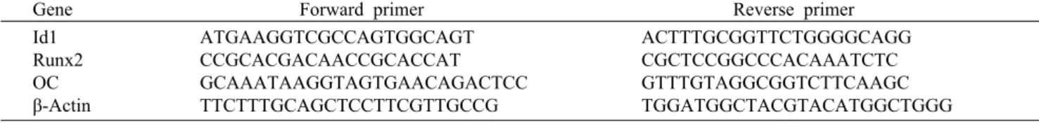
Fig. 3. Diosmin induces mRNA expression of osteoblast marker gene and recover TNF-α reduced osteoblast differentiation marker gene expression. MC3T3-E1 cells were incubated with 50 μg/mL A.A, 5 mM β-GP, 10 ng/mL TNF-α, and diosmin (+: 0.5 μM, ++: 1 μM) for 2 days, after which cells were harvested for total RNA isolation. RT-PCR was carried out using mouse β-actin, Id1, Runx2, ALP, and OC primers. **P<0.01, ***P<0.005 compared to the untreated control, ###P<0.005 compared to A.A+β-GP treated group, ϯP<0.05, ϯϯϯP<0.005 compared to A.A+β-GP and TNF-α treated group, respectively. Results were presented as the mean±SEM (n≥3).
징을 갖고 있기 때문에 골 형성과 관련된 연구에서 사용되고 있다(31-33). MTT 시험법을 통해 MC3T3-E1 세포에서 diosmin의 독성 여부를 확인하였다. 0.1, 0.2, 0.5, 1, 2 μM 의 농도에서 세포독성 여부를 알아본 결과 0.1, 0.2, 0.5,
1 μM의 농도에서는 독성을 나타내지 않았지만 2 μM의 농도 에서 독성을 나타내었다(Fig. 2). 따라서 이후 실험에서는 1 μM의 농도를 사용하여 실험을 진행하였다.
Diosmin이 조골세포 표지 유전자에 미치는 영향 조골세포 분화 표지 유전자 발현량 차이를 통해 diosmin 의 조골세포 분화 효과를 확인하고자 조골세포 분화의 조절 유전자인 inhibitors of DNA binding/differentiation(Id1), Runx2, alkaline phosphatase(ALP), osteocalcin(OC)의 유전자 발현을 확인하였다. 그 결과 diosmin 단독처리에 의 해서 Id1, Runx2, OC의 발현이 높게 나타났지만 ALP는 증 가시키지 못했다. 그뿐만 아니라 조골세포 분화 유도 조건에 diosmin을 함께 처리했을 때 조골세포 분화 마커 유전자에 서 상승효과가 나타났다(Fig. 3A). Id1은 조골세포 분화 초 기 단계에서 중요한 역할을 하는 것으로 알려져 있다(34, 35). Runx2는 runt domain에 속하는 유전자로 조골 전구세 포에서 ALP, OC, bone sialoprotein(BSP)과 같은 유전자 의 발현을 증가시켜 조골세포 분화를 유도하는 유전자이다 (36-38). TNF-α는 파골세포의 분화를 증가시켜 뼈의 양을
A B
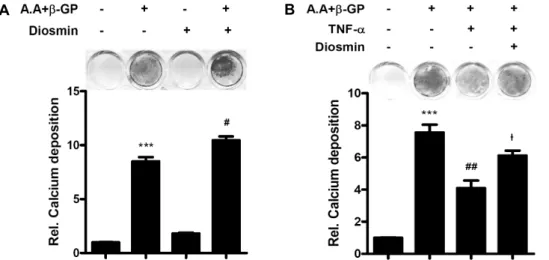
Fig. 4. Diosmin increases calcium deposition on MC3T3-E1. MC3T3-E1 cells were incubated with 50 μg/mL A.A, 5 mM β-GP, 10 ng/mL TNF-α, and 1 μM diosmin for 3 weeks. ***P<0.005 compared to the untreated control, ##P<0.01 compared to A.A+β-GP treated group, ϯP<0.05 compared to A.A+β-GP and TNF-α treated group, respectively. Results were presented as the mean±SEM (n≥3).
감소시키는 역할을 하며, 조골세포 분화를 억제한다(39). 조 골세포 분화유도 중에 TNF-α가 분화 마커 유전자를 감소시 키는 데 diosmin에 의해 감소하는 유전자 발현이 회복되었 다(Fig. 3B).
Diosmin이 칼슘 침착에 미치는 영향
Bone nodule 형성은 조골세포 분화에 있어서 중요한 표 지 인자이다. 이를 확인하기 위해 Alizarin Red Solution을 이용하는데 이는 식물성 염료로 칼슘에 특이적으로 결합을 한다. 이것은 세포 외 기질에 침착되어 있는 칼슘에 특이적 으로 결합력이 높다. 이를 통해 세포 외 기질에 침착된 칼슘 의 양을 알 수 있다(40,41). 칼슘 침착 정도를 알아본 결과, diosmin 단독 처리에서는 효과가 없으나 조골세포 분화 조 건에 함께 처리하였을 때 상승효과가 나타났다(Fig. 4A).
그리고 TNF-α에 의해 저해되는 칼슘 침착을 회복시키는 효과도 있었다(Fig. 4B).
요 약
본 연구에서는 diosmin이 조골세포 분화에 미치는 영향에 대해서 알아보았다. 먼저 세포독성 여부를 확인하기 위해 MTT assay를 수행하였고 독성이 없다고 확인된 1 μM의 농도에서 실험을 진행하였다. 그리고 조골세포로 분화할 수 있는 조골 전구세포인 MC3T3-E1에 조골세포 분화를 유도 하면서 diosmin을 함께 처리하여 표지 유전자인 Id1, Runx 2, OC의 발현을 확인하였다. 그리고 세포 외 기질의 무기질 화를 확인하였다. 확인한 결과 표지유전자들의 발현이 대조 군에 비교해서 증가한 것을 확인하였고, 특히 조골세포 분화 조건에 함께 처리하였을 때 상승효과가 있었다. 그뿐만 아니 라 TNF-α에 의해 감소하는 조골세포 분화를 회복시키는 효과를 나타내기도 하였다. 이러한 결과들을 토대로 dio-
smin은 기존의 조골세포 분화를 촉진한다는 것을 알게 되었 고, 골 질환 관련 제제로서 diosmin이 이용 가능할 수 있다 고 생각된다.
감사의 글
이 논문은 2016학년도 대구대학교 학술연구비 지원에 의한 논문임을 밝히며 이에 감사드립니다.
REFERENCES
1. Seo J, Hwang ES, Kim GH. 2011. Antioxidative and differ- entiation effects of Artemisia capillaris T. extract on hydro- gen peroxide-induced oxidative damage of MC3T3-E1 os- teoblast cells. J Korean Soc Food Sci Nutr 40: 1532-1536.
2. Kim MJ, Im NK, Yu MH, Kim HJ, Lee IS. 2011. Effects of extracts from sarcocarp, peels, and seeds of avocado on osteoblast differentiation and osteoclast formation. J Korean Soc Food Sci Nutr 40: 919-927.
3. Shin JM, Park CK, Shin EJ, Jo TH, Hwang IK. 2008. Effects of Scutellaria radix extract on osteoblast differentiation and osteoclast formation. Korean J Food Sci Technol 40: 674- 679.
4. Yamaguchi A, Komori T, Suda T. 2000. Regulation of osteo- blast differentiation mediated by bone morphogenetic pro- teins, hedgehogs, and Cbfa1. Endocr Rev 21: 393-411.
5. Komori T. 2006. Regulation of osteoblast differentiation by transcription factors. J Cell Biochem 99: 1233-1239.
6. Canalis E. 1985. Effect of growth factors on bone cell repli- cation and differentiation. Clin Orthop Relat Res 193: 246- 263.
7. Lee MH, Javed A, Kim HJ, Shin HI, Gutierrez S, Choi JY, Rosen V, Stein JL, van Wijnen AJ, Stein GS, Lian JB, Ryoo HM. 1999. Transient upregulation of CBFA1 in response to bone morphogenetic protein-2 and transforming growth factor beta1 in C2C12 myogenic cells coincides with sup- pression of the myogenic phenotype but is not sufficient for osteoblast differentiation. J Cell Biochem 73: 114-125.
8. Ducy P, Zhang R, Geoffroy V, Ridall AL, Karsenty G. 1997.
Osf2/Cbfa1: a transcriptional activator of osteoblast differ- entiation. Cell 89: 747-754.
9. de Jong DS, Vaes BL, Dechering KJ, Feijen A, Hendriks JM, Wehrens R, Mummery CL, van Zoelen EJ, Olijve W, Steegenga WT. 2004. Identification of novel regulators as- sociated with early-phase osteoblast differentiation. J Bone Miner Res 19: 947-958.
10. Yang X, Matsuda K, Bialek P, Jacquot S, Masuoka HC, Schinke T, Li L, Brancorsini S, Sassone-Corsi P, Townes TM, Hanauer A, Karsenty G. 2004. ATF4 is a substrate of RSK2 and an essential regulator of osteoblast biology; im- plication for Coffin-Lowry Syndrome. Cell 117: 387-398.
11. Hsieh TP, Sheu SY, Sun JS, Chen MH, Liu MH. 2010. Icariin isolated from Epimedium pubescens regulates osteoblasts anabolism through BMP-2, SMAD4, and Cbfa1 expression.
Phytomedicine 17: 414-423.
12. Xiao HH, Fung CY, Mok SK, Wong KC, Ho MX, Wang XL, Yao XS, Wong MS. 2014. Flavonoids from Herba epi- medii selectively activate estrogen receptor alpha (ERα) and stimulate ER-dependent osteoblastic functions in UMR-106 cells. J Steroid Biochem Mol Biol 143: 141-151.
13. Tang DZ, Yang F, Yang Z, Huang J, Shi Q, Chen D, Wang YJ. 2011. Psoralen stimulates osteoblast differentiation through activation of BMP signaling. Biochem Biophys Res Commun 405: 256-261.
14. Kim MB, Song Y, Hwang JK. 2014. Kirenol stimulates os- teoblast differentiation through activation of the BMP and Wnt/β-catenin signaling pathways in MC3T3-E1 cells. Fito- terapia 98: 59-65.
15. Srinivasan S, Pari L. 2012. Ameliorative effect of diosmin, a citrus flavonoid against streptozotocin-nicotinamide gen- erated oxidative stress induced diabetic rats. Chem Biol Interact 195: 43-51.
16. Kastrup J, Petersen P, Dejgård A, Angelo HR, Hilsted J.
1987. Intravenous lidocaine infusion-a new treatment of chronic painful diabetic neuropathy?. Pain 28: 69-75.
17. Tanrikulu Y, Sahin M, Kismet K, Kilicoglu SS, Devrim E, Tanrikulu CS, Erdemli E, Erel S, Bayraktar K, Akkus MA.
2013. The protective effect of diosmin on hepatic ischemia reperfusion injury: an experimental study. Bosn J Basic Med Sci 13: 218-224.
18. Silambarasan T, Raja B. 2012. Diosmin, a bioflavonoid re- verses alterations in blood pressure, nitric oxide, lipid per- oxides and antioxidant status in DOCA-salt induced hyper- tensive rats. Eur J Pharmacol 679: 81-89.
19. Tahir M, Rehman MU, Lateef A, Khan R, Khan AQ, Qamar W, Ali F, O’Hamiza O, Sultana S. 2013. Diosmin protects against ethanol-induced hepatic injury via alleviation of in- flammation and regulation of TNF-α and NF-κB activation.
Alcohol 47: 131-139.
20. Tanrikulu Y, Kismet K, Serin Kilicoglu S, Devrim E, Erel S, Sen Tanrikulu C, Dinc S, Edebal OH, Erdemli E, Akkus MA. 2011. Diosmin ameliorates intestinal injury induced by hepatic ischemia reperfusion in rats. Bratisl Lek Listy 112:
545-551.
21. Golbabapour S, Hajrezaie M, Hassandarvish P, Abdul Majid N, Hadi AH, Nordin N, Abdulla MA. 2013. Acute toxicity and gastroprotective role of M. pruriens in ethanol-induced gastric mucosal injuries in rats. Biomed Res Int 2013: 974185.
22. Tahir M, Rehman MU, Lateef A, Khan AQ, Khan R, Qamar W, O’Hamiza O, Ali F, Hasan SK, Sultana S. 2013. Diosmin abrogates chemically induced hepatocarcinogenesis via alle- viation of oxidative stress, hyperproliferative and inflamma-
tory markers in murine model. Toxicol Lett 220: 205-218.
23. Rehman MU, Tahir M, Khan AQ, Khan R, Lateef A, Hamiza OO, Ali F, Sultana S. 2013. Diosmin protects against tri- chloroethylene-induced renal injury in Wistar rats: plausible role of p53, Bax and caspases. Br J Nutr 110: 699-710.
24. Algate K, Haynes DR, Bartold PM, Crotti TN, Cantley MD.
2016. The effects of tumour necrosis factor-α on bone cells involved in periodontal alveolar bone loss; osteoclasts, os- teoblasts and osteocytes. J Periodontal Res 51: 549-566.
25. Dosseva-Panova VT, Popova CL, Panov VE. 2014. Subgin- gival microbial profile and production of proinflammatory cytokines in chronic periodontitis. Folia Med (Plovdiv) 56:
152-160.
26. Shaker OG, Ghallab NA. 2012. IL-17 and IL-11 GCF levels in aggressive and chronic periodontitis patients: relation to PCR bacterial detection. Mediators Inflammation 2012:
174764.
27. Ritchlin CT, Haas-Smith SA, Li P, Hicks DG, Schwarz EM.
2003. Mechanisms of TNF-α- and RANKL-mediated osteo- clastogenesis and bone resorption in psoriatic arthritis. J Clin Invest 111: 821-831.
28. Wei S, Kitaura H, Zhou P, Ross FP, Teitelbaum SL. 2005.
IL-1 mediates TNF-induced osteoclastogenesis. J Clin Invest 115: 282-290.
29. Kitajima I, Soejima Y, Takasaki I, Beppu H, Tokioka T, Maruyama I. 1996. Ceramide-induced nuclear translocation of NF-κB is a potential mediator of the apoptotic response to TNF-α in murine clonal osteoblasts. Bone 19: 263-270.
30. Kaneki H, Guo R, Chen D, Yao Z, Schwarz EM, Zhang YE, Boyce BF, Xing L. 2006. Tumor necrosis factor pro- motes Runx2 degradation through up-regulation of Smurf1 and Smurf2 in osteoblasts. J Biol Chem 281: 4326-4333.
31. Pols HA, Felsenberg D, Hanley DA, Stenpάn J, Muñoz- Torres M, Wilkin TJ, Qin-sheng G, Galich AM, Vandormael K, Yates AJ, Stych B. 1999. Multinational, placebo-con- trolled, randomized trial of the effects of alendronate on bone density and fracture risk in postmenopausal women with low bone mass: results of the FOSIT study. Fosamax International Trial Study Group. Osteoporos Int 9: 461-468.
32. Centrella M, McCarthy TL, Canalis E. 1987. Transforming growth factor β is a bifunctional regulator of replication and collagen synthesis in osteoblast-enriched cell cultures from fetal rat bone. J Biol Chem 262: 2869-2874.
33. Letton RW, Fanti P, Malluche HH. 1990. Regulation of 25- hydroxyvitamin D3 metabolism in cultures of osteoblastic cells. J Bone Miner Res 5: 815-823.
34. Maeda Y, Tsuji K, Nifuji A, Noda M. 2004. Inhibitory he- lix-loop-helix transcription factors Id1/Id3 promote bone formation in vivo. J Cell Biochem 93: 337-344.
35. Katagiri T, Yamaguchi A, Komaki M, Abe E, Takahashi N, Ikeda T, Rosen V, Wozney JM, Fujisawa-Sehara A, Suda T. 1994. Bone morphogenetic protein-2 converts the differ- entiation pathway of C2C12 myoblasts into the osteoblast lineage. J Cell Biol 127: 1755-1766.
36. Franceschi RT, Ge C, Xiao G, Roca H, Jiang D. 2007. Tran- scriptional regulation of osteoblasts. Ann N Y Acad Sci 1116:
196-207.
37. Komori T. 2005. Regulation of skeletal development by the Runx family of transcription factors. J Cell Biochem 95:
445-453.
38. Marie PJ. 2008. Transcription factors controlling osteoblas- togenesis. Arch Biochem Biophys 473: 98-105.
39. Redlich K, Hayer S, Ricci R, David JP, Tohidast-Akrad M, Kollias G, Steiner G, Smolen JS, Wagner EF, Schett G.
2002. Osteoclasts are essential for TNF-α-mediated joint destruction. J Clin Invest 110: 1419-1427.
40. Maeda T, Matsunuma A, Kawane T, Horiuchi N. 2001. Sim- vastatin promotes osteoblast differentiation and mineraliza- tion in MC3T3-E1 cells. Biochem Biophy Res Commun 280:
874-877.
41. Schiller PC, D’Ippolito G, Balkan W, Roos BA, Howard GA.
2001. Gap-junctional communication is required for the ma- turation process of osteoblastic cells in culture. Bone 28:
362-369.