의학 석사학위 논문
제대혈 유래 Mesenchymal Stem Cell
배양을 통한 골아세포의 분화 유도
아 주 대 학 교 대 학 원
의 학 과
제대혈 유래 Mesenchymal Stem Cell
배양을 통한 골아세포의 분화 유도
지도교수 임 호 영
이 논문을 의학 석사학위 논문으로 제출함.
2002년 8월
아 주 대 학 교 대 학 원
의 학 과
김 영 진
김영진의 의학 석사학위 논문을 인준함.
심 사 위 원 장 인
심 사 위 원 인
심 사 위 원 인
아 주 대 학 교 대 학 원
2002년 6월 21일
감사의 글 이 논문이 완성되기까지 자상하게 저를 이끌어 주신 종양혈액내과 김효철 선생님, 임호영 선생님께 깊이 감사 드립니다. 바이오 벤처 기업에 몸 담고 있으면서 대학원 과정을 병행한다는 것이 힘들 것이라 예상했지만, 그래도 LifeCord & 아주대 의과대학 산학협력 연구소를 통해 많은 배움의 길을 열어 주신데 대해 종양혈액내과학 교 실원 모두에게 지면을 빌어 감사 드립니다. 그리고 끝까지 저를 도와 이 논문을 완성해준 LifeCord Biomedical Research Institute 의 윤희훈 연 구원께 정말 고맙다는 인사를 드립니다. 대학원 석사과정에 응시한다는 말에 선뜻 동의해준 애들 엄마에게 고 맙고, 피곤한 나를 힘솟게 하고 항상 곁에서 세상 사는 재미를 듬뿍 주 는 우리 딸 승원이와 승주에게 사랑한다는 말을 전하고 싶습니다. 세상에 서로 의지하며 살아갈 수 있는 형제가 있다는 것에 항상 하느님 께 감사 드리며, 저 세상에서 우리 가족을 위해 기도하고 계실 어머니 와 항상 온화함과 성실함으로 저희를 이끌어 주신 아버님의 은혜에 깊 이 감사 드립니다. 세상을 살아가며 늘 힘이 되어 주셨던 모든 분께 감 사와 사랑의 마음을 전합니다.
2002년 5월
김 영 진
-국문요약-
제대혈 유래 Mesenchymal Stem Cell 배양을 통한
골아 세포의 분화유도
목 적: 골수내 존재하는 중간엽 줄기세포(mesenchymal stem cell, MSCs)는
자기복제 능력과 적절한 환경하에서 다른 조직으로 분화 가능한 잠재력 이 있음이 밝혀졌다. 본 저자는 폐기물로 취급되고 있는 태반(placenta) 과 제대혈(umbilical cord blood) 안에도 중간엽 줄기세포가 존재하는지를 알아보고, 이러한 세포들이 골아세포 (osteoblast)로 분화 유도가 되는지 알아보고자 하였다. 재 료 및 방 법: 출산후 통상적으로 폐기되는 제대혈(또는 탯줄 혈액) 안 의 단핵구 세포들을 원심분리를 통해 얻고, 이들이 골수 중간엽 줄기세 포처럼 골아세포로 분화 되는지를 알아보기 위해 적절한 환경하에서 배 양하였다. 배양된 세포가 중간엽 줄기세포의 특성을 지녔는지를 알아보 기 위해 여러 확인 검사등을 시행하였다.
결 과: 배양 일주일 후 plastic adherent layer를 얻을 수 있었으며, 이들은
줄기세포의 특징적인 섬유아세포 형상(fibroblastoid morphology)을 나타냈 다. 적절한 환경하에서 분화시켜 골아 세포의 특성을 나타내는 alkaline phosphatase, Von Kossa 염색에서 양성 반응을 보였다.
이지 않았다. 이러한 결과를 통해 다른 조직으로의 분화능력 (transdifferentiation, differentiation plasticity)이 있는 중간엽 줄기세포의 특 성을 지닌 세포들이 골수 뿐만 아니라 제대혈 안에도 존재함을 알 수 있었다. 결 론: 제대혈 유래 중간엽 줄기세포는 골아세포로 분화 유도됨이 밝혀 짐에 따라 다양성(plasticity) 능력이 증명되었으며, 이러한 세포들은 조 직공학의 발달과 더불어 재생의학에서 치료제로서의 역할을 담당할 수 있으리라 사료된다. __________________________________________________________________ 핵심되는 말 : 제대혈, 중간엽 줄기세포, 골아세포, 분화, 다양성
차 례
국 문 요 약 ...1
차 례 ...3
그 림 차 례 ...4
I. 서 론 ...5
II. 재료 및 방 법 ...8
1. 제대혈 채취...8 2. 단핵세포 분리 및 배양...8 3. 세포 표면 항원 검사...9 4. 골아세포로의 분화...9 5. 콜라젠 지지체 내 세포 접종 및 골아세포 분화...10 6. 세포 염색...10 6-1. Alkaline phosphatase 염색 ...10 6-2. Von Kossa 염색 ... 11III. 결 과 ...12
1. 제대혈 유래 부착성 세포 배양...12 2. 제대혈 유래 부착성 세포의 표면 항원 검사...14 3. 골아세포(osteoblast)로의 분화 ...16IV. 고 찰 ...19
V. 결 론 ...27
참 고 문 헌 ...29
영 문 요 약 ...38
그 림 차 례
Fig. 1. Photomicrograph of plastic-adherent cells in microscopic fields after 7 days in culture ………..……….. 14
Fig. 2. Cell proliferation according to serum concentration …..………… 14
Fig. 3. Immunocharacterization of umbilical cord blood-drived plastic-adherent cells ………. 16
Fig. 4. Alkaline phosphatase staining after osteogenic differentiation activity ……… 18
Fig. 5. Von Kossa staining after osteogenic differentiation activity …….. 18
Fig. 6. Alkaline phosphatase staining after osteogenic differentiation activity by inoculation plastic-adherent cells into collagen scaffold ……… 19
I. 서 론
골수(bone marrow)내의 기질 조직(stromal tissues)은 미세한 혈관들과 여러 종류의 세포들이 세포외 기질에 의해 서로 연결되어 조직망을 형 성하고 있으며, 주위 뼈조직과 밀착되어 있다. 골수에 존재하는 세포들 로는 조혈세포(hematopoietic cell), 내피세포(endothelial cell), 기질세포 (stromal cell) 등이 있으며, 이들 세포들은 출생 후부터 조직발생학적으 로 다른 조상세포(progenitor)로부터 유래된다.1-2 이중 기질세포는 골수이식에 절대적인 조혈모세포의 유지 및 분화에 관여하는 것으로만 여겨져 왔으나,2 골,3-5 연골,6 지방,1 근육,7 혈액1,8 그 리고 신경세포9-10 등으로 분화될 수 있는 다양성(plasticity)7,11-13을 가지 고 있는 것이 1974년 Friedenstein등에14 의해 밝혀진 이래 “colony
forming unit- fibroblast (CFU-F)” “marrow stromal fibroblast(MSF)” 그후 “골 수기질 간세포(bone marrow stromal stem cell) 혹은 중간엽 간세포, 간엽성 간세포, 간엽성 줄기세포(mesenchymal stem cell, MSC)등으로 부르게 되었 다.10-12, 15-17 이러한 세포의 성질 및 그 역할에 대해서는 현재까지도 아 직 많은 부분이 알려져 있지 않다. 하지만 이러한 중간엽 줄기세포를 이용하여 골격계의 손상을 재생하려는 시도가 계속되고 있으며,17-18 그 예로 스스로의 재생이 불가능한 것으로 알려진 관절 연골의 재생에 이 용될 수 있다고 보고하고 있고, 뼈 조직 손상, 인대의 제작 등을 위한 연구가 활발하게 진행되고 있다.4 이외에도 Muscular dystrophy나
치료에도 이식법을 통한 치유를 시도하고 있다.19,20 그러나 이러한 연구 에는 정상 골수조직을 얻기가 쉽지 않다는 제한점이 있다. 동물의 골수 조직으로 연구를 하는 것도 인체에의 적용을 위해서는 반드시 사람의 조직으로 연구를 지속하여야 하기 때문이다. 한편 최근 골수이식의 대체 치료법으로 각광 받고 있는 제대혈 조혈 모세포 이식(umbilical cord blood hematopoietic stem cell transplantation)21은
출산 후 통상적으로 폐기되고 있는 태반(placenta)과 탯줄(umbilical cord) 에 들어있는 조혈모세포(hematopoietic stem cell)를 이용한 치료법인데, 만 약 그런 풍부한 조혈모세포 공급원인 태반과 제대혈안에 중간엽 줄기세 포가 동시에 존재한다면 무한한 공급원이 될 수 있어서 줄기세포 연구 에 박차를 가하는 계기를 제공할 수 있을 것이다. 더욱이 제대혈안에 존재하는 단핵구 자체는 미성숙(immature)하여 항원 표현(antigen expression)이 약하므로 타인의 면역체계에 대한 거부반응이 적다. 그래 서 이러한 장점이 실제 임상에 이용되어 동종이식(allogeneic transplantation)을 통해 손상된 조직의 복원을 이룰 수 있다면,22 소중한 생명을 위한 자원의 재활용 측면에서도 가치를 지닌다 할 수 있다. 이에 본 저자는 현재 골수 조혈모세포 이식의 대체 치료로 인식되고 있는 제대혈 조혈모세포이식의 공급원인 태반과 제대혈에서 중간엽 줄 기세포의 특징적인 표현형을 발현하는 세포들이 존재함을 밝히고, 지속 배양을 통하여 더욱 분화된 형태인 골아세포의 유도를 증명하고 골절 (fracture)이나 골 결함(bone defects)등 임상적 적용의 기초 자료로 삼고자 한다. 또한 이를 위해 생 분해성 지지체(biodegradable scaffold)인 콜라젠
(collagen)을 사용하여 지지체 내에서의 플라스틱 부착성 세포들의 활성 을 알아보고자 하였다.
II. 재료 및 방법
1. 제대혈 채취 임신 38주 이상의 산모에게서 동의를 얻고, 정상분만 직후 제대(탯줄)를 결찰하고, 제대정맥을 통해 항응고제가 포함된 혈액백이나 주사기로 제대 혈을 채취하였다. 이때 산모혈액이 오염(contamination) 되지 않도록 주의를 기울였다. 세포 생존율을 높이고 세포 손상을 막기 위해 채취 후 24시간 내에 실험과정을 시작하였다. 2. 단핵세포 분리 및 배양 항응고제가 포함된 약 50 ml의 제대혈을 10 ml 5개로 분주하였다. 다음 10 ml의 제대혈 당 30 ml의 PBS(phosphate buffered saline, Gibco BRL)을 첨가하고, 각각의 20 ml의 혼합용액을 10 ml의 Ficoll-PlaqueT M
plus(1.077 g/ml, Amersham Pharmacia Biotech.) 용액 위로 천천히 흘려보낸 후 2000 rpm으로 20 분간 원심분리(density gradient centrifugation)를 하였다. 다음 맨 아래층인
적혈구 층과 맨 위층인 PBS 층의 사이인, Ficoll-PlaqueT M plus 계면에 있는 단핵세포 층을 회수하여 다시 1800 rpm으로 5 분간 원심분리를 하여 단핵 세포를 모았다. 이렇게 분리된 단핵세포를 1×10⁶ cells/cm2로 배양 플라스크(75 T flask, Nunc)에 접종한 다음 10% 또는 20% 우태아 혈청(fetal bovine serum, FBS, Gibco BRL)이 포함된 α-MEM(alpha minimum essential medium, Gibco BRL)
배지로 배양하였다. 모든 세포수 측정은 trypan blue dye exclusion 방법을 이 용하였다. 3. 세포 표면 항원 검사 배양된 세포를 0.25% trypsin/0.02% EDTA로 떼어낸 다음 0.5×10⁶ cells/ml 로 농도를 맞추고 여러 가지 항체로 20 분간 반응시켰다. 이때 사용한 항 체는 Negative control로 FITC, PE(BD Pharmingen)를, 단핵구 및 백혈구 항체 로는 anti-CD14, CD45(이하 BD Pharmingen)를, 면역세포 항체로는 anti-CD3, CD4, CD19, CD25를, 조혈세포 항체로는 anti-CD34를, 그리고 내피세포 항 체로는 anti-CD54를 사용하였다. 위와 같이 항체와 반응한 세포 용액을 2 배 부피의 PBS로 세척한 다음 flow buffer(1% paraformaldehyde, 0.1% sodium azide, and 0.5% bovine serum albumin in PBS)로 고정시킨 후 이 세포들을 FACScan(Becton-Dickinson)으로 분석하였다.
4. 골아 세포로의 분화
배양된 제대혈 유래 부착성 세포를 0.25% trypsin/0.02% EDTA(Gibco BRL)
을 이용하여 계대배양한 후 1×10⁴
cells/cm2로 6 well-plate에 분주하였다. 다음 10% 우태아 혈청이 포함된 DMEM(Dulbecco modified Eagle's medium, high glucose, Gibco BRL)에 10-8 M dexamethasone(Sigma), 10-4 M ascorbate-2-phosphate(Sigma), 100 ng/ml β-glycerophosphate(Sigma)를 첨가하여 4주에서 8주간 배양하였다.
5. 콜라젠 지지체 내 세포 접종 및 골아세포 분화 2 ml의 0.3% 제 1형 콜라젠 용액(Nittagelatin)을 12 well-plate에 분주한 다 음 -20 ℃에서 냉동하고 동결건조기(lyophilizer)를 이용하여 동결건조하였 다. vacuum oven에서 105℃로 24시간동안 처리하여 스폰지 형태의 다공성 콜라젠 지지체를 제조하였다. 이렇게 제조된 콜라젠 지지체에 제대혈 유래 부착성 세포를 떼어서 1×10⁵ cells/ml의 세포 용액을 만들어 지지체와 함께
spinner flask(Belco)에서 교반하여 동적 세포 접종(dynamic cell seeding)을 실 시하였다. 다음 위와 동일한 배지를 이용하여 골아세포로 분화시켰다.
6. 세포 염색
6-1. Alkaline phosphatase 염색
배양된 세포를 alkaline phosphatase buffer로 가볍게 세척하였다. 그 다음 10 mg naphthol AS-TR phosphate/10 ml ALP buffer와 2 mg Fast red violet/1 ml ALP buffer 혼합용액을 12 well plate의 well 당 1-1.5 ml 씩 분주한 후 37℃ 에서 20-30 분간 반응시켰다. 반응시킨 후 염색시약을 제거하고 바닥에 부 착한 세포가 떨어지지 않도록 PBS로 가볍게 3회 세척하였다. 10 분간 고 정한 다음 PBS로 3회 세척하였다. 그 다음 냉동실에 보관한 100% methyl alcohol을 1-1.5 ml 씩 분주한 다음 30 분간 냉장실에 방치하였다. 이 methyl alcohol을 버리고 증류수로 3회 세척한 후 well-plate를 filter paper에 뒤집어 얹어서 건조 시켰다.
6-2. Von Kossa 염색
배양된 세포를 4% paraformaldehyde로 10 분간 고정한 다음 PBS로 3회 세척하였다. 그 후 5% silver nitrate 수용액을 조직과 방치시켰다. 이때 자외 선 램프를 조사시켜 60분간 방치하였다. 증류수로 3회 세척한 다음 5% sodium thiosulfate 용액으로 2-3 분간 처리하여 반응하지 않고 남은 silver 이온을 제거시키고 흐르는 물에 5 분간 세척하였다. 그 다음 Nuclear fast red 염색용액으로 5 분간 염색하고 증류수로 세척한 후 탈수과정을 거쳐 고정하였다.
III. 결 과
1. 제대혈 유래 부착성 세포 배양
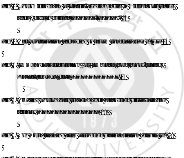
제대혈에서 분리한 단핵세포(mononuclear cell)를 10% 또는 20% 우태아 혈청(fetal bovine serum, FBS)을 포함한 α-MEM 배지로 7일간 배양한 결과, Figure 1에서 보는 바와 같이 10% 우태아 혈청에서 배양한 경우가 더 많은 부착성 세포가 나타났다. 또한 배양된 부착성 세포를 보면 세포의 형태가 비균일하며 크기가 상당히 작은 것을 확인할 수 있다. 혈청 농도에 따른 부착성 세포의 증식 능력을 검사하기 위하여 7일과 14일간 배양한 후 세 포 수를 측정한 결과, 이 경우에도 역시 10% 우태아 혈청에서 배양한 경 우가 세포수가 더 많았다(Figure 2). 이는 제대혈 유래 부착성 세포의 혈청 의존성을 나타내는 것으로 생체 내 제대혈에서 이러한 세포가 증식하지 않는 이유와 일치하는 것으로 사료된다.
(A): 10% FBS containing media (B): 20% FBS containing media
Figure 1. Photomicrograph of plastic-adherent cells in microscopic fields after 7 days in culture (×100).
7 14
Total cell number ( X10
5 ) 0.0 0.5 1.0 1.5 2.0 2.5 3.0 10% FBS 20% FBS
Figure 2. Cell proliferation according to serum concentration.
7
2. 제대혈 유래 부착성 세포의 표면 항원 검사 제대혈 유래 부착성 세포가 어떤 특성을 지니고 있으며, 어느 세포 유래 인지를 검사하기 위하여 FACScan을 실시하였다. 그 결과 Figure 3에서 보 는 바와 같이 단핵구 및 백혈구 항체(anti-CD14, CD45), 면역세포 항체 (anti-CD3, CD4, CD19, CD25), 조혈세포 항체(anti-CD34)에 대하여 음성인 것 으로 보아 조혈세포 계통에서 유래한 세포는 아닌 것으로 판단된다. 또한 내피세포 항체(anti-CD54)에 대해서도 음성의 결과가 나왔다. 따라서 이 부 착성 세포들은 제대혈 내 조혈모세포나 내피세포가 아닌 전혀 다른 세포 군에서 유래된 것으로 사료된다.
Figure 3. Immunocharacterization of cord blood-derived plastic-adherent cells.
Negative control, X (Axis): FITC, Y(Axis): PE, (B) X: CD45, Y: CD14, (C) X : CD3, Y: CD4, (D) X: CD3, Y : CD19, (E) X: CD3, Y : CD25, (F) CD11a, (G) CD34,
3. 골아세포(osteoblast)로의 분화
배양된 부착성 세포를 골아세포로 분화시킬 수 있는 10-8
M dexamethasone, 10-4 M ascorbate-2-phosphate, 100 ng/ml β-glycerophosphate로 처리한 결과, Figure 4에서 보이는 바와 같이 alkaline phosphatase 염색에 양 성임을 확인하였고, Von Kossa 염색을 실시한 Figure 5 에서는 칼슘 침착 (calcium deposit s)과 결절 형성이 활발히 일어나는 것으로 보아 이 부착성 세포가 골(bone)을 형성하는 골아 세포로 분화함을 확인하였다.
더구나 제 1형 콜라젠 지지체에 접종하여 단층 배양과 동일한 방법으로 분화를 유도하였을 경우에도 Figure 6에서 보이는 바와 같이 alkaline phosphatase 염색에 전반적으로 양성의 소견을 보였다.
(a) (b)
Figure 4. Alkaline phosphatase staining after osteogenic differentiation activity.
In comparison with negative controls(a), cells in the osteogenic differentiation group(b) show varying degrees of positive stain for alkaline phosphatase (X 100).
Figure 6. Alkaline phosphatase staining after osteogenic differentiation activity by inoculating plastic-adherent cells into collagen scaffold.
IV. 고 찰
출산후 통상적으로 폐기되어온 태반과 탯줄에 혈액세포를 생산하는 조혈모세포가 풍부하게 들어있다는 것은 오래 전부터 알려져 왔으며, 1988년 프랑스에서 Fanconi’s anemia환자에게 형제의 제대혈 조혈모세포 이식이 성공함으로써, 기존의 골수이식에 의존하던 조혈모세포 이식의 대체 치료법으로 중요하게 인식되고 있다. 이러한 제대혈 이식의 장점으로는 조직적합성이 완전히 일치하지 않아도 이식후 생착율이 골수이식과 비슷하며, 그 이유로는 제대혈안의 단핵구가 미성숙(immature)하여 항원 표현(antigen expression)이 약하므 로 타인의 면역체계에 노출되더라도 거부반응이 적게 나타나기 때문이 다. 또한 조직적합성이 4/6만 부분 일치하더라도 이식 성공율이 높아 적합한 공여 유니트(units)를 구하기 쉽기 때문에 이식까지의 기간이 단 축되는 장점이 있다. 하지만 이러한 장점에도 불구하고 제한된 조혈모 세포의 양으로 인해 골수 생착(bone marrow engraftment)이 지연되고, 특 히 성인에 있어서는 주입되는 총 세포수(total cell count)가 부족하다는 문제점이 대두되었다. 이러한 한계점을 극복하고자 많은 연구가 이루어 지고 있는데, 그중 대표적인 분야가 체외증폭(ex vivo expansion)과 서로다른 제대혈 혼합 주입 등을 들 수 있다.23
또한 골수내 존재하는 중간 엽 줄기세포를 조혈모세포와 동시에 주입함으로써 공여자와 수혜자의 조직적합성이 일치하지 않는 경우에 골수 생착률을 높일 수 있다는 보
이렇듯이 태반과 제대혈이 조혈모세포 공급원외에 다른 의학적 용도가
있는지에 대한 많은 임상 연구가 시도되고 있다.24
최근 골수의 비조혈 세포 성분(non-hematopoietic cell components) 중에 기질세포(stromal cell)는 중간엽(mesoderm) 뿐만 아니라 외배엽(ectoderm) 성상을 보이는 세포로 배양하거나 분화되는 연구 결과를 보여주고 있다. 그와 함께 조직공학에의 응용을 통하여 윤리적인 논란이 되고 있는 배 아 줄기세포(embryonic stem cell)연구에 병행하여 성체 줄기세포 연구에 박차를 가하는 계기를 제공하였다. 1974년에는 Friedenstein이 중간엽 줄기세포가 뼈 형성 잠재력이 있다 고 처음으로 보고한 이래 많은 다른 연구자들에 의해 확인되고 증명되 어 졌고,3 더욱 발전시켜 중간엽 줄기세포를 이용한 치료법들이 임상 시험 수준까지 도달하게 되었다.25 21세기 초에 골수 중간엽 줄기세포와 조직공학을 이용한 연구성과가 배아 간세포 연구와 함께 여러 문헌에 소개되었다.26 또한 성체 줄기세포의 연구도 활발하여 많은 임상시험의 결과가 보고되기도 했으며,22, 27, 28 이에 따라 성체 줄기세포의 근원 (source)에 대한 관심이 고조되었다. 제대혈 안에서도 이러한 성상을 보이는 세포 존재를 증명하기 위해 여러 연구가 시도되었지만,29-32 최근에야 비로소 그러한 특징을 보이는 세포의 존재를 보고하기 시작하였다.33 골수내 중간엽 줄기세포는 배양 플라스크내에서 쉽게 부착된 균등한 단 일층(plastic adherent layer)을 얻어 쉽게 증폭시키기 쉬운 반면 제대혈에 서는 그렇치 못한 것의 설명으로 여러 이유가 제기 되고 있다.
저자도 연구 초기에 경험했듯이 골수에서처럼 plastic adherent layer를 얻 는 것이 쉽지 않았다. 따라서 시행착오를 거치면서 혈청의 농도, 동일 제작 lot 번호의 배지 사용 등이 영향을 미치는 중요한 요소임을 알 수 있었다. 또 많은 연구진들이 사용한 방법에서, 검사를 위해 배양에 쓰이는 혈청 의 lot 번호가 각기 다른 것은 매우 의미 있는 차이를 드러낸다. 왜냐하 면 사용하는 혈청의 batch들이 잘 특징지어져 있지 않고, 사용할 때마다 일정치가 않아서 실험결과도 다르게 나오는데 이것은 세포의 부착, 확 산, 성장 그리고 분화에 각기 다른 영향을 주게 되는 때문이다.34 중간엽 줄기세포의 실험실내 회수율은 골수 세포 배양인 경우가 훨 씬 높다고 보고 되고 있으며, Phinney등도 플라스틱 부착성 골수세포 배 양에서 중간엽 줄기세포는 전체 세포군의 10-20% 정도로 나타났다고 하였다.35 어떤 경우에는 플라스틱 부착성 세포들이 파골세포(osteoclast) 의 특성을 나타내는 경우를 보고하기도 하였고, 또한 회수율도 25% ~ 75% 까지로 다양한 결과를 보이고 있다.36 미숙아(Pre-term< 36 weeks) 제 대혈의 서로 다른 유니트를 섞은 pool을 이용한 연구 보고서 결과에서 는 75%에서 중간엽 성상을 보여주었고, 다핵성 파골 세포의 특징을 나 타냈다. 그리고 이 연구자들은 만삭아의 제대혈에 비해 미숙아의 제대 혈에 중간엽 줄기세포가 더 풍부히 들어있다는 결과를 보고하였다. 1974년 Friedenstein 등이 plastic에 부착되는 성질을 이용, 골수에서 섬 유아세포양 세포(fibroblastoid cells)를 분리한 이후 많은 연구의 진전이 있어왔으며,14
세포군으로 알려져 있다. 최근 보고에 의하면3 저농도(10 cells/cm2)에서 배양된 MSC이 단일층으로 계대배양 것에 비해 복제 능력이 뛰어나다 는 의미있는 보고를 하였다. 중간엽 줄기세포가 0.01-0.001% 정도 존재한다고 보고되어진 골수와 비교 해 보면 상대적으로 상당히 적은 수의 부착성 세포가 나타나는 것으로 보 이는데 이는 아마도 제대혈 유래 부착성 세포는 생체 내에서 "circulating" 세포이기 때문인 것으로 생각된다. 하지만 성인 골수에서 유래된 중간엽 줄기세포와는 다르게, 배양하였을 경 우 이 세포들의 발현 빈도가 낮으며 제대혈이 발생학적 측면에서 먼저 선 행되는 것으로 보아 이 두 가지 세포 집단이 완전히 일치하는 것으로는 여겨지지 않는다. 현재까지 중간엽 줄기세포를 특징짓는 정리된 criteria는 없을 뿐더러 중간엽 줄기세포로 세포를 동정할 만한 표현형 표지자(phenotypic marker)의 single set은 없는 것으로 알려져 있다.
일부 주장으로는 Flow cytometry에서 관찰되는 Side Population(SP) 세포들 은 Transdifferentiation potential이 있는 중간엽 줄기세포라고 언급하고 있 다.37 MSCs가 표현하는 특이항원으로는 SH-2, SH-3, SH-4, STRO-1,38 MAB 1470등이 있으며, 단클론 항체중 SH series가 골수세포로부터 중간엽 줄기세포를 분리하는데 이용되고 있다.39 또 다른 항체로는 골수 섬유아세포에 대해 만들어진 STRO-1로서 in vitro상 기질세포와 조혈모세포의 상호작용을 간섭함으로써 조혈작용을 억제하기도 한다.
골수에 존재하는 중간엽 줄기세포는 그 분화 정도가 각기 다른 세포 들의 복합체이다. 그러므로 이들 세포들의 분화 정도에 따라 세포 표면 에 발현되는 항원 표현들도 변화하게 될 것이다. 그러나 가장 미분화된 중간엽 줄기세포에서만 발현되는 표면 항원은 현재까지 알려져 있지 않 다. 지금까지 보고된 표면 항원 중 Simmons등40에 의해 개발된 STRO-1 단클론 항체가 가장 근접하는 것으로 인식되고 있지만 일부 조혈세포에 서도 양성을 보이고, 고정된 조직에서 염색이 불가능한 문제점을 가지 고 있다. 중간엽 줄기세포 표면에 CD34, CD90 (Thy-1) 항원의 존재에 대해서는 아직 정확히는 알려져 있지 않다.41 다만 중간엽 줄기세포는 Hematopoietic marker인 CD34, CD 45를 표현하지 않는다는 것이 정설로 받아들여지고 있다. Negative marker는 Positive marker와는 대조적으로 11 개 이상의 항원이 세포 표면에 존재하지 않은 것으로 알려져 있다. Negative staining for CD marker로는 CD3, CD4, CD8, CD 11c, CD33, CD36,
CD 38, CD 45, CD117, glycophorin-A 그리고 HLA-II(DR) 등 이다.42 본 연구에서 적용한 CD marker(CD 3, CD 4, CD 19, CD 25, CD 34, CD45) 모두에서 음성을 보여 다른 연구 보고들과 동일한 결과를 보였다(Figure 3). 중간엽 줄기세포는 형태학적, 표현형별, 기능적으로 heterogeneous 한 것이 특징이다. Friedenstein은 배양된 중간엽 줄기세포는 크기, 형태, 증 식력, alkaline phosphatase 를 표현하는 정도, 생체내에서 뼈 생성 능력등 을 다양하게 나타낸다고 하였다. 반면에 Pittenger 등은 균일한 집단의 중간엽 줄기세포를 배양-증폭, 분리되는 조건을 보고한 바 있다.11
alkaline phosphatase의 활성도를 염색하거나 생화학적으로 측정하는 것, 둘째, osteocalcin의 발현을 mRNA나 단백질 단위에서 측정하는 것이다. 셋째, hydroxyapatite에 의하여 형성되는 mineralized nodule을 관찰하는 것 인데, 이를 위하여서는 organic 또는 inorganic phosphate 를 배양액에 첨 가하여 하며, Von Kossa 염색에 의하여 확인이 가능하다. Osteoblast임을 증명하는 terminal phenotype identification marker로는 cbfa-1, alkaline phosphatase, bone sialoprotein, osteopontin, osteocalcin, collagen type I 등이 있 다. 본 연구에서는 그 중에서 alkaline phosphatase activity와 Von Kossa 염 색 2가지 방법으로 골아세포로의 분화를 증명하였다(Figure 4 & 5).
더구나 제 1형 콜라젠 지지체에 접종하여 단층배양과 동일한 방법으로 분 화를 유도하였을 경우에도 Figure 6에서 보이는 바와 같이 alkaline phosphatase 염색에 전반적으로 양성의 소견을 보이는 것으로 보아 골절 같은 질환에 적용할 수 있는 콜라젠 지지체와 같은 scaffold를 이용한 조직 공학적 골(tissue engineered bone) 제조에도 유용하게 쓰일 수 있을 것으로 보인다.
참고로, 세포를 접종할 경우에는 주사기 접종이나 원심분리 등을 이용하는 것보다 spinner flask를 이용한 dynamic 세포 접종 방법을 이용할 경우 세포 접종율이 거의 95% 이상(4 시간 후)이고 지지체 전반에 걸쳐 고르게 부착 되는 것을 확인하였다.
Osteoblast로의 분화를 증진시키는 modulator로 작용할 수 있는 1.25-Dihydroxyvit D3, Prostaglandin E2, GH, IL-6, Leptin, TGF-β3, BMP-4
등이 있다.43
는 골아 세포로의 급속한 분화를 유도한다. 또한 골수내 구조적 환경으 로 인하여 골아세포는 여러 인자 즉 G-CSF, GM-CSF, IL-6 , IL-1β와 같 은 자극인자, TGF-β, TNF 등의 억제인자를 분비함으로써 조혈 세포의 분화에 영향을 준다. 그리고 dexamethasone, TGF-β, 2, 4, BMP-7 그리고 성장호르몬(GH) 같은 유도물질이 존재하면 줄기세포에서 골 아세포로의 분화가 더욱 강화된다고 보고하였다.20,43 특히 dexamethasone 과 β-glycerophospha te는 골아세포로의 분화를 가능케 하며 mineralized bone matrix를 생산한다고 보고한 바 있다. 한편 분화 조건과 환경에 대한 연구와 함께 다량의 중간엽 줄기세포 를 얻을 수 있는 근원과 방법에 대한 노력이 있어 왔으며 더 나아가 골 수내 중간엽 줄기세포뿐만 아니라 제대혈 중간엽 줄기세포를 임상에 적 용하여 치료법으로 개발하려는 노력이 많은 과학자에 의해 시도되었 고,52-53 상당 부분 진전된 연구 성과를 보고하기도 하였다.44-46
특히 Chen Jieli등51은 rat에서 stroke를 유발시킨 후 인간 제대혈을 정맥
내 주입을 통해 행동 결함을 개선시켰다는 보고를 하였고, Sanchez-Ramos등은 인간 제대혈에서 retinoic acid(RA)와 nerve growth factor(NGF)
를 처리한 결과 독특한 neural marker를 표현하였다는 보고를 하였다.45 또한 다량의 항암 요법을 요하는 암환자에게 자가 조혈모세포와 체외 증폭된 중간엽 줄기세포를 동시 이식함으로써 신속히 조혈기능이 회복 된다는 고무적인 결과를 보고하기도 하였다.47,49,50 골수에 비해 채취가 용이하고, 충분한 공급이 가능한 제대혈은 치료적 목적 외에 또 하나의 가능성을 추가할 수 있을 것이라 사료된다.
앞으로 중간엽 줄기세포에 대한 연구는 조직 공학, 재생 의학적인 측 면 뿐만 아니라,24,27,48 이식을 위한 적절한 투여방법,54 정확한 투여량 등 으로 확대되어 나갈 것이다. 따라서 연구에 활용될 연구재료의 공급원 으로서 제대혈은 많은 관심을 받게 된다. 많은 수의 중간엽 줄기세포 확보 방안, 골수에서 처럼 플라스틱 부착기간을 단축하는 방법 모색, 여러 다른 제대혈을 섞은 후 중간엽 줄기세포 분리 시도, pre-term & term 제대혈과의 광범위한 비교, 적절한 지지체(scaffold)와의 조직공학적 접근등의 연구가 이루어진다면 향후 제대혈의 임상적 응용은 확대될 것 으로 사료된다. 제대혈 유래 중간엽 줄기세포가 주위환경 신호 (environmental signals)나 진정한 원시세포들에 의해 유발되는 유전자 발
현 (gene expression)의 다양성(plasticity)55을 나타낼 것인지에 대해서는
더 많은 연구가 필요하다. 또한 제대혈 유래 중간엽 줄기세포 분리를 위한 적절한 환경(조건)에 대한 연구가 계속되어야 하고, 세포 표면에 특징적으로 발현되는 표면항원(surface antigen) 혹은 표현형 표지자 (phenotypic marker) 개발 등이 필요하다고 사료된다.
V. 결 론
최근 들어 Transdifferentiation이 증명된 성체 중간엽 줄기세포(adult mesenchymal stem cell)를 이용하여 필요한 장기나 조직으로 분화를 유도, 임상적으로 이용하려는 적극적인 시도가 이루어지고 있다.. 그러나 이러 한 임상적 시도와 연구는 정상인의 골수 천자라는 과정을 통해 골수조 직을 얻을 때 만이 가능하기 때문에 많은 제한이 따르게 된다. 이에 저 자는 출산후 통상적으로 폐기되고 있는 제대혈안에 중간엽 줄기세포의 존재 유무를 밝히고, 이의 임상적 응용을 시도하였다. 제대혈의 단핵구 세포들을 원심분리를 통해 얻고, 이들이 골수 중간 엽 줄기세포처럼 골아세포(osteoblast)로 분화 되는지를 알아보기 위해 적절한 환경하에서 배양하였다. 배양된 세포가 중간엽 줄기세포의 특성 을 지녔는지를 알아보기 위해 여러 확인 검사등을 시행하였다.
배양 일주일 후 plastic adherent layer를 얻을 수 있었으며, 이들은 특징적 인 섬유아세포 형상(fibroblastoid morphology)을 보였다. 적절한 환경하에 서 분화시켜 골아 세포의 특성을 나타내는 alkaline phosphatase, Von Kossa 염색에서 양성 반응을 보였다.
같은 조건하에서 배양한 CD 34 양성 세포에서는 위와 같은 반응을 보 이지 않았다. 이러한 결과를 통해 다른 조직으로의 분화능력 (transdifferentiation, differentiation plasticity)이 있는 중간엽 줄기세포의 특 성을 지닌 세포들이 골수 뿐만 아니라 제대혈안에도 존재함을 알 수 있 어서 태반과 탯줄의 재활용 가능성을 다양하게 제시하였으며, 이러한
세포들은 조직공학(tissue engineering)의 발달과 더불어 재생의학 (regenerative medicine)에서 치료제로서의 역할을 담당할 수 있으리라 사 료된다.
참 고 문 헌
1. Darwin J. Prockop: Marrow Stromal Cells as Stem Cells for Non-Hematopoietic Tissues. Science 276(Apr):71-74, 1997
2. M. Dominicl, T.J. Hofmann and E.M. Horwitz: Bone marrow mesenchymal cells: biological properties and clinical applications. J Biol Regul Homeost Agents 15:28-37, 2001
3. Scott P. Bruder, Neelam Jaiswal and Stephen E. Haynesworth: Growth Kinetics, Self-Renewal, and the Osteogenic Potential of Purified Human Mesenchymal Stem Cells During Extensive Subcultivation and Following Cryopreservation. Journal of Cellular Biochemistry 64:278-294, 1997
4. Neelam Jaiswal, Stephen E. Haynesworth, Arnold L. Caplan and Scott P. Bruder: Osteogenic differentiation of purified, culture-expanded human mesenchymal stem cells in vitro. Journal of Cellular Biochemistry 64(2):295-312, 1997
5. Paul H. Krebsbach, Keni Gu, Renny T. Franceschi, and R. Bruce Rutherford: Gene Therapy-Directed Osteogenesis: BMP-7-Transduced Human Fibroblasts Form Bone in vivo. Human Gene Therapy 11:1201-1210, 2000 6. Manas K. Majumdar, Valerie Banks, Diane P. Peluso and Elisabeth A.
Morris: Isolation, Characterization, and Chondrogenic Potential of Human Bone Marrow-Derived Multipotential Stromal Cells. Journal of Cellular Physiology 185:98-106, 2000
Floyd, Kristina Hawkinds and Karen Thomas et al.: Human Reserve Pluripotent Mesenchymal Stem Cells Are Present in the Connective Tissues of Skeletal Muscle and Dermis Derived from Fetal, Adult, and Geriatric Donors. The Anatomical Record 264:51-62, 2001
8. Surasit Issarachai, Gregory V. Priestley, Betty Nakamoto and Thalia Papayannopoulou: Cells with hemopoietic potential residing in muscle are itinerant bone marrow-derived cells. Experimental Hematology 30:366-373, 2002
9. Dale Woodbury, Emily J. Schwarz, Darwin J. Prockop and Ira B. Black: Adult Rat and Human Bone Marrow Stromal Cells Differentiate Into Neurons. Journal of Neuroscience Research 61:364-370, 2000
10. C.P. Hofstetter, E.J. Schwarz, D. Hess, J. Widenfalk, A. EI. Manira, Darwin J. Prockop and L. Olson.: Marrow stromal cells form guiding strands in the injured spinal cord and promote recovery. PNAS 99(4):2199-2204, 2002
11. Roland Mertelsmann: Plasticity of Bone Marrow-Derived Stem Cells. Journal of Hematotherapy & Stem Cell Research 9:957-960, 2000
12. Robert J. Deans and Annemarie B. Moseley: Mesenchymal stem cells: Biology and potential clinical uses. Expermental Hematology 28:875-884, 2000
13. Mark F. Pittenger, Alastair M. Mackay, Stephen C. Beck, Rama K. Jaiswal, Robin Douglas, Joseph D. Mosca, Mark A. Moorman et al.: Multilineage Potential of Adult Human Mesenchymal Stem Cells. Science 284(Apr
2):143-147, 1999
14. Friedenstein AJ, Deriglasova UF and Kulagina NN: Precursors for fibroblasts in different populations of hematopoietic cells as detected by the in vitro colony assay method. Exp Hematol 2:83-92, 1974
15. DJ Prockop, I Sekiya and DC Colter: Isolation and characterization of rapidly self- renewing stem cells from cultures of human marrow stromal cells. Cytotherapy 3(5):393-396, 2001
16. Manas K. Majumdar, Mark A. Thiede, Joseph D. Mosca, Mark Moorman and Stanton L. Gerson: Phenotypic and Functional Comparison of Cultures of Marrow-Derived Mesenchymal Stem Cells (MSCs) and Stromal Cells. Journal of Cellular Physiology 176:57-66, 1998
17. Scott P. Bruder, Karl H. Kraus, Victor M. Goldberg and Sudha Kadiyala: The Effect of Implants Loaded with Autologous Mesenchymal Stem Cells on the Healing of Canine Segmental Bone Defects. The Journal of Bone and Joint Surgery 80-A(July):985-996, 1998
18. Sujata Kale, Sybil Biermann, Claire Edwards, Catherine Tarnowski, Michael Morris and Michael William Long: Three-dimensional cellular development is essential for ex vivo formation of human bone. Nature Biotechnology 18(Sep):954-958, 2000
19. JT Triffitt, ROC Oreffo, AS Virdi and Z Xia: Osteogenic stem-cell characterization and development: potentials for cytotherapy. Cytotherapy
20. Hajime Ohgushi, Arnold I. Caplan: Stem Cell Technology and Bioceramic: From Cell to gene Engineering. J Biomed Mater Res 48:913-927, 1999
21. Suradej Hongeng, Sawang Petvises, Budsaba Rerkamnuaychoke, Surapon Worapongpaiboon, Pimpan Tardtong, Suntaree Apibal and Artit Ungkanont: Host Origin of Marrow Mesenchymal Stem Cells Following Allogeneic Cord-Blood Stem-Cell Transplantation. Int J Hematol. 74:235-236, 2001
22. Joseph P Vacanti, Robert Langer: Tissue engineering; the design and fabrication of living replacement device for surgical reconstruction and transplantation. The Lancet 354(suppl): 32-34, 1999
23. G. Dravid, S.G.A. Rao: Ex Vivo Expansion of Stem Cells from Umbilical Cord Blood: Expression of Cell Adhesion Molecules. Stem Cells 20:183-189, 2002
24. Robert Langer and Joseph P. Vacanti: Tissue Engineering. Science 260:920-926, 1993
25. Jose J. Minguell, Alejandro Erices and Paulette Conget: Mesenchymal Stem Cells. Exp Biol Med Vol. 226(6): 507-520, 2001
26. James A. Allay, James E. Dennis, Stephen E. Haynesworth, Manas K. Majumdar, D. Wade Clapp, Leonard D. Shultz, Arnold I. Caplan et al.: LacZ and Interleukin-3 Expression In Vivo after Retroviral Transduction of Marrow-Derived Human Osteogenic Mesenchymal Progenitors. Human Gene Therapy 8(Aug 10):1417-1427, 1997
subpopulation of rapidly self- renewing and multipotential adult stem cells in colonies of human marrow stromal cells. PNAS 98(14): 7841-7845, 2001
28. David H. Kohn, Mojga n Sarmadi, Joseph I. Helman and Paul H. Krebsbach: Effects of pH on human bone marrow stromal cells in vitro: Implications for tissue engineering of bone. J Biomed Mater Res 60:292-299, 2002
29. Margarita Gutierrez-Rodriguez, Elba Reyes-Maldonado and Hector Mayani: Characterization of the Adherent Cells Developed in Dexter-Type Long-Term Cultures from Human Umbilical Cord Blood. Stem Cells 18:46-52, 2000
30. Hector Mayani, Margarita Gutierrez-Rodriguez, Laura Espinoza, Edith Lopez-Chalini, Alejandra Huerta-Zepeda, Eugenia Flores, Elizabeth Sanchez-Valle et al.: Kinetics of Hematopoiesis in Dexter-Type Long-Term Cultures Established from Human Umbilical Cord Blood Cells. Stem Cells 16:127-135, 1998
31. Ralf Huss, Claudia Lange, Eva M. Weissinger, Hans-Jochem Kolb and Karin Thalmeier: Evidence of Peripheral Blood-Derived, Plastic-Adherent CD34 -/low Hematopoietic Stem Cell Clones with Mesenchymal Stem Cell Characteristics. Stem Cells 18:252-260, 2000
32. Ralf Huss: Isolation of Primary and Immortalized CD34- Hematopoietic and Mesenchymal Stem Cells from Various Sources. Stem Cells 18:1-9, 2000
Haematology 109:235-242, 2000
34. Donald G. Phinney: Building a Consensus Regarding the Nature and Origin of Mesenchymal Stem Cells. Journal of Cellular Biochemistry Supplement 38:7-12, 2002
35. Donald G. Phinney, Gene Kopen, Rivka L. Isaacson and Darwin J. Prockop: Plastic Adherent Stromal Cells From the Bone Marrow of Commonly Used Strains of Inbred Mice: Variations in Yield, Growth, and Differentiation. Journal of Cellular Biochemistry 72:570-585, 1999
36. Katia Mareschi, Eleonora Biasin, Wanda Piacibello, Massimo Aglietta, Enrico Madon and Franca Fagioli: Isolation of human mesenchymal stem cells: bone marrow versus umbilical cord blood. Haematologica 86:1099-1100, 2001
37. E.A. de Wynter, N.G. Testa: Interest of cord blood stem cells. Biomed Pharmacother 55:195-200, 2001
38. Karina Stewart, Susan Walsh, Joanne Screen, Carolyn M. Jefferiss, Jonathan Chainey, Grant R. Jordan and Jon N.: Further Characterization of Cells Expressing STRO-1 in Cultures of Adult Human Bone Marrow Stromal Cells. Journal of Bone And Mineral Research 14(Aug):1345, 1999
39. Scott P. Bruder, Nancy S. Ricalton, Raymond E. Boynton, Timothy J. Connolly, Neelam Jaiswal, Joseph Zaia and Frank P. Barry: Mesenchymal Stem Cell Surface Antigen SB-10 Corresponds to Activated Leukocyte Cell Adhesion Molecule and Is Involved in Osteogenic Differentiation. Journal of
Bone and Mineral Research 13(4): 655-663, 1998
40. Simmons, P.J., Torok-Strob, B.: Identification of stromal cell precursors in human bone marrow by a novel monoclonal antibody, STRO-1. Blood 78:55-62, 1991
41. Henry E. Young, Cecile Duplaa, T. Michele Young, Julie A. Floyd, Michelle L. Reeves, Kathryn H. Davis and Greg J. Mancini: Clonogenic Analysis Reveals Reserve Stem Cells in Postnatal Mammals: I. Pluripotent Mesenchymal Stem Cells. The Anatomical Record 263:350-360, 2001
42. Steven M. Devine: Mesenchymal Stem Cells: Will They Have a Role In the Clinic?. Journal of Cellular Biochemistry Supplement 38:73-79, 2002
43. Richard O.C. Oreffo, Vesna Kusec, Silke Romberg, and James T. Triffitt: Human Bone Marrow Osteoprogenitors Express Estrogen Receptor-Alpha and Bone Morphogenetic Proteins 2 and 4 mRNA During Osteoblastic Differentiation. Journal of Cellular Biochemistry 75:382-392, 1999
44. Andrea Banfi, Anita Muraglia, Beatrice Dozin, Maddalena Mastrogiacomo, Ranieri Cancedda and Rodolfo Quarto: Proliferation kinetics and differentiation Potential of ex vivo expanded human bone marrow stromal cells:Implications for their use in cell therapy. Experimental Hematology 28: 707-715, 2000
45. Juan R. Sanchez-Ramos, Shijie Song, Siddharth G. Kamath, Tanja Zigova, Alison Willing, Fernando Cardozo-Pelaez, Todd Stedeford and et al.:
Experimental Neurology 171: 109-115, 2001
46. Luisa Bertolini: Characteristics of Adherent, Fibroblast-Like Hematopoietic Stem Cells. Journal of Hematotherapy & Stem Cell Research 9:607-609, 2000
47. Steven M. Devine, Amelia M. Bartholomew, Nadim Mahmud, Mary Nelson, Sheila Patil, Wayne Hardy, Dord Sturgeon et al.: Mesenchymal stem cells are capable of homing to the bone marrow of non-human primates following systemic infusion. Experimental Hematology 29:244-255, 2001
48. 최윤혁: 장기복원을 위한 간세포 응용연구 동향. 보건산업기술동향 (여름) 11-18, 2001
49. Omer N. Koc, Stanton I. Gerson, Brenda W Cooper, Stephanie M. Dyhouse, Stephen E. Hayesworth, Arnold I. Caplan and Hillard M. Lazarus: Rapid Hematopoietic Recovery After Coinfusion of Autologous-Blood Stem Cells and Culture-Expanded Marrow Mesenchymal Stem Cells in Advanced Breast Cancer Patients Receiving High-Dose Chemotherapy. Journal of clinical oncology 18(2): 307-316, 2000
50. 강석윤, 김재홍, 김현수, 박준성, 임호영, 김영진, 김효철 등: 동종 조혈모세포 및 배양확장된 간엽모세포 동시이식의 가능성. 대한 조 혈모세포이식학회지 제6권 제2호: 167-177, 2001
51. Jieli Chen, Paul R. Sanberg, Yi Li, Lei Wang, Mei Lu, Allison E. Willing, Juan Sanchez-Ramos et al.: Intravenous Administration of Human Umbilical Cord Blood Reduces Behavioral Deficits After Stroke in Rats.
Stroke:32:2682-2688, 2001
52. Judicael Toquet, Ramin Rohanizadeh, Jerome Guicheux, Severine Couillaud, Norbert Passuti, Guy Daculsi and Dominique Heymann: Osteogenic potential in vitro of human bone marrow cells cultured on macroporous biphasic calcium phosphate ceramic. J Biomed Mater Res 44:98-108, 1999
53. Donald G. Phinney, Gene Kopen, Willian Righter, Stephen Webster, Nicola Tremain and Darwin J. Prockop: Donor Variation in the Growth Properties and Osteogenic Potential of Human Marrow Stromal Cells. Journal of Cellular Biochemistry 75:424-436, 1999
54. E. Kon, A. Muraglia, A. Corsi, P. Bianco, M. Marcacci, I. Martin and A. Boyde: Autologous bone marrow stromal cells loaded onto porous hydroxyapatite ceramic accelerate bone repair in critical-size defects of sheep long bones. J Biomed Mater Res 49:328-337, 2000
55. Gerald G. Wulf, Kathyjo A. Jackson and Margaret A. Goodell: Somatic stem cell plasticity: Current evidence and emerging concepts. Experimental Hematology 29:1361-1370, 2001
-Abstract-
Osteogenic Differentiation Activity by Cells
Isolated from Umbilical Cord Blood
Young Jin Kim
Department of Medical Science The Graduate School, Ajou University (Supervised by Associate Professor Ho Yeong Lim)
Purpose: Bone marrow mesenchymal stem cells(MSCs) have the capacity for
renewal and the potential to differentiate into multiple lineages of mesenchymal tissues. In the laboratory, MSCs have tendency to adhere to plastic culture flask and are characterized by fibroblastic morphology. I have cultured cord blood cells under osteogenic conditions with the aim of investigating the “trans-differentiation” potential like bone marrow MSCs.
Materials & Methods: Mononuclear cells and CD34+ cells were cultured in
serum containing medium and osteogenic supplements. Media was changed at 24 hrs and then weekly with a 50% exchange. Cells were incubated at 370 C in 5% CO2 for at least 6 weeks. After 1 week, colonies of adherent fibroblastoid cells were apparent in the plastic flasks. Once 50-60% confluent, cells were passaged
Results: Those plastic-adherent cells were strongly positive for alkaline
phosphatase activity. Also, calcium(mineral) deposits were detected by Von Kossa staining. No cells with osteoblastic characteristics were detected when CD 34+ cord blood cells were plated in the same conditions.
Conclusion: These findings indicate the presence of osteogenic precursor cells in
human umbilical cord blood which are much more potent in their differentiation plasticity as the so called MSCs observed in human bone marrow. These cord blood-derived MSCs appear to be candidates for the development of regenerative therapeutics.
__ ________________________________________________________
Key Words: Umbilical Cord Blood, Mesenchymal Stem Cell, Osteoblast,
Transdifferentiation, Plasticity, Regenerative Therapeutics