https://doi.org/10.15204/jkobgy.2018.31.4.001
MC3T3-E1 조골세포주와 RAW 264.7 파골세포주에서 길경을 함유한 한약재 추출물의 항골다공증 효과
1 한림대학교 식의약품 효능평가 및 기능성소재개발센터, 2 동서대학교 식품영양학과
3 세명대학교 한의과대학, 4 세명대학교 화장품과학과, 5 구운식품 기업부설연구소 정재인 1 , 이현숙 2 , 김형준 3 , 김용민 4 , 김수현 5 , 유동진 5 , 김은지 1
ABSTRACT
Anti-osteoporotic Activity of Mixed Herbal Extract Involving Platycodon Grandiflorum Root in Osteoblastic MC3T3-E1 and Osteoclastic RAW 264.7 Cells
Jae-In Jung 1 , Hyun-Sook Lee 2 , Hyung-Joon Kim 3 , Yong-Min Kim 4 , Soo-Hyun Kim 5 , Dong-Jin Yoo 5 , Eun-Ji Kim 1
1 Center for Efficacy Assessment and Development of Functional Foods and Drugs, Hallym University
2 Dept. of Food Science & Nutrition, Dong-Seo University
3 Korean Medicine College, Se-Myung University
4 Dept. of Cosmetic Sciences, Se-Myung University
5 Research Institute, Guwoon Food
Objectives: Osteoporosis is considered a serious human disease. We developed an extract of mixed herbs containing root of Platycodon grandiflorum (ExMH-PGR), which is expected to be effective in preventing or treating osteoporosis. The aim of this study was to investigate the anti-osteoporotic effect of ExMH-PGR in osteoblastic MC3T3-E1 cells and osteoclastic RAW 264.7 cells.
Methods: To examine the anti-osteoporotic effect of ExMH-PGR, osteoblast and osteoclast differentiation were induced and cultured with various concentrations of ExMH-PGR. Alkaline phosphatase (ALP) activity, collagen synthesis, osteocalcin production, and mineralization in MC3T3-E1 cells were analyzed. Tartrate-resistant acid phosphatase (TRAP) activity and the formation of actin ring in RAW 264.7 cells were analyzed.
Results: ExMH-PGR at concentration up to 25 μg/mL significantly increased ALP activity, collagen synthesis, osteocalcin production, and mineralization in MC3T3-E1 cells. ExMH-PGR at 50 to 200 μg/mL significantly inhibited TRAP activity and the formation of actin ring in RAW 264.7 cells.
Conclusions: These results demonstrate that ExMH-PGR stimulates osteoblastic activities and inhibits osteoclastic activities in in vitro systems, suggesting that ExMH-PGR might be considered as an anti-osteoporotic candidate for treatment of osteoporosis disease.
Key Words: Osteoporosis, Osteoblast, Osteoclast, Medicinal Herb, Platycodon Grandiflorum
1 )
“This study was supported by the Ministry of Trade, Industry and Energy (MOTIE) and Korea Institute for Advanced of Technology (KIAT) through the Research and
Promoting Specialized Industry (R0005131), Korea.”
Conflict of Interest : The authors declare no potential of interests.
Corresponding author(Eun-Ji Kim) : Center for Efficacy Assessment and Development of Functional Food and Drugs, Hallym University, 1 Hallymdaehak-gil, Chuncheon, Gangwon 24252, Korea
Tel : 82-33-248-3106 Fax : 82-33-248-3107 E-mail : [email protected]
Ⅰ. Introduction
Osteoporosis is a systemic skeletal disorder characterized by decreased bone density and microstructural degradation. It results in increased bone fragility and fracture risk, consequently causing serious complications 1) . Osteoporosis is seriously affects human health, and there is no safe and effective method to restore lost bone. Since the number of osteoporosis patients is increasing rapidly as elderly population grows globally, the market for bone disease treatment is expected to grow. Thus, global research institutes and pharmaceutical companies are heavily investing in the development of bone disease treatments. Drugs currently available for prevention and treatment of osteoporosis act by inhibiting osteoclastic bone resorption. Current drugs used for treating osteoporosis include hormone replacement therapy drugs, bisphosphonates, calcitonin, selective estrogen receptor modulators (SERMS), and parathyroid hormone analogs 2) . Unfortunately, long-term treatment with these drugs can induce serious side effects. Hormone therapy can increase the risk of breast cancer, endothelial carcinoma, heart attack, blood clots, and stroke. The use of bisphosphonates can increase the risk of jaw osteonecrosis, atypical femoral fractures, and atrial fibrillation. Although the use of SERMS reduces the risk of fracture, it increases the risk of thrombosis. The use of parathyroid hormone increases the risk of nausea,
headache, dizziness, hypercalcemia, and hypercalciuria while the use of calcitonin poses oncogenic risk 3,4) . However, osteoporosis cannot be treated by only short-term administration of a drug. Long-term administration of the drug is essential.
Therefore, there is a need to develop new substance with excellent drug efficacy that has no side effects even with long-term administration.
The root of Platycodon grandiflorum (PGR) commonly known as kilkyong or doraji in Korean, kikyo in Japanese, jiegeng in Chinese, and balloon flower in English, is used in both medicinal and edible in Asia. In Korea, PGR has been used for food such as salad, soup, sauces, dried fried vegetables, and mixed vegetables of spices. It is also traditionally used as a treatment for bronchia, asthma, pulmonary tuberculosis, diabetes, and inflammatory diseases 5) . Inflatable flowers are used in both medicinal and edible in Asia.
The major component of PGR is platycodin
D, a triterpenoid saponin with various
pharmacological activities such as antioxidant
activity 6) , anti-inflammatory activity 7) ,
anti-allergy activity 8) , anti-obesity and
hypolipidemic effect 9,10) , antiproliferative
effect on tumor cells 11,12) , immune response
augmenting ability 13) , anti-hepato toxicity 14) ,
anti microbial activity 15) , and neuroprotective
activity 16) . Recently, it has been reported that
platycodin D can also stimulate osteoblast
differentiation and inhibit osteoclastogenesis 17,18) .
This means that PGR may be useful as
an anti-osteoporotic candidate. However,
their bone-related effects have not been fully elucidated yet.
We developed an aqueous extract of mixed herbs containing PGR (ExMH-PGR) during our search for useful ingredient to develop dietary supplement or beverage that could be effective in preventing or treating osteoporosis using natural foods and ingredients. This study was conducted to investigate the anti-osteoporotic effect of ExMH-PGR by measuring its effect on bone metabolism related biomarkers in in vitro systems using osteoblastic MC3T3-E1 cells and osteoclastic RAW 264.7 cells.
Ⅱ. Methods
1. Materials
The reagents and materials in this study were purchased from the indicated suppliers:
α-minimal essential medium (α-MEM), fetal bovine serum (FBS), penicilin/streptomycin, trypsin-ethylenediaminetetraacetic acid (Trypsin-EDTA), and Alexa Fluor 594 Phalloidin from Thermo Fisher Scientific Inc.
(Waltham, MA, USA); Dulbecco’s Modified Eagle’s Medium (DMEM) from Welgene (Daegu, Korea); 3-(4,5-dimethylthiazol-2-yl) -2,5-diphenyl tetrazolium bromide (MTT), 2-(4-amidinophenyl)-6-indolecarbamidine
dihydrochloride (DAPI), ascorbic acid, β-glycerophosphate, and RANKL from Sigma-Aldrich Co. (St. Louis, MO, USA);
TRACP & ALP assay kit and osteocalcin ELISA Kit from Takara Bio Inc. (Kusatsu, Japan); Sirius red collagen detection kit from Chondrex Inc. (Redmond, WA, USA);
Osteogenesis assay kit from Millipore (Burlington, MA, USA); PD98059 from Santa Cruz Biotechnology (Santa Cruz, CA, USA).
2. Preparation of ExMH-PGR
PGR and other herbs were purchased
from a local herbal medicine drugstore
(Wonju, Korea). Compositions and contents
of herbs used in ExMH-PGR production
are shown in Table 1. All herbs were cut
into 5 cm each, washed with water, and
dried. Five times water was added to these
herbs and the mixture was extracted at
95℃ for 6 h using an ultra-high vacuum
low temperature extractor (COSMOS-660,
Kyungseo machine, Incheon, Korea). The
extract was naturally dipped for 2 h and
then filtered through a filter paper to
separate precipitate and extract. Filtered
extract was concentrated under reduced
pressure followed by freeze-drying to obtain
powder. The resulting powder was used
as ExMH-PGR and stored at -20℃.
Botanical name Weight (g)
Platycodon grandiflorum (桔梗) root 100
Angelica gigas Nakai (當歸) root 150
Atractylodes macrocephala Koidz (白朮) rhizome 150 Gardenia jasminoides Ellis (梔子) fruit 100
Glycyrrhiza uralensis (甘草) root 100
Menthe arvensis var piperascens (薄荷) leaf 50
Paeonia japonica (白芍藥) root 150
Poria cocos (Schw.) Wolf (茯苓) sclerotium 150
Zingiberis siccatum Rhizoma (乾薑) 50
Total 1,000
*ExMH-PGR : mixed herbs containing root of Platycodon grandiflorum
Table 1. Used to Prepare the Extract of Herbs Mixture [ExMH-PGR*]
3. Cell culture and differentiation MC3T3-E1 cells were obtained from the American Type Culture Collection (ATCC, Manassas, VA, USA) and cultured in α-MEM supplemented with 10% FBS, 100,000 U/L penicillin, and 100 mg/L streptomycin at 37℃ in a humidified atmosphere of 5% CO 2 . When cells were 90% confluent, they were further incubate in α-MEM supplemented with 10% FBS, 10 mmol/L β-glycerophosphate, and 50 mg/L ascorbicacid (osteoblast differentiation inducing medium, OB-DIM) to induce osteoblast differentiation. Fresh osteoblast differentiation inducing medium was changed every 3 days.
Mouse-derived macrophage RAW 264.7 cells were obtained from the ATCC and cultured in DMEM supplemented with 10% FBS, 100,000 U/L penicillin, and 100 mg/L streptomycin at 37℃ in a humidified atmosphere of 5% CO 2 . To induce differentiation of RAW 264.7 cells into osteoclast, cells were incubated in DMEM supplemented with 10% FBS, 50 μg/L RANKL, and 10 μmol/L PD98059
(osteoclast differentiation inducing medium, OC-DIM) for 5 days. Fresh osteoclast differentiation inducing medium was changed every 2 days.
4. Cell viability assay
MC3T3-E1 cells and RAW 264.7 cells were seeded into a 24-well plate at 5×10 4 cells/well. After incubation for 24 h, cells were treated with various concentrations of ExMH-PGR and incubated for 24 or 48 h. Cell viability was measured by MTT assay, as previously described 19) .
5. Measurement of alkaline phosphatase (ALP) activity
MC3T3-E1 cells were plated into 48-well plates at 2×10 4 cells/well and incubated for 24 h. After 24 h of incubation, cells were incubated in OB-DIM containing ExMH-PGR at various concentrations (0, 1, 10, 25 μg/mL) for 5 days. ALP activity was measured using TRACP &
ALP assay kit (Takara Bio Inc.) in accordance
with the manufacturer's instructions.
6. Measurement of collagen synthesis The degree of collagen synthesis was measured by staining cells using Sirius Red, an anionic dye that strongly binds to collagen molecules. MC3T3-E1 cells were plated and treated with ExMH-PGR as described above. Cells were incubated for 8 days. Collagen synthesis was measured using Sirius Red collagen detection kit (Chondrex) according to the manufacturer's instructions.
7. Measurement of mineralization Alizarin Red S, an anthraquinonoid derivative, was used to measure the amount of calcium deposited in osteoblasts to investigate mineralization of osteoblasts.
MC3T3-E1 cells were plated and treated with ExMH-PGR as described above. Cells were incubated for 14 days to induce mineral deposition in cells. Mineral deposition of cells was measured by the method described in osteogenesis assay kit (Millipore).
8. Measurement of osteocalcin production MC3T3-E1 cells were plated and treated with ExMH-PGR as described above.
Cells were incubated for 14 days. Media conditioned for 24 h were collected. Levels of osteoblast-secreted osteocalcin in 24 h-conditioned media were measured using osteocalcin ELISA Kit (Takara Bio Inc.) according to the manufacturer's instructions.
9. Measurement of tartrate-resistant acid phosphatase (TRAP) activity
RAW 264.7 cells were seeded into 24-well
plates at 5×10 4 cells/well and incubated for 24 h. Following 24 h, cells were incubated for 5 days in OC-DIM containing ExMH-PGR at various concentrations (0, 50, 100, 200 μg/mL). After 5 days, TRAP activity of the cells treated with various concentration of ExMH-PGR was measured using TRACP
& ALP assay kit (Takara Bio Inc.) in accordance with the manufacturer's instructions.
10. Morphological observation of actin ring in RAW 264.7 cells
RAW 264.7 cells were seeded into 24-well plates containing cover glass at 1×10 4 cells/well and treated with ExMH-PGR as described above. After 5 days, cells were washed with PBS, fixed with 4%
paraformaldehyde containing 0.1% Triton X-100, and stained with Alexa Fluor 594 Phalloidin and DAPI to stain actin and nuclei, respectively. Morphological changes of actin ring in cells were observed via microscopy. Photographs were taken with a Carl Zeiss AxioImager microscope (Carl Zeiss, Jena, Germany).
11. Statistical analysis
All statistical analysis was performed with SAS software version 9.4 (SAS Institute, Cary, NC, USA). Data are expressed as mean±SE. Statistical significance was determined using ANOVA followed by Duncan’s multiple range test to express the difference between experimental groups.
Statistical significance was set at P<0.05.
Ⅲ. Results
1. Effect of ExMH-PGR on cell viability in MC3T3-E1 and RAW 264.7 cells
In order to determine the concentration of ExMH-PGR that could affect the viability of MC3T3-E1 and RAW 264.7 cells, we incubated cells for 48 h in culture media containing 0~800 μg/mL ExMH-PGR followed by MTT assay. No remarkable cytotoxicity was observed when MC3T3-E1 cells were exposed to ExMH-PGR at concentration up to 800 μg/mL for 48 h.
In RAW 264.7 cells, no notable cytotoxicity was observed after exposure to ExMH-PGR at concentration of 0 to 200 μg/mL. However, cell viability was significantly decreased when the concentrations of ExMH-PGR was above 200 μg/mL (data not shown).
To exclude cytotoxic effects, we used ExMH-PGR at concentrations below 200 μg/mL in subsequent experiments.
2. ExMH-PGR promotes early osteoblast differentiation in MC3T3-E1 cells
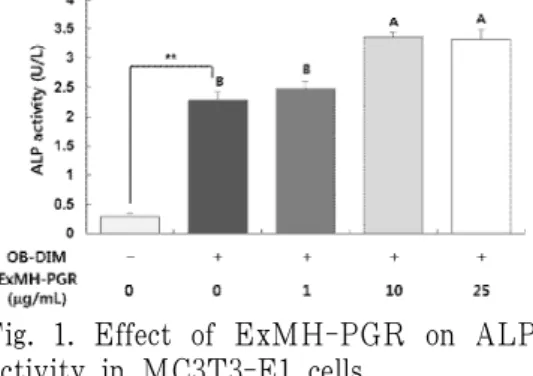
Based on preliminary experiments, the appropriate concentration range of ExMH-PGR to treat MC3T3-E1 cells was chosen to be 0~5 μg/mL. We first examined the effect of ExMH-PGR on ALP activity as an early-stage marker of osteoblast differentiation in MC3T3-E1 cells. The activity of ALP was dramatically increased by OB-DIM. Treatment of cells with 1 μg/mL ExMH-PGR did not significantly affect OB-DIM-induced ALP activity.
Treatment of cells with 10 or 25 μg/mL ExMH-PGR significantly increased OB-DIM -induced ALP activity. However, there was no significant difference in ALP activity between 10 and 25 μg/mL ExMH-PGR treatment groups (Fig. 1).
Collagen synthesis was measured to investigate the effect of ExMH-PGR on osteoblast differentiation because collagen is another early-stage marker of osteoblast differentiation. Similar to ALP activity, collagen synthesis was greatly increased by OB-DIM. OB-DIM-induced collagen synthesis was not significantly affected by treatment with 1 μg/mL ExMH-PGR.
However, treatment of cells with 10 or 25 μg/mL ExMH-PGR significantly increased OB-DIM-induced collagen synthesis (Fig. 2).
These results indicate that ExMH-PGR can promote early stage osteoblast differentiation of MC3TC-E1 cells.
Fig. 1. Effect of ExMH-PGR on ALP activity in MC3T3-E1 cells.
MC3T3-E1 cells were plated at 2×10
4
cells/well and incubated for 24 h. After 24 h of incubation, cells were incubated for 5 days in OB-DIM containing various concentrations of ExMH-PGR.ALP activity was measured. Each bar represents the mean±SE. **P<0.01 significantly different from OB-DIM untreated group. Means without a common letter differ significantly at P<0.05.
Fig. 2. Effect of ExMH-PGR on collagen synthesis in MC3T3-E1 cells.
MC3T3-E1 cells were plated at 2×10
4
cells/well and incubated for 24 h. After 24 h of incubation, cells were incubated for 8 days in OB-DIM containing various concentrations of ExMH-PGR. Collagen synthesis was measured. Each bar represents the mean±SE. ***P<0.001 significantly different from OB-DIM untreated group. Means without a common letter differ significantly at P<0.05.3. ExMH-PGR encourages late osteoblast differentiation in MC3T3-E1 cells
To investigate whether ExMH-PGR affects late process of osteoblast differentiation, we measured the production of osteocalcin and the degree of mineralization as late-stage markers of osteoblast differentiation in MC3TC-E1 cells. As shown in Fig. 3, OB-DIM markedly stimulated the production of osteocalcin and ExMH-PGR significantly promoted OB-DIM-induced osteocalcin production in MC3TC-E1 cells.
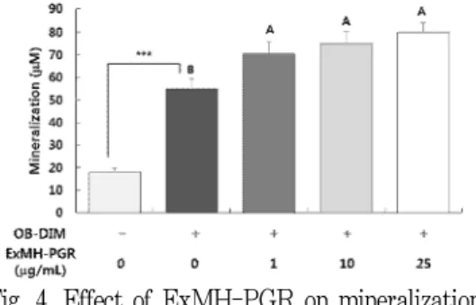
Alizarin Red has been used in biochemical assay to quantitatively determine the presence of calcific deposition by cells of osteogenic lineage by colorimetry. The amount of calcified is proportional to the degree of Alizarin Red staining 20) . Therefore, Alizarin red staining was performed in this study to determine the effect of ExMH-PGR on mineralization in MC3TC-E1 cells.
As demonstrated in Fig. 4, the degree of mineralization in MC3TC-E1 cells was
increased with induction of differentiation by OB-DIM. OB-DIM induced mineralization was effectively promoted by treatment with 1, 10, and 25 μg/mL ExMH-PGR.
However, there was no significant difference in mineralization among ExMH-PGR treatment groups at different concentrations (1, 10, and 25 μg/mL).
Fig. 3. Effect of ExMH-PGR on osteocalcin production in MC3T3-E1 cells.
MC3T3-E1 cells were plated at 2×10
4
cells/well and incubated for 24 h. After 24 h of incubation, cells were incubated for 14 days in OB-DIM containing various concentrations of ExMH-PGR.Media conditioned for 24 h were collected. Levels of osteoblast-secreted osteocalcin in 24 h-conditioned media were measured. Each bar represents the mean±SE. ***P<0.001 significantly different from OB-DIM untreated group. Means without a common letter differ significantly at P<0.05
Fig. 4. Effect of ExMH-PGR on mineralization in MC3T3-E1 cells.
MC3T3-E1 cells were plated at 2×10
4
cells/well and incubated for 24 h. After 24 h of incubation, cells were incubated for 14 days in OB-DIM containing various concentrations of ExMH-PGR. The degree of mineralization was measured. Each bar represents the mean±SE. ***P<0.001 significantly different from OB-DIM untreated group. Means without a common letter differ significantly at P<0.05.4. ExMH-PGR inhibits osteoclast differentiation in RAW 264.7 cells
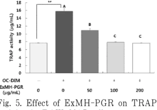
To investigate the effect of ExMH-PGR on osteoclast differentiation, we incubated RAW 264.7 cells in OC-DIM to induce osteoclast differentiation and treated cells with various concentrations of ExMH-PGR.
We then measured TRAP activity as osteoclast differentiation marker. OC-DIM markedly increased TRAP activity in RAW 264.7 cells.
OC-DIM-induced TRAP activity was significantly decreased with increasing ExMH-PGR concentration (Fig. 5).
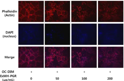
Next, we performed experiments to determine the role of ExMH-PGR in the formation of actin ring by immunofluorescence analyses.
Actin ring structure on osteoclast surface is essential for bone resorption process of osteoclast, while osteoclast can absorb bone matrix through this structure. When osteoclast is differentiated, its size becomes large and the surface forms the actin ring, which is involved in the bone resorption process. Thus, the formation of actin rings in osteoclasts is an important indicator
of the ability of cells to absorb bone 21) . In ExMH-PGR untreated control group, actin rings were the most vivid and cells were dense. These actin rings were markedly decreased with the concentration of ExMH-PGR used for treatment was increased (Fig. 6). This result shows that treatment with ExMH-PGR can inhibit the differentiation of osteoclast to a stage where bone resorption is possible.
Fig. 5. Effect of ExMH-PGR on TRAP activity in RAW 264.7 cells.
RAW 264.7 cells were plated at 5×10
4
cells/well and incubated for 24 h. After 24 h of incubation, cells were incubated for 5 days in OC-DIM containing various concentrations of ExMH-PGR.TRAP activity was measured. Each bar represents the mean±SE. **P<0.01 significantly different from OC-DIM untreated group. Means without a common letter differ significantly at P<0.05.
Fig. 6. Effect of ExMH-PGR on actin ring formation in RAW 264.7 cells.
RAW 264.7 cells were seeded into 24-well plates containing cover glass at 1×10
4
cells/well and incubated for 24 h. After 24 h of incubation, cells were incubated for 5 days in OC-DIM containing various concentrations of ExMH-PGR. After 5 days, cells were fixed with 4% paraformaldehyde containing 0.1% Triton X-100 and stained with Alexa Fluor 594 Phalloidin and DAPI to stain actin and nuclei, respectively. Morphological changes of actin ring in cells were observed via microscopy (n=3; magnification, 200×).Ⅳ. Discussion
Phytochemicals, or phytonutrients, are plant-based compounds that have a low possibility of side effects and a fairly high effectiveness in preventing and treating chronic diseases such as cardiovascular diseases, cancer, and osteoporosis 22) . PGR contains various phytochemicals such as polysaccharides, flavonoids, polyphenolic, and polyacetylene as well as saponin.
Triterpenoid saponins isolated from PGR have been reported to be the major class of its bioactive compounds 5) . Most of the PGR saponin is Platycosin D, which is known to have a variety of physiological effects such as obesity, inflammation and cancer prevention. It was recently reported
that platycodin D can increase bone mineral density 23) . Administration of PGR reduced ovariectomy-induced bone loss and alleviated the reduction of plasma levels of alkaline phosphatase, calcium, and phosphorus observed in ovariectomized mice. This nutritional approach to the treatment of osteoporosis could be seen as a feasible alternative to pharmaceutical ones 18) .
According to the review article of Ann
et al. 24) , there were Hominis Placenta (紫
河車), Phlomis Radix (續斷), Eucommiae
Cortex (杜冲), Psoraleae Fructus (補骨
脂), Allii Tuberosi Semen (韭子), Cervi
Pantotrichum Cornu (鹿茸), Cervi Cornu
(鹿角), Nokgyonggol (鹿脛骨), Morindae
Radix (巴戟天), Trigonellae Semen (葫蘆
巴), Drynariae Rhizoma (骨碎補), Cynomorii Herba (鎖陽), Dendrobii Herba (石斛), Pelodiscis Carapax (鱉甲), Lycii Fructus (枸杞子), Ganodermae Polyporus (靈芝), Ginseng Radix (人蔘), Dioscoreae Rhizoma (山藥), Ginseng Radix Rubra (紅蔘), Rehmanniae Radix Preparata (熟地黃), Polygoni Multiflori Radix (何首烏), Carthami Fructus (紅花子), Sambuci Lignum (接 骨木), Caraganae Radix (骨擔草), Corni Fructus (山茱萸), Mori Folium (桑), Loranthi Ramulus (寄生), and Platycodon grandiflorum (桔梗) as medicinal herbs used in the trial of osteoporosis efficacy. The extract of PG have been reported to be effective in improving bone metabolism in ovariectomized rats 25) . An example of the use of PGR as an oriental gynecologist has been reported to be effective for the apoptosis of uterine leiomyoma cells as a material of Hyulbuchukeo-tang (血府逐瘀湯) 26) .
We prepared herbal extract from a mixture of ordinary medicinal herbs with various concentrations of PGR. We conducted preliminary experiments to determine the most effective concentrations of PGR that could activate osteoblasts and inactivate osteoclasts. Compositions of ExMH-PGR are shown in Table 1.
Ingredients used with PGR for the preparation of ExMH-PGR are: Angelica gigas Nakai (root), Atractylodes macrocephala Koidz (rhizome), Gardenia jasminoides Ellis (fruit), Glycyrrhiza uralensis (root), Menthe arvensis var piperascens (leaf), Paeonia japonica (root), Poria cocos Wolf (sclerotium), and Zingiberis siccatum
Rhizoma. They have tonic, digestive, diuretic, antihydrotic, antiphlogistic, laxative, choleretic, hemostatic, antiphlogistic, mucolytic, expectorant, analgesic, stomachic, antiemetic, antidiarrheal, antiasthmatic, hemostatic, and cardiotonic effects. They have traditionally been used in Korea and China to treat gynecological disorders such as menopausal disorders, dysmenorrhea, digestive and respiratory diseases, and herbal toxicity detoxification. In other words, these components of ExMH-PGR are not related to osteoporosis except for PGR 27,28) . In this in vitro study, we investigated the efficacy of ExMH-PGR as a dietary supplement for prevention and treatment of osteoporosis.
In our present study, we analyzed ALP activity, collagen synthesis, mineralization, and osteocalcin concentration as biomarkers related to bone formation using MC3T3-E1 cells as an osteoblast cell line. ALP is present in almost all tissues, especially in the bone, kidney, intestinal mucosa, placenta, liver and bile duct. ALP level in bone tissue increases when active bone formation is occurring, because ALP is a byproduct of osteoblast activity 29) . Collagen is an important protein distributed in each connective tissue. It is present in various parts of the body. Up to now, 28 kinds of collagen have been identified.
However, over 90% of human collagen
is type 1 collagen which is abundant in
the skin, bone and ligaments. It is mainly
synthesized by osteoblasts 30) . The ability
to form bone calcification is an important
biomarker for osteoblast differentiation 31) . Osteocalcin is a protein secreted by osteoblasts. It is involved in bone mineralization and calcium ion homeostasis as well as bone formation. Since osteocalcin is produced by osteoblasts, it is often used as an index of the bone formation process. It has been reported that higher serum-osteocalcin levels are relatively strongly correlated with increases in bone mineral density while using anabolic bone formation drugs to treat osteoporosis. In many studies, osteocalcin is used as a primary biomarker to determine the effect of the drug to help bone formation 32) . In this study, we showed that PGR has the ability to increase bone formation in osteoblasts cells. ExMH-PGR treatment increased ALP activity, collagen synthesis, mineralization, and osteocalcin production.
Numerous studies have reported that anti-osteoporotic agents can increase osteoblast differentiation and suppress osteoclast differentiation at the same time 33-5) . PGR-derived saponins appear to stimulate osteoblast differentiation by regulating RUNX2, the principle osteogenic master gene for bone formation 17) .
According to cellular and molecular-level osteoporosis studies, the mechanism of bone loss is usually caused by osteoclast differentiation and bone resorption beyond osteoblast bone formation 36) . To investigate the effect of ExMH-PGR on the function of osteoclast, we analyzed TRAP activity and actin ring formation in RAW 264.7 cells. Osteoclasts are the cells involved
in bone matrix resorption and bone and calcium homeostasis. Since TRAP is a specific marker of osteoclast and its secretion increases during bone resorption, TRAP activity has been used to confirm osteoclast differentiation 37) . An actin ring is a unique actin structure essential for bone resorption by osteoclasts. Activation of osteoclasts begins with recognition of the matrix, adhesion to the bone surface, and rearrangement of the cytoskeleton.
The osteoclast cytoskeleton can polarize the osteoclast to create a ruffled border with a separate environment of the resorptive microenvironment of the bone-cell interface and form a gasket-like structure. This is called actin ring or sealing zone. Actin rings separate the resorptive microenvironment from the normal extracellular space 21,38) . Osteoclasts synthesize and secrete lytic enzymes in the cytoplasm, which acidifies the lacunae underneath osteoclasts by exocytosis.
Osteoclasts then bring in the degraded
material through endocytosis. These exocytic
and endocytic processes are essential
sequences for effective bone resorption
by osteoclasts 21,39) . In this study, RAW
264.7 cells treated with ExMH-PGR showed
decreased TRAP activity and actin ring
dose-dependently (Fig. 5, 6). The inhibitory
effect of PGR and its major component,
platycodin D, on osteoclast activity have
also been reported in other studies. Lee
et al. 40) have reported that oral administration
of platycodin D inhibits osteoclast formation
and blocks bone destruction caused by
breast cancer. Choi et al. 18) have also reported that platycodin D inhibits ERK/p38 MAPK signaling pathway as well as NF-κB signaling and thus inhibits osteoclast differentiation.
These results suggest that ExMH-PGR promotes osteoblast activity and inhibits osteoclast activity. However, in our study, PGR was not as a single medicinal product, but was a part of a combination. Thus, it cannot be guaranteed that these results were entirely caused by PGR. Further research on many components of PGR and their osteoporosis-related mechanisms needs conducting.
V. Conclusion
In this study, we conducted an in vitro
study to determine whether ExMH-PGR, an extract of mixed herbs containing root of Platycodon grandiflorum, was effective in preventing or treating osteoporosis.
ExMH-PGR treatment significantly increased ALP activity, collagen synthesis, mineralization, and osteocalcin production in osteoblasts and inhibited TRAP activity and actin ring formation in osteoclast.
These results indicate that ExMH-PGR can be regarded as an anti-osteoporotic candidate for treatment of osteoporosis disease. In the future, it will be necessary to confirm its effect through follow-up study in animal and human and clarify its action mechanism.
□ Received : Sep 19, 2018
□ Revised : Sep 24, 2018
□ Accepted : Nov 26, 2018
국 문초록
![Table 1. Used to Prepare the Extract of Herbs Mixture [ExMH-PGR*]](https://thumb-ap.123doks.com/thumbv2/123dokinfo/5246652.363001/4.799.137.702.148.362/table-used-prepare-extract-herbs-mixture-exmh-pgr.webp)