• 검색 결과가 없습니다.

A Case of Pulmonary Sarcoidosis with Endobronchial Nodular Involvement

N/A
N/A
Protected

Academic year: 2021

Share "A Case of Pulmonary Sarcoidosis with Endobronchial Nodular Involvement"

Copied!
6
0
0

로드 중.... (전체 텍스트 보기)

전체 글

(1)

Tuberc Respir Dis 2013;74:274-279

CopyrightⒸ2013. The Korean Academy of Tuberculosis and Respiratory Diseases. All rights reserved.

A Case of Pulmonary Sarcoidosis with Endobronchial Nodular Involvement

Kyung Hwa Cho, M.D.1, Jeong Hyun Shin, M.D.1, Seong Hoon Park, M.D.2, Heon Soo Kim, M.D.3, Sei Hoon Yang, M.D.1

Departments of 1Internal Medicine, 2Radiology, and 3Pathology, Wonkwang University College of Medicine, Iksan, Korea

Sarcoidosis is a multisystemic disorder of unknown cause that is characterized pathologically by noncaseating granulomas. Diagnosis is based on the exclusion of other infectious, interstitial, and neoplastic diseases and on the typical pathology. Although the lungs and mediastinal lymph nodes are almost involved, endobronchial nodular lesions of sarcoidosis with lung involvements are rare. We report a case of sarcoidosis with lung involvements and endobronchial nodules as confirmed by bronchial biopsy.

Key Words: Sarcoidosis; Bronchi; Biopsy

Address for correspondence: Sei Hoon Yang, M.D.

Department of Internal Medicine, Wonkwang University Hospital, Wonkwang University College of Medicine, 895 Muwang-ro, Iksan 570-749, Korea

Phone: 82-63-859-2582, Fax: 82-63-855-2025 E-mail: [email protected]

Received: Sep. 4, 2012 Revised: Sep. 24, 2012 Accepted: Oct. 23, 2012

CCIt is identical to the Creative Commons Attribution Non-Commercial License (http://creativecommons.org/licenses/by-nc/3.0/).

Introduction

Sarcoidosis is a cryptogenic general granulomatosis that typically affects adults up to 50 years of age.

Sarcoidosis infiltrates all organs, particularly the bilateral hilar lymph nodes, lungs, peripheral lymph nodes, skin, eyes, liver, and spleen

1

. Boek first used the term 'sar- coid' in 1899 in regard to a condition of multiple tumors and rashes. In Korea, sarcoidosis has occurred in ap- proximately 0.13 out of 100,000 individuals since the first case was reported here in 1984 (The 2nd Nation- wide Actual Condition Survey, 2000), but it has recently been on an increasing trend

2

. The clinical symptoms of sarcoidosis are usually nonspecific and generalized, and include fever, fatigue, anorexia, weight loss, and mala- ise. The symptoms vary depending upon the organs that

are infiltrated. Given that sarcoidosis mostly infiltrates the lungs, respiratory symptoms such as exertional dyspnea, cough, and chest pain are observed in most cases
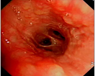
3

. In our experience, it is rare that sarcoidosis nod- ularly infiltrates the bronchus. This is a report of a case of endobronchial nodular sarcoidosis diagnosed by bronchoscopy and a biopsy.

Case Report

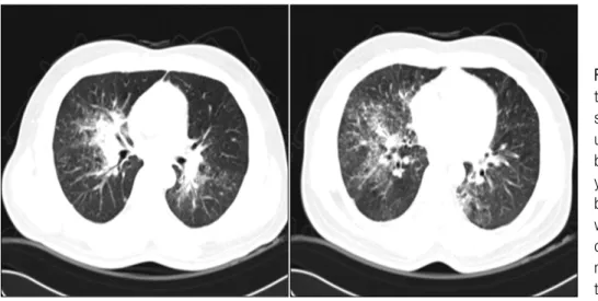
A 42-year-old Indian male who was employed as an automotive engineer was referred to our hospital with complaints of a month-long fever and cough. The pa- tient had been diagnosed with pulmonary tuberculosis 5 years previously in India, and he had returned from Thailand a month before visiting our hospital complain- ing of fever, persistent cough, and general malaise.

Before he was referred to our center, he had undergone

computed tomography (CT) at another hospital because

his symptoms had persisted even after medical treat-

ment, whereon abnormalities had been detected. As

noted, the patient had been previously diagnosed with

pulmonary tuberculosis by positive mycobacterium tu-

berculosis culture in April 2003, in India. He completed

a 7-month course of anti-tuberculosis medications was

completely recovered, and he had no history of dia-

(2)

Figure 1. Chest radiograph showing ill-defined nodular peribronchial lesions in both lungs, most prominently in the mid right and lower lung zones.

Figure 2. Chest computed tomography showed con- solidation along the right upper and intermediate bronchus and small peril- ymphatic, centrilobular, and bronchovascular nodules with interlobular septal thi- ckening in the upper right, mid, and lower lobes and the lower left lobe.

betes, hypertension, or hepatitis. Blood pressure, pulse, respiratory rate, and temperature were 110/70 mm Hg, 80/min, 20/min, and 36.4

o

C, respectively. He was alert and oriented to person, place, and time. The cervical lymph nodes and thyroid were not palpable, nor were any noteworthy abnormalities observed on chest, ab- dominal, or neurologic examinations. On complete blood count hemoglobin level, platelets, and white blood cells count were 14.3 g/dL (rane, 13–18 g/dL), 192,000/mm

3

(range, 150,000–450,000/mm

3

), and 5,880/

mm

3

(range, 4,000–10,000/mm

3

) (neutrophils 54.2%, lymphocytes 31.5%, monocytes 9.0%, eosinophils 4.8%).

Blood chemistry revealed the following: total protein 7.2 g/dL (range, 6.5–8.3 g/dL), aspartate amino-trans- ferase 23 U/L (range, 5–35 U/L), alanine amino-trans- ferase 17 U/L (range, 5–40 U/L), alkaline phosphatase 201 U/L (range, 66–220 U/L), blood urea nitrogen 11.4 mg/dL (range, 8–20 mg/dL), creatinine 0.97 mg/dL (range, 0.5–1.3 mg/dL), sodium 137 mEq/L (range, 135 –150 mEq/L), potassium 4.4 mEq/L (range, 3.3–5.5 mEq/L), calcium 9.24 mg/dL (range, 8.4–10.2 mg/dL), C-reactive protein 5.87 mg/dL (range, 0–5 mg/dL), an- giotensin converting enzyme (ACE) 67 IU/L (range, 8–

52 IU/L), and urine calcium 4.6 mg/dL. There were no notable abnormalities detected on urinalysis, sputum smear, sputum culture for bacteria and fungus, tubercle bacilli test, or viral and immune serum tests. There was no foreign body in sputum or bronchoalveolar lavage (BAL) fluid.

The pulmonary function tests showed a restrictive

ventilatory defect with forced vital capacity (FVC) 3.06

L (66% of the predicted value), forced expiratory vol-

ume in one second (FEV

1

) 2.44 L (68% of the predicted

value), FEV

1

/FVC 80%, and DLco 18.6 mL/mm Hg/min

(74% of the predicted value). On a plain radiography

performed before the patient visited this hospital, nod-

ular lesions were found to be distributed along the

bronchi in the right middle and lower lobes (Figure 1),

and on the chest CT, thickened lobules and multiple

peribronchovascular nodules were observed throughout

the entire right lung and in the left lower lobe, in addi-

(3)

Figure 3. Follow-up chest radiography and computed tomography showed an increased extent of peribronchial infiltration and air space consolidation, interlobular septal thickening, and ill-defined nodules in both lungs, along with increased size of the multiple enlarged hilar and interlobar lymph nodes.

Figure 4. Bronchoscopy showed multiple variable small nodular endobronchial lesions.

Figure 5. Biopsy of endobronchial nodules showed diffuse granulomatous inflammation with multinucleated giant cells (H&E stain, ×100).

tion to lymphadenopathy around the bronchi and the right pulmonary hilum (Figure 2). The patient's symp- toms were relieved to some extent after symptomatic treatment, but they were not eliminated, even during the follow-up. Repeat chest radiography and CT were performed, and the results showed that the known mul- tiple nodules had increased considerably and the hilar lymph nodes had become more enlarged (Figure 3).

The patient underwent bronchoscopy, and multiple

nodules of different sizes were observed throughout the

bronchi (Figure 4). An endobronchial biopsy was per-

formed, whereon multiple non-caseating chronic gran-

ulomatous lesions were diagnosed (Figure 5). No

acid-fast bacilli and fungus figured out on tissue stain-

ing. On BAL, macrophages, lymphocytes, neutrophils,

CD4, and CD8 cells were counted as 31%, 68%, 1%,

(4)

Figure 6. Follow-up chest radiography and computed tomography after treatment with oral steroid showed decreased extent of the peribronchial infiltration and air space consolidation in both lungs, along with a significantly decreased extent of the interlobar septal thickening and perilymphatic or ill-defined nodules, and decreased sizes of the multiple enlarged hilar and interlobar lymph nodes.

75.1%, and 16.6%, respectively. Bacteria, fungi, virus, and tubercle bacillus were not detected in the BAL fluid.

Repeat blood chemistry results included ACE and cal- cium levels of 94 IU/L and 9.7 mg/dL, respectively.

Ophthalmic and dermatologic abnormalities were not detected, and there were no notable abnormalities on echocardiography or gallium-67 scan. Finally, the pa- tient was diagnosed with endobronchial sarcoidosis in- volving lungs and lymph nodes, and oral prednisolone was prescribed (70 mg; 1 mg/kg/day). The symptoms continued to recede with administration of predni- solone, and the dose was gradually reduced to 5 mg for 7 months. The chest radiography and CT performed at 7 months' follow-up confirmed a remission (Figure 6). Currently, the patient is being monitored with regu- lar follow-up and is not receiving steroids.

Discussion

Sarcoidosis is a cryptogenic disease that is charac- terized by non-caseating chronic granulomas, which in- filtrate all organs, including lungs, skin, eyes, bones, nerves, and lymph nodes. The symptoms vary depend-

ing upon the organ system affected, but pulmonary in- filtration actually comprises more than 90% of cases, and thus, respiratory symptoms such as cough, exer- tional dyspnea, and chest pain are observed in most cases. Bilateral symmetric hilar or paratracheal lympha- denopathy are cited as salient findings. Approximately 60% of patients will also show an increase in ACE lev- els, and 10% develop hypercalcemia. Arthritis may oc- cur in 5% to 10% of patients

1,4

.

It is uncommon that pulmonary sarcoidosis infiltrates the bronchi. When it involves airways, the disease is manifested as mucosal erythema, edema, granularity, and cobblestoning, with plaques, nodules, and bron- chial stenosis, airway distortion, traction bronchiectasis, and bronchiolitis. If the disease infiltrates the trachea, main carina, or major bronchi alone, it will occasionally produce significant obstructive symptoms or airway dysfunction.

The diagnosis of sarcoidosis is made based on symp-

toms, radiographic findings, and tissue biopsy

5

. It is

standard practice to rule out other causes when non-

caseating granuloma is found on biopsy. The challenge

is that it is difficult to diagnose sarcoidosis in as much

(5)

as it is similar in clinical, radiological, and histological characteristics to tuberculous and non-tuberculous my- cobacterial lung diseases. When it occurs in the bron- chus, as in the present case, a biopsy should be per- formed to distinguish it from malignant diseases such as lung cancer.

Nodules in the upper lobes of the both lungs and bi- lateral intrathoracic lymphadenopathy are typical radio- logical findings, but there may be atypical cases, and in such cases, it is difficult to distinguish sarcoidosis from the other similar diseases. In addition, there have been reports of bilateral and multiple peripheral or peri- bronchovascular non-homogeneous shadows, pulmo- nary micronodules, or indurated micronodules asso- ciated with pulmonary sarcoidosis

6,7

.

The stages of sarcoidosis are classified by radiological findings. Stage 0 is defined by absence of abnormalities on chest radiography. Stage I includes bilateral hilar lymphadenopathy. In stage II, there is bilateral hilar ad- enopathy and pulmonary infiltration, and in stage III, the pulmonary infiltration is observed but there is no lymphadenopathy

8

. Thus, our patient was diagnosed with stage II sarcoidosis, because both adenopathy and pulmonary infiltration were observed.

History and physical exam are crucial to check for sarcoid infiltration of other organs after the sarcoidosis diagnosis. Pulmonary function tests, ophthalmologic ex- am, gallium scan, and BAL are also necessary, as well as measurements of serum and urine calcium levels and serum ACE

9

.

Sarcoidosis has a favorable prognosis, and it is re- ported that 80% to 90% of patients with acute-phase sar- coidosis recover spontaneously

10

. On the other hand, sarcoidosis will have a poor prognosis when it occurs in patients aged 40 and older, when it lasts for 6 months and longer, when it is not accompanied by eryth- ematous nodules, when lymphadenopathy occurs, when it infiltrates at least 3 organ systems, and when it causes only pulmonary disease

11

. Explicit uniform treatment guidelines for sarcoidosis have not yet been established. Oral steroids or methotrexate maybe used if sarcoidosis has infiltrated the eyeballs, the nervous

system, or the heart at a serious level, or if the patient has severe hypercalcemia, stage II pulmonary disease, or stage II or III symptomatic pulmonary disease, but there are no widely accepted recommendations for dose or duration

12

.

This patient visited the hospital with a chief com- plaint of a month-long fever and cough, pulmonary sar- coidosis was suspected on chest radiography, and he was definitively diagnosed with endobronchial nodular sarcoidosis by bronchoscopy with a bronchoscopic biopsy. The patient has achieved complete remission following an extend course (7 months) of oral steroid therapy.

Acknowledgements

This paper was supported by a grant from Won- kwang University in 2011.

References

1. Iannuzzi MC, Rybicki BA, Teirstein AS. Sarcoidosis. N Engl J Med 2007;357:2153-65.

2. Kim DS, Ahn JJ. Sacoidosis in Korea. Tuberc Respir Dis 2000;49:274-80.

3. Newman LS, Rose CS, Maier LA. Sarcoidosis. N Engl J Med 1997;336:1224-34.

4. Choi HH, Hong YA, Choi JK, Kim JS, Kim SJ, Kim SC, et al. A case of sarcoidosis that was initially mis- diagnosed as nontuberculous mycobacteria pulmonary disease. Tuberc Respir Dis 2009;66:309-13.

5. Hanno R, Callen JP. Sarcoidosis: a disorder with prom- inent cutaneous features and their interrelationship with systemic disease. Med Clin North Am 1980;64:

847-66.

6. Sharma OP. Sarcoidosis: a worldwide phenomenon.

Sarcoidosis 1984;1:11-5.

7. Nam JE, Ryu YH, Park JG, Choe KO, Im JG, Lee KS, et al. High resolution CT findings of pseudoalveolar sarcoidosis. J Korean Radiol Soc 2002;47:191-6.

8. Tay HL, Vaughan-Jones R, Qureshi SS. Ethmoidal sarcoidosis. J Laryngol Otol 1994;108:682-4.

9. Nishimura K, Itoh H, Kitaichi M, Nagai S, Izumi T.

Pulmonary sarcoidosis: correlation of CT and histo- pathologic findings. Radiology 1993;189:105-9.

10. Lee SJ, Kim JY, Lee JC, Kim GS, Yoo CG, Kim YW,

(6)

et al. A case of sarcoidosis involving bone marrow, skin, uvea, joints, liver. Korean J Med 1997;53:580-5.

11. Chung SG, Park CH. Axillary lymph node sarcoidosis.

J Korean Surg Soc 2001;61:220-3.

12. Lee BH, Kim JM, Kim DW, Kim JH, Bang KT, Lee KY,

et al. A case of sarcoidosis with cavitation. Tuberc

Respir Dis 2005;59:546-50.

수치

Figure  2.  Chest  computed  tomography  showed   con-solidation  along  the  right  upper  and  intermediate  bronchus  and  small   peril-ymphatic, centrilobular, and bronchovascular  nodules  with  interlobular  septal   thi-ckening  in  the  upper  rig
Figure 5. Biopsy of endobronchial nodules showed diffuse granulomatous  inflammation  with  multinucleated  giant  cells  (H&E  stain,  ×100).
Figure  6.  Follow-up  chest  radiography  and  computed  tomography  after  treatment  with  oral  steroid  showed  decreased extent  of  the  peribronchial  infiltration  and  air  space  consolidation  in  both  lungs,  along  with  a  significantly  de

참조

관련 문서

Initial chest radiograph showed diffuse patchy and nodular densities in the left lower lung (Figure 1A) and a low dose computed tomography of chest revealed variable sized

A chest computed tomography scan performed 2 years later in the studied patient showing an increased extent of the nod- ules, particularly in the right middle lobe and

After the discontinuation of quetiapine therapy and 14 days of steroid therapy, a high-resolution computed tomography scan of the chest revealed a decreased extent

Chest computed tomography showed diffuse nodular pleural thickening, and microscopic finding disclosed diffuse thickening of visceral pleura with infiltrations of

The chest X-ray and chest CT(computed tomography) showed a nodular lesion obstructing the proximal portion of the left lower lobar bronchus and atelectasis of

(A) Chest computed tomography showing aggravated wall thickening with stenosis in the main pulmonary artery and newly developed filling defect in the anterior

Follow-up contrast-enhanced chest computed tomography obtained after embolization and three courses of methotrexate shows that multi- ple metastatic nodules with residual

Axial CT scan (lung window) shows small right hemithorax with mild interlobular septal thickening in right lower lobe and right middle lobe.. Contrast-enhanced axial CT