236
Address for Correspondence: Eun-Young Kang, M.D.
Department of Radiology, Korea University Guro Hospital, 97 Guro-dong, Guro-gu, Seoul 152-703, Korea
Phone: 82-2-2626-1342, Fax: 82-2-863-9282 E-mail: [email protected]
Received: Jan. 31, 2008 Accepted: Mar. 11, 2008
A Pulmonary Sarcoidosis Manifesting as a Rare Atypical Pattern and Distribution
Departments of
1Radiology,
2Pathology, and
3Internal Medicine, Korea University Guro Hospital, Korea University College of Medicine, Seoul, Korea
Sun-Hwa Hong, M.D.
1, Eun-Young Kang, M.D.
1, Ok Hee Woo, M.D.
1, Hwan Seok Yong, M.D.
1, Yu-Whan Oh, M.D.
1, Bong Kyung Shin, M.D.
2, Jae Jeong Shim, M.D.
3드문 비전형적인 형태와 분포를 보인 폐 유육종증 1예
홍선화1, 강은영1, 우옥희1, 용환석1, 오유환1, 신봉경2, 심재정3
고려대학교 의과대학
1영상의학교실,
2병리학교실,
3내과학교실
A unique case of atypical pulmonary sarcoidosis in a 62-year-old man complaining of dyspnea is presented. Chest CT scan showed an unusual pattern and distribution of pulmonary sarcoidosis manifesting mainly as reticular densities, interlobular septal thickening, and ground-glass opacities, in the subpleural and lower lung predominancy.
However, a surgical lung biopsy revealed classical findings of sarcoidosis. Knowledge of this atypical pulmonary involvement may improve understanding sarcoidosis as the great masquerader. (Tuberc Respir Dis 2008;64:236-239) Key Words: Sarcoidosis, Pulmonary disease, CT
Introduction
Sarcoidosis is a systemic granulomatous disease of unknown etiology and can affect any organ system. The thorax is one of the most commonly affected areas, and thoracic involvement is reported in up to 90% of patients with sarcoidosis1-4. Thoracic sarcoidosis usually affects the lymph nodes and the lung parenchyma. Radiologic manifestation of thoracic sarcoidosis is well established, and symmetric bilateral hilar and mediastinal lympha- denopathy with or without concomitant lung paren- chymal infiltrates is the most common manifestation of thoracic sarcoidosis1,3. Although 60% to 70% of patients with thoracic sarcoidosis have characteristic radiologic findings, the remaining patients may have a variety of radiologic findings5,6. Furthermore, the appearance of pulmonary sarcoidosis is occasionally very unusual,
nonspecific, and atypical. Therefore, pulmonary sarcoi- dosis often resembles many other lung diseases.
A rare, atypical case of thoracic sarcoidosis manifest- ing with an unusual pattern and distribution of lung in- volvement is reported in this paper.
Case Report
A 62-year-old man visited the hospital with a com- plaint of NIHA class II dyspnea. On auscultation, his breathing sound was normal and on a thorough physical examination, no abnormalities were found. The patient had a past history of hypertension and was an ex-smoker of 40-pack years. In laboratory tests, collagen vascular disease markers were negative.
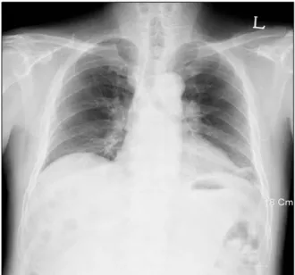
Simple chest radiography was performed as a routine check-up. Lymph node enlargement was suspected in both hilum (Figure 1). A chest CT scan taken 4 days later showed fine reticular densities, interlobular septal thickenings, and faint ground-glass opacities on both lungs. The lesions were distributed on both lungs sym- metrically, but predominant lower and subpleural lung involvement was noted (Figure 2A, B). Discrete lymph node enlargements without internal calcifications were
Tuberculosis and Respiratory Diseases Vol. 64. No. 3, Mar. 2008
237
Figure 1. Chest PA view shows bilateral hilar lympha-denopathies.
Figure 2. Thin-section CT with lung window setting (A, B) shows fine reticular densities, faint ground-glass opacities, and interlobular septal thickenings on both lungs, predominantly the lower subpleural lung zone. Enhanced chest CT scan with a mediastinal window setting (C) shows bilateral mediastinal and hilar lymphadenopathies.
Figure 3. Surgical lung biopsy specimen shows extensive infiltration by noncaseating granulomas distributed along the pleura, interlobular septa, and bronchovascular bun- dles (H&E stain, ×40).
observed in the prevascular, subaortic, subcarinal, and both paratracheal and hilar areas (Figure 2C). Thus, with the presumptive diagnosis of idiopathic interstitial pneumonia such as nonspecific interstitial pneumonitis and lymphocytic interstitial pneumonia, or lymphoma, a surgical lung biopsy was performed on the left upper and lower lobes. A mediastinoscopic biopsy was also performed on the right paratracheal lymph nodes.
However, the result of the lung biopsy revealed ex- tensive infiltration by noncaseating granulomas distributed along the pleura, interlobular septa, and bronchovas- cular bundles, which are characteristic findings of pul- monary sarcoidosis (Figure 3). A mediastinal lymph node
biopsy also revealed noncaseating chronic granulomatous inflammation consisting of sarcoidosis.
Discussion
The CT features of pulmonary sarcoidosis include mi- cronodules and nodules, central peribronchovascular thickening, alveolar or pseudoalveolar consolidations, septal and nonseptal lines, ground-glass opacity, con- glomerate masses, lung architectural distortion, bron- chiectasis, honeycombing or other types of cyst, emphy- sema, and thickening of the pleural surface1. Pulmonary sarcoidosis is typically bilateral and symmetric, and in-
SH Hong et al: A pulmonary sarcoidosis manifesting as a rare atypical pattern and distribution
238
volves mainly the central rather than peripheral lungs.
The upper lobes, particularly the apical and posterior segments, are the most severely affected2.
The most characteristic CT appearance of pulmonary sarcoidosis consists of small nodules in a perilymphatic distribution1,2,6. Small nodules usually have irregular margins and range from 1 to 10 mm in size. They are typically scattered throughout the interstitium of the lungs, along the lymphatics, particularly in the peri- bronchovascular and subpleural spaces, and along the interlobular septa. Pathologically, noncaseating gran- ulomas distribute mostly along the lymphatics in the peribronchovascular sheath and, to a lesser extent, in the subpleural and interlobular septal lymphatics6,7. However, the diseases in lymphatic and perilymphatic distribution include lymphangitic carcinomatosis, lym- phoproliferative disease, silicosis, and amyloidosis as well as sarcoidosis. Therefore, CT findings of these dis- eases can be similar to that of sarcoidosis3,8. Sarcoidosis is more significantly involved bilaterally and in the up- per lungs than lymphangitic carcinomatosis and malig- nant lymphoma. Thickened interlobular septa observed in lymphangitic carcinomatosis are rare in sarcoidosis8. Lymphoma may present with predominant peribroncho- vascular nodules but are typically larger and less profuse than sarcoidosis. Silicosis also shows perilymphatic nod- ules, but the distinction can be made easily by clinical history. Thus, sarcoidosis can usually be differentiated from these diseases.
Sarcoid granulomas can coalesce and become con- fluent to produce large nodules and alveolar opacities.
The alveolar opacities vary in size from 1∼10 cm, are bilateral with ill-defined borders, and may show internal air bronchograms. This peripheral nonsegmental airspace consolidation is called alveolar sarcoidosis. Pulmonary sarcoidosis sometimes shows patchy areas of ground- glass opacity, which may be superimposed on a back- ground of interstitial nodules or fibrosis. Other CT find- ings include solitary or multiple pulmonary masses, ate- lectasis, focal or asymmetric alveolar or interstitial disease.
Pulmonary sarcoidosis can show miliary nodules located in a random distribution. This miliary pattern can show
similar findings of miliary tuberculosis, miliary fungal in- fection, and hematogenous metastasis.
Pulmonary sarcoidosis may resolve spontaneously or progress to fibrosis. As fibrosis progresses, irregular re- ticular densities, lung parenchymal distortions with large conglomerate mass, and lung volume loss are predo- minant. These findings can be associated with traction bronchiectasis and peripheral honeycombing2.
In the present case, fine reticular densities and inter- lobular septal thickening with basal subpleural distri- bution, and faint ground-glass opacity without a back- ground of visible interstitial nodules rendered a diag- nosis of sarcoidosis difficult. Furthermore, areas of ground- glass attenuation and reticular opacities distributed pe- ripherally in both lower lung zones were contrary to the upper and central lung distribution pattern of classic pulmonary sarcoidosis. These are the dominant CT find- ings among patients with nonspecific interstitial pneu- monia, and they are similar to lymphocytic interstitial pneumonia, which manifests with diffuse ground-glass attenuation, poorly defined centrilobular nodules, thick- ening of perilymphatic interstitium, septal thickening, and scattered thin-walled cysts with lower lung zone predominance9. Hence, an initial diagnosis was made of idiopathic interstitial pneumonia, such as nonspecific interstitial pneumonia and lymphocytic interstitial pneu- monia, rather than sarcoidosis in this case. Therefore, a surgical lung biopsy was performed in the left upper and lower lobes to make a diagnosis. However, histo- logically, the pathologic specimen showed classical findings of pulmonary sarcoidosis.
Summary
In this report, a rare atypical case of pulmonary sar- coidosis manifesting mainly as reticular densities, inter- lobular septal thickening, with ground-glass opacities, in subpleural and lower lung predominancy is presented.
Pulmonary sarcoidosis can mimic other lung diseases radiologically and cause radiologic diagnostic uncer- tainties. Awareness of the diverse manifestations as well as typical manifestations can help prevent unnecessary
Tuberculosis and Respiratory Diseases Vol. 64. No. 3, Mar. 2008
239
procedures in patients with sarcoidosis.References
1. Nunes H, Brillet PY, Valeyre D, Brauner MW, Wells AU. Imaging in sarcoidosis. Semin Respir Crit Care Med 2007;28:102-20.
2. Vagal AS, Shipley R, Meyer CA. Radiological manifes- tations of sarcoidosis. Clin Dermatol 2007;25:312-25.
3. Koyama T, Ueda H, Togashi K, Umeoka S, Kataoka M, Nagai S. Radiologic manifestations of sarcoidosis in various organs. Radiographics 2004;24:87-104.
4. Hennebicque AS, Nunes H, Brillet PY, Moulahi H, Valeyre D, Brauner MW. CT findings in severe thoracic sarcoidosis. Eur Radiol 2005;15:23-30.
5. Hamper UM, Fishman EK, Khouri NF, Johns CJ, Wang KP, Siegelman SS. Typical and atypical CT manifes-
tations of pulmonary sarcoidosis. J Comput Assist Tomogr 1986;10:928-36.
6. Müller NL, Kullnig P, Miller RR. The CT findings of pul- monary sarcoidosis: analysis of 25 patients. AJR Am J Roentgenol 1989;152:1179-82.
7. Nishimura K, Itoh H, Kitaichi M, Nagai S, Izumi T.
Pulmonary sarcoidosis: correlation of CT and histo- pathologic findings. Radiology 1993;189:105-9.
8. Honda O, Johkoh T, Ichikado K, Yoshida S, Mihara N, Higashi M, et al. Comparison of high resolution CT findings of sarcoidosis, lymphoma, and lymphangitic carcinoma: is there any difference of involved inter- stitium? J Comput Assist Tomogr 1999;23:374-9.
9. Johkoh T, Müller NL, Pickford HA, Hartman TE, Ichikado K, Akira M, et al. Lymphocytic interstitial pneumonia: thin-section CT findings in 22 patients.
Radiology 1999;212:567-72.