A Case of Segniliparus rugosus Pulmonary Infection in an Immunocompetent Patient with Non-cystic Fibrosis
Jung Yeon Lee, M.D.1, Gyu Rak Chon, M.D.1, tae-young Jung, M.D.2, Heungsup Sung, M.D.3, Tae Sun Shim, M.D.4 and Kyung-Wook Jo, M.D.4
1Division of Pulmonary and Critical Care Medicine, Department of Internal Medicine, Konkuk University School of Medicine, Chungju, 2Department of Internal Medicine, Konkuk University Chungju Hospital, Konkuk University School of Medicine, Chungju, 3Department of Laboratory Medicine and 4Division of Pulmonary and Critical Care Medicine, Asan Medical Center, University of Ulsan College of Medicine, Seoul, Korea
Segniliparus species is a novel genus that is reported to be the new emerging respiratory pathogens. Here, we report a very rare case of S. rugosus pulmonary infection in an immunocompetent patient with non-cystic fibrosis. The organism was identified by 16S rRNA gene sequencing. The patient was successfully treated with antibiotics.
Keywords: Respiratory Tract Infections; Nontuberculous Mycobacteria; RNA Sequencing
unusual case of S. rugosus pulmonary infection in an immu- nocompetent, non-cystic fibrosis patient.
Case Report
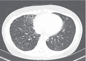
A 47-year-old woman was referred to our hospital for a re- cently aggravating cough lasting for 3 months. About 20 years earlier, she had been treated for pulmonary tuberculosis. The patient had experienced chronic coughing for the previous 3 years. A chest X-ray and chest computed tomography (CT) taken 2 years previously revealed centrilobular nodules and bronchiectatic changes (Figure 1). Although AFB cultures yielded Mycobacterium abscessus growth on several oc- casions at that time, she did not undergo treatment for M.
abscessus because her symptoms were mild and CT did not reveal severe changes. She therefore underwent regular follow-up visits. Although she had been relatively well and her chest X-ray did not show significant changes during the ap- proximately 2-year follow-up, she experienced an aggravation of her symptoms.
On physical examination, the patient was alert and in no distress. Her body temperature was 36.3oC, blood pressure was 106/70 mm Hg, pulse was 72 beats per minute with a regular rhythm, and respiratory rate was 20 breaths per min- ute. Bronchial breathing sounds and inspiratory rhonchi were Copyright © 2014
The Korean Academy of Tuberculosis and Respiratory Diseases.
All rights reserved.
Introduction
Segniliparus is a novel genus consisting of two species, S. rugosus and S. rotundus, that was first described in 2005 as a distinct genus isolated from human sources as a group of rapidly growing acid-fast bacilli (AFB)1. Since then, cases of Segniliparus infection have been reported in bronchiec- tasis patients with2,3 or without4 cystic fibrosis. In addition, Segniliparus isolation was recently even reported in a non- bronchiectasis patient in South Korea5. Here, we report a very
CASE REPORT http://dx.doi.org/10.4046/trd.2014.77.5.227
ISSN: 1738-3536(Print)/2005-6184(Online) • Tuberc Respir Dis 2014;77:227-229
227
Address for correspondence: Kyung-Wook Jo, M.D.
Division of Pulmonary and Critical Care Medicine, Asan Medical Center, University of Ulsan College of Medicine, 88 Olympic-ro 43-gil, Songpa- gu, Seoul 138-736, Korea
Phone: 82-2-3010-5783, Fax: 82-2-3010-6968 E-mail: [email protected] Received: Jul. 3, 2014
Revised: Aug. 4, 2014 Accepted: Aug. 19, 2014
cc It is identical to the Creative Commons Attribution Non-Commercial License (http://creativecommons.org/licenses/by-nc/3.0/).
JY Lee et al.
228 Tuberc Respir Dis 2014;77:227-229 www.e-trd.org
heard in the bilateral anterior chest. A complete blood count revealed a white blood cell count of 6,500/mm3 (64% neutro- phils), hemoglobin of 13.7 g/dL, and platelets of 239,000/mm3. Her C-reactive protein concentration was 1.7 mg/dL. Routine chemical laboratory data were all within normal ranges. The patient was negative for antibodies to human immunodefi- ciency virus.
Compared with a scan taken 2 years previously, her chest CT showed an increased amount and extent of multifocal small nodules as well as newly developed consolidation (Fig- ure 2). Multiple specimens were examined for mycobacteria.
Although AFB stains were positive in sputum examination us- ing both auramine-rhodamine fluorescent and Ziehl-Neelsen
methods, no organism was identified using an ACE detection kit (Seegene, Seoul, Korea) that can detect both Mycobacteri- um tuberculosis (MTB) and non-tuberculosis mycobacterium (NTM).
To identify a source of infection other than MTB or NTM, 16S rRNA polymerase chain reaction (PCR) was performed using primers specific for the 9−806 bp (8FPL: 5′-AGT TTG ATC CTG GCT CAG-3′, 806R: 5′-GGA CTA CCA GGG TAT CTA AT-3′) and 515−1,390 bp (515FPL: 5′-TGC CAG CAG CCG CGG TAA-3′, 13B: 5′-AGG CCC GGG AAC GTA TTC AC-3′) segments according to previously published methods6. Purified PCR products were directly sequenced using the Big- Dye Terminator v3.1 Cycle Sequencing kit (Applied Biosys- tems, Foster City, CA, USA). According to a search using the Basic Local Alignment Search Tool (BLAST) database (http://
www.ncbi.nlm.nih.gov/blast/) and EzTaxon-e (http://eztaxon- e.ezbiocloud.net/), the sequence of this isolate exhibited 100%
homology (1,316 of 1,316 bp) with that of S. rugosus ATCC BAA-974T and 98.8% homology (1,300 of 1,316 bp) with that of S. rotundus DSM 44985T. In line with the Clinical Labora- tory Standard Institute guidelines, the organism was identified as S. rugosus7.
Drug susceptibility testing for S. rugosus failed several times due to contamination and inadequate growth. Antibiotic treat- ment for S. rugosus was initiated with oral clarithromycin at 1,000 mg/day and moxifloxacin at 400 mg/day for 6 months, intravenous amikacin 15 mg/kg/day for 3 months, and intra- venous imipenem/cilastatin 2,250 mg/day for 1 month. The patient’s symptoms improved rapidly after initiation of treat- ment and culture conversion was seen after 1 month. Treat- ment was completed after 6 months at which time a CT scan was taken and showed an improvement in lung lesions (Figure 3). The patient has since been on a regular follow-up for 1 year Figure 1. An initial chest computed tomography scan of the studied
patient showing bronchiectatic changes with small centrilobular nodules in the lingular division of the left upper lobe, the right middle lobe, the right lower lobe, and the left lower lobe.
Figure 2. A chest computed tomography scan performed 2 years later in the studied patient showing an increased extent of the nod- ules, particularly in the right middle lobe and right lower lobe, and newly developed patchy consolidation in the right lower lobe.
Figure 3. A chest computed tomography (CT) scan after comple- tion of treatment for Segniliparus rugosus infection showing a decreased extent of the nodules compared with that in the previous CT scan and the disappearance of consolidation.
Segniliparus rugosus pulmonary infection
http://dx.doi.org/10.4046/trd.2014.77.5.227 229
www.e-trd.org at our hospital without.
Discussion
We have here described here a very rare case of S. rugosus infection in an immunocompetent patient with non-cystic fibrosis. Only a few studies to date have reported S. rugosus infection in patients with cystic fibrosis2,3 or simple S. rugosus isolation from a patient with radiographic features mimicking those of NTM5. To our knowledge, our current report is the first to describe a case of infection due to S. rugosus in a non- cystic fibrosis patient that was successfully treated with antibi- otics.
In our current patient, S. rugosus infection seemed to have occurred in damaged and inflamed bronchus caused by pre- vious infection with M. abscessus. The patient was a 47-year- old woman with underlying bronchiectasis, which coincides with the age and gender predilection for the nodular bron- chiectatic type of NTM lung disease. Given that Segniliparus can be easily confused with nonchromogenic rapid-growing mycobacteria (RGM) under the microscope due to their simi- lar acid-fast staining properties, our case could have simply been mislabeled as aggravated M. abscessus infection. Thus, along with the other case reports2-5, our case indicates that physicians should be aware of the possibility of infection with this rare but emerging organism if Mycobacterium species is not detected with AFB culture or PCR methods from positive AFB smear specimens. Molecular diagnostic methods using sequencing analysis of 16S rRNA genes enable the accurate identification of S. rugosus.
Regarding the treatment of Segniliparus species, only lim- ited information is available. For the first case report of the S.
rugosus in cystic fibrosis patient, successful treatment was done using imipenem (1 g IV, every 6 to 8 hours for 6 months), oral rifampin (150 mg once daily), and oral sulfamethoxazole- trimethoprim (80 mg) according to the susceptibility test re- sults2. And, in case of S. rotundus in Korea, favorable outcome were noted using oral clarithromycin (1,000 mg/day) and ci- proproxacin (1,000 mg/day) for 2 months of treatment4. Even effective treatment for RGM has not been established, 2007 American Thoracic Society (ATS)/Infectious Disease Society of America (IDSA) guideline suggested that clarithromycin combined with amikacin, cefoxitin or imipenem for 2 to 4 months usually produced clinical and microbiologic improve- ment8. Since we were unable to perform drug susceptibility testing, we treated our patient empirically in accordance with
ATS/IDSA guideline for RGM and considering previous two case study regimen with a successful response2,4,8. Further studies are required to establish the optimal regimen for this organism and its duration.
Conflicts of Interest
No potential conflict of interest relevant to this article was reported.
References
1. Butler WR, Floyd MM, Brown JM, Toney SR, Daneshvar MI, Cooksey RC, et al. Novel mycolic acid-containing bacteria in the family Segniliparaceae fam. nov., including the genus Seg- niliparus gen. nov., with descriptions of Segniliparus rotun- dus sp. nov. and Segniliparus rugosus sp. nov. Int J Syst Evol Microbiol 2005;55(Pt 4):1615-24.
2. Butler WR, Sheils CA, Brown-Elliott BA, Charles N, Colin AA, Gant MJ, et al. First isolations of Segniliparus rugosus from patients with cystic fibrosis. J Clin Microbiol 2007;45:3449-52.
3. Hansen T, Van-Kerckhof J, Jelfs P, Wainwright C, Ryan P, Coul- ter C. Segniliparus rugosus infection, Australia. Emerg Infect Dis 2009;15:611-3.
4. Koh WJ, Choi GE, Lee SH, Park YK, Lee NY, Shin SJ. First case of Segniliparus rotundus pneumonia in a patient with bron- chiectasis. J Clin Microbiol 2011;49:3403-5.
5. Choi SM, Kang HJ, Jeong YJ, Lim JH, Choe WS, Hwang SH, et al. First isolation of Segniliparus rugosus from a patient with radiologic features similar to non-tuberculous mycobacterio- sis. Tuberc Respir Dis 2012;72:82-7.
6. Relman DA. Universal bacterial 16sRNA amplification and sequencing. In: Persing DH, Smith TF, Tenover FC, White TJ, editors. Diagnostic molecular microbiology: principles and applications. Rochester: Mayo Foundation; 2013. p. 489-95.
7. Petti CA, Bosshard PP, Brandt ME, Clarridge JE, Feldblyum TV, Foxall P, et al. Interpretive criteria for identification of bac- teria and fungi by DNA target sequencing. approved guide- line. MM 18-A. Wayne: Clinical and Laboratory Standards Institute; 2008.
8. Griffith DE, Aksamit T, Brown-Elliott BA, Catanzaro A, Daley C, Gordin F, et al. An official ATS/IDSA statement: diagnosis, treatment, and prevention of nontuberculous mycobacterial diseases. Am J Respir Crit Care Med 2007;175:367-416.