• 검색 결과가 없습니다.

Delayed Diagnosis of Tuberculosis Mistaken for Tinea Corporis in a Healthy Adult

N/A
N/A
Protected

Academic year: 2021

Share "Delayed Diagnosis of Tuberculosis Mistaken for Tinea Corporis in a Healthy Adult"

Copied!
2
0
0

로드 중.... (전체 텍스트 보기)

전체 글

(1)

245 Mycobacterium tuberculosis infection is a serious health

problem in Korea

1,2

. Cutaneous tuberculosis is a relatively rare manifestation of tuberculosis infection, comprising <2% of all extrapulmonary tuberculosis cases

3,4

. In recent years, this infection has become rare, and frequent misdiagnosis leads to delayed diagnosis

5

. General physicians are more familiar with cutaneous fungal diseases such as tinea corporis than with tuberculosis verrucosa cutis. Therefore, clinicians should be aware of this disease for timely diagnosis. We have reported a case of concurrent tuberculosis in the lung and on the skin of the buttock.

A 41-year-old man presented to our clinic with a 4-year his- tory of itching and mild tenderness in the perianal area. He was previously diagnosed with tinea corporis at a dermatol-

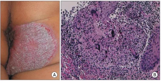
ogy clinic and had received topical antifungal treatment for four years. On presentation, he denied experiencing fever, chills, non-productive and productive cough, weight loss, and night sweats. Physical examination revealed elevated coales- cent erythematous scaling plaques (8 cm×8 cm) on the right buttock (Figure 1A). Laboratory examination revealed hu- man immunodeficiency virus negativity. Biopsy of the lesion revealed pseudoepitheliomatous hyperplasia with granulo- matous inflammation in the superficial dermis (Figure 1B);

periodic acid-Schiff and acid-fast bacilli stains were negative.

Chest radiography revealed focal haziness in the right upper lobe, and chest computed tomography showed nodules and peribronchial infiltration in the right upper lobe (Figure 2).

M. tuberculosis was isolated from the cutaneous tissue and

Delayed Diagnosis of Tuberculosis Mistaken for Tinea Corporis in a Healthy Adult

Jae-Wang Kim, M.D., Ph.D.

1

, Jeong Rae Yoo, M.D.

2

, Hyunjoo Oh, M.D.

2

, Misun Kim, M.D.

2

and Sang Taek Heo, M.D., Ph.D.

2

Departments of

1

Dermatology and

2

Internal Medicine, Jeju National University College of Medicine, Jeju, Republic of Korea

Address for correspondence: Sang Taek Heo, M.D., Ph.D.

Department of Internal Medicine, Jeju National University College of Medicine, 102 Jejudaehang-ro, Jeju 54987, Republic of Korea Phone: 82-64-717-1296, Fax: 82-64-717-1131, E-mail: [email protected]

Received: Mar. 12, 2021, Revised: Apr. 2, 2021, Accepted: Apr. 14, 2021, Published online: Apr. 15, 2021

cc

It is identical to the Creative Commons Attribution Non-Commercial License (http://creativecommons.org/licenses/by-nc/4.0/).

IMAGES OF INTEREST

https://doi.org/10.4046/trd.2021.0041

ISSN: 1738-3536(Print)/2005-6184(Online) • Tuberc Respir Dis 2021;84:245-246

Copyright © 2021

The Korean Academy of Tuberculosis and Respiratory Diseases.

A B

Figure 1. (A) Elevated coalescent ery- thematous scaling plaques on the right buttock. (B) Histopathological findings showing pseudoepitheliomatous hyper- plasia with granulomatous inflammation in the superficial dermis (H&E stain,

×100). The results of the acid-fast bacilli

strain and polymerase chain reaction for

M. tuberculosis were negative.

(2)

JW Kim et al.

246 Tuberc Respir Dis 2021;84:245-246 www.e-trd.org

sputum; it was not resistant to anti-tuberculosis agents. Thus, treatment with anti-tuberculosis agents such as isoniazid, rifampicin, ethambutol, and pyrazinamide was initiated for 2 months, which was followed by isoniazid, and rifampicin therapy for 4 months. Subsequently, the lesion completely re- solved without recurrence.

Authors’ Contributions

Conceptualization: Kim JW, Heo ST, Yoo JR. Methodology:

Yoo JR, Kim JW. Formal analysis: Kim M, Oh H. Data curation:

Kim M, Oh H. Software: Yoo JR. Validation: Heo ST, Kim JW. In- vestigation: Kim M, Oh H. Writing - original draft preparation:

Kim JW, Heo ST. Writing - review and editing: Kim JW, Heo ST.

Approval of final manuscript: all authors.

Conflicts of Interest

No potential conflict of interest relevant to this article was reported.

Funding

This work was supported by the 2021 education, research and student guidance grant funded by Jeju National Univer- sity.

References

1. Cho KS. Tuberculosis control in the Republic of Korea. Epide- miol Health 2018;40:e2018036.

2. Kwon OY. Public health center on tuberculosis management in Korea: from 1945 to the late 1970s. Uisahak 2019;28:721- 54.

3. van Zyl L, du Plessis J, Viljoen J. Cutaneous tuberculosis over- view and current treatment regimens. Tuberculosis (Edinb) 2015;95:629-38.

4. Santos JB, Figueiredo AR, Ferraz CE, Oliveira MH, Silva PG, Medeiros VL. Cutaneous tuberculosis: epidemiologic, etio- pathogenic and clinical aspects - part I. An Bras Dermatol 2014;89:219-28.

5. Chen Q, Chen W, Hao F. Cutaneous tuberculosis: a great imi- tator. Clin Dermatol 2019;37:192-9.

A B

Figure 2. (A) Chest radiography showing

focal consolidation in the right upper

lobe. (B) Chest computed tomography

showing a few nodular lesions and peri-

bronchial infiltrates in the right upper

lobe.

수치

Figure 1. (A) Elevated coalescent ery- ery-thematous scaling plaques on the right  buttock
Figure 2. (A) Chest radiography showing  focal consolidation in the right upper  lobe

참조

관련 문서

Computed tomography revealed nodules on the right and left upper lobes of the lung and multiple cystic liver lesions.. Based on pathologic findings and culture from

Computed tomography revealed nodules on the right and left upper lobes of the lung and multiple cystic liver lesions.. Based on pathologic findings and culture from

Chest CT showed irregular mass with central calcification and surrounding centrilobular nodules and branching lesions in superior segment of right lower lobe.. Figure

Materials and Methods: Consecutive patients with suspected sarcoidosis (stage I and II) on chest radiography and chest computed tomography were includ- ed. All 33 patients

Positron emission tomogra- phy/computed tomography (PET/CT) scan revealed an in- creased fluorodeoxyglucose (FDG) uptake in the mass involving the right upper lobe, which

Contrast-enhanced computed tomography (CT) of the chest revealed a 5.6×2.5-cm-sized, well-defined, cystic lesion with peripheral enhancement in the right middle lobe,

(A) Chest radiography shows interstitial pneumonia in right upper lobe at the presentation.. (B) Chest CT shows consolidation and bronchiectasis with cystic formation in

(B) High resolution com- puted tomography of the chest shows cystic lesions and ground grass opacities at left upper lobe..