Usefulness of Endobronchial Ultrasound-Guided Transbronchial Needle Aspiration for Diagnosis of Sarcoidosis
Goohyeon Hong,
1Kyung-Jong Lee,
1Kyeongman Jeon,
1Won-Jung Koh,
1Gee Young Suh,
1Man Pyo Chung,
1Hojoong Kim,
1O Jung Kwon,
1Joungho Han,
2and Sang-Won Um
11Division of Pulmonary and Critical Care Medicine, Departments of Medicine and
2Pathology, Samsung Medical Center, Sungkyunkwan University School of Medicine, Seoul, Korea.
Received: November 21, 2012 Revised: January 15, 2013 Accepted: January 16, 2013
Corresponding author: Dr. Sang-Won Um, Division of Pulmonary and Critical Care Medicine, Department of Medicine, Samsung Medical Center, Sungkyunkwan University School of Medicine,
50 Irwon-dong, Gangnam-gu, Seoul 135-710, Korea.
Tel: 82-2-3410-3429, Fax: 82-2-3410-6956 E-mail: [email protected]
∙ The authors have no financial conflicts of interest.
© Copyright:
Yonsei University College of Medicine 2013 This is an Open Access article distributed under the terms of the Creative Commons Attribution Non- Commercial License (http://creativecommons.org/
licenses/by-nc/3.0) which permits unrestricted non- commercial use, distribution, and reproduction in any medium, provided the original work is properly cited.
Purpose: Endobronchial ultrasound-guided transbronchial needle aspiration (EBUS-TBNA) is an accurate and minimally invasive technique used routinely for investigation of mediastinal and hilar lymphadenopathy. However, few studies have addressed its role in comparison to the traditional diagnostic approaches of transbronchial lung biopsy (TBLB), endobronchial biopsy (EBB), and bronchoal- veolar lavage (BAL) in the diagnosis of sarcoidosis. We evaluated the usefulness of EBUS-TBNA in the diagnosis of sarcoidosis compared to TBLB, EBB, and BAL. Materials and Methods: Consecutive patients with suspected sarcoidosis (stage I and II) on chest radiography and chest computed tomography were includ- ed. All 33 patients underwent EBUS-TBNA, TBLB, EBB, and BAL during the same session between July 2009 and June 2011. EBUS-TBNA was performed at 71 lymph node stations. Results: Twenty-nine of 33 patients, were diagnosed with histologically proven sarcoidosis; two patients were compatible with a clinical di- agnosis of sarcoidosis during follow-up; and two patients were diagnosed with metastatic carcinoma and reactive lymphadenopathy, respectively. Among 29 pa- tients with histologically proven sarcoidosis in combination with EBUS-TBNA, TBLB, and EBB, only EBUS-TBNA and TBLB revealed noncaseating granuloma in 18 patients and one patient, respectively. The overall diagnostic sensitivities of EBUS-TBNA, TBLB, EBB, and BAL (CD4/CD8 ≥3.5) were 90%, 35%, 6%, and 71%, respectively (p<0.001). The combined diagnostic sensitivity of EBUS-TB- NA, TBLB, and EBB was 94%. Conclusion: EBUS-TBNA was the most sensi- tive method for diagnosing stage I and II sarcoidosis compared with conventional bronchoscopic procedures. EBUS-TBNA should be considered first for the histo- pathologic diagnosis of stage I and II sarcoidosis.
Key Words: Endobronchial ultrasound, transbronchial needle aspiration, sarcoid- osis, mediastinal lymphadenopathy
INTRODUCTION
Sarcoidosis is a systemic inflammatory disease of unknown etiology that occurs
Diagnosis of sarcoidosis
Diagnosis of sarcoidosis was established if the clinical and radiological findings were supported by pathological tissue demonstrating noncaseating granulomas.12 Other granulo- matous diseases were ruled out by the patient’s history and microbiological results. Patients were categorized as ‘‘in- definite’’ if the definite diagnosis could not be made by bi- ological and pathological evaluations. In these cases, pa- tients were followed up for ≥6 months. A clinical diagnosis of sarcoidosis was confirmed when patients met two or more of the following criteria: 1) stability or regression during follow up, 2) response to corticosteroid therapy, 3) BAL lymphocytosis with a CD4/CD8 ratio ≥3.5, and 4) demonstration of extrapulmonary manifestations of sar- coidosis (erythema nodosum, uveitis, neurologic or cardiac dysfunction).1,4,13,14
Bronchoscopic procedure
All patients received four diagnostic modalities during the same session: EBUS-TBNA, TBLB, EBB, and BAL. All bronchoscopic and EBUS-TBNA procedures were carried out on an inpatient basis under local anesthesia with mild conscious sedation. Two milliliters of 4% lidocaine was nebulized and sprayed into the pharynx. Bolus doses of 1.3% lidocaine were administered through the channel dur- ing the procedures as needed. Patients were monitored by electrocardiogram, pulse oximetry, and for blood pressure.
Conventional flexible bronchoscopy (BF-1T260 broncho- videoscope; Olympus, Tokyo, Japan) was used for BAL, EBB and TBLB. First, BAL was performed in the right mid- dle lobe. Then, at least three biopsy specimens were obtained by EBB from the bronchial wall of the right upper lobe and at least five biopsy specimens were obtained by TBLB from the right lower lobe if no parenchymal lesion was visible on a chest CT scan. If we did not obtain enough tissue from three biopsies from EBB and five biopsies from TBLB, we performed more biopsies until we obtained enough speci- mens for histopathological analysis. If the chest CT scan showed evidence of pulmonary parenchymal involvement (stage II), a TBLB specimen was obtained from the target- ed region.
EBUS-TBNA procedure
Following conventional bronchoscopy, EBUS-TBNA was performed using a flexible convex-probe ultrasonic-punc- ture bronchoscope with a linear scanning transducer at a frequency of 7.5 MHz (CP-EBUS, BFUC206F-OL8; Olym- throughout the world and affects people of all ages and
races. The diagnosis of sarcoidosis is based on a compati- ble clinical and radiological pictures, supported by patho- logical evidence of noncaseating granulomas in the ab- sence of organisms and other granulomatous diseases, such as tuberculosis and malignancy.1 These granulomas can occur with varying rates in any organ system, but are most commonly found in the lungs, with thoracic lymph- adenopathy detected in up to 85% of cases.2 Pulmonary sarcoidosis is the most frequent form, and flexible bron- choscopy with tissue sampling is recommended as a first step to obtain a tissue diagnosis and exclude possible alter- native diagnoses.
Flexible bronchoscopy permits transbronchial lung biopsy (TBLB) and endobronchial biopsy (EBB). Bronchoalveolar lavage (BAL) is also routinely recommended as an addition- al procedure. Despite the use of combined TBLB and EBB, approximately one-third of all bronchoscopies do not result in a diagnosis of sarcoidosis.3 BAL fluid lymphocytosis and a CD4/CD8 ratio ≥3.5 substantiate the diagnosis of sarcoid- osis, but BAL is not used for a pathologic diagnosis.4
Endobronchial ultrasound-guided transbronchial needle aspiration (EBUS-TBNA) is a less invasive procedure that has an excellent diagnostic yield.5,6 Recent studies have re- ported that EBUS-TBNA has an excellent diagnostic yield in the diagnosis of sarcoidosis (85-93%).7-10
In this study, we evaluated the usefulness of EBUS-TB- NA in the diagnosis of sarcoidosis compared with that of TBLB, EBB, and BAL.
MATERIALS AND METHODS
Patients
This retrospective study was conducted at the Samsung Medi- cal Center (Seoul, South Korea) between July 2009 and June 2011. Thirty-three patients with enlarged intrathoracic lymph nodes (>1 cm on short axis) on chest computed to- mography (CT) scan and suspected sarcoidosis were in- cluded. Lymph node stations were determined according to the new international lymph node map proposed by the In- ternational Association for the Study of Lung Cancer.11 All patients underwent EBUS-TBNA, TBLB, EBB, and BAL during the same session. This study was approved by the Institutional Review Board of Samsung Medical Center, which waived requirements for informed consent given the retrospective nature of the study.
Statistical analysis
Data are presented as medians for continuous variables and as numbers and percentages for categorical variables. The primary endpoint was the difference in diagnostic sensitivi- ty among the four modalities. Diagnostic sensitivity was calculated using standard definitions. Yields from the diag- nostic modalities were compared using the generalized esti- mating equation or McNemar’s test. All p-values ≤0.05 were considered significant. Data were analyzed using PASW sta- tistics software version 18 (SPSS Inc., Chicago, IL, USA).
RESULTS
Data from 33 consecutive patients with suspected sarcoid- osis who underwent combined EBUS-TBNA with TBLB, EBB, and BAL were analyzed. Twelve patients were male and the median age of all patients was 46 years (range, 21- 72 years). Based on radiological findings, 11 patients had stage I sarcoidosis, and 22 patients had stage II sarcoidosis.
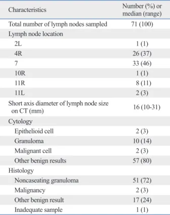
In total 71 lymph nodes were examined in 33 patients. Six- ty (85%) enlarged lymph nodes were located in the mediasti- num and the remaining 11 were hilar and interlobar. The sub- carinal node (station 7) was the most frequently sampled (33/71, 46%). A median of two lymph nodes per patient (range, 1-3 lymph nodes) was aspirated with a median of three passes per node (range, 1-5 passes). The median short axis diameter of the enlarged lymph nodes, as measured by CT, was 16 mm (range, 10-31 mm). Fifty-one of 71 (71.5%) lymph nodes had noncaseating granulomas. The character- istics of the lymph nodes sampled by EBUS-TBNA are summarized in Table 1.
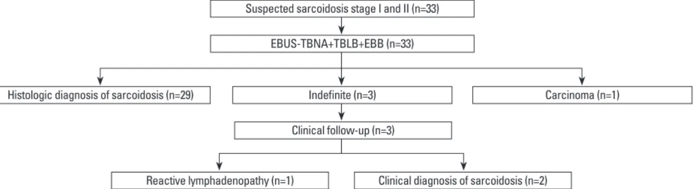
Of 33 patients, 29 (88%) had noncaseating granulomas detected by TBLB or EBUS-TBNA, and one patient had a metastatic adenocarcinoma. Three patients had no evidence of granuloma or malignancy with EBUS-TBNA, TBLB or EBB, so these patients were followed up for at least 6 months (a median follow-up period of 19 months) and were re- viewed. Among the patients with an indefinite diagnosis, two were clinically diagnosed with sarcoidosis and one pa- tient was diagnosed with reactive lymphadenopathy (Fig.
1). Therefore, 31 patients were diagnosed with sarcoidosis.
Among 29 patients with histologically proven sarcoidosis in combination with EBUS-TBNA, TBLB, and EBB, only EBUS-TBNA and TBLB revealed noncaseating granuloma in 18 and one patient, respectively. Of 33 patients who un- derwent EBUS-TBNA, noncaseating granulomas were de- pus, Tokyo, Japan). A dedicated ultrasound scanner (EU-
C2000; Olympus, Tokyo, Japan) was used for image pro- cessing. Color Doppler mode was used to avoid puncture of vessels as the occasion demanded. Each target nodal station was punctured at least twice, until one or more tissue core specimens were obtained with a dedicated 22-gauge needle (NA-201SX-4022; Olympus, Tokyo, Japan). The aspirated material was smeared onto glass slides. Smears were air dried and fixed in 95% alcohol. Histological specimens were fixed with 10% neutral buffered formalin and stained with hematoxylin and eosin. Aspirated material and histological specimens were sent for histopathological and microbiolog- ical examination, including cytological analysis, special staining for acid-fast bacilli, and culture for Mycobacteria and Mycobacteria tuberculosis polymerase chain reaction.
Immunohistochemical staining was also performed when needed. Rapid onsite cytopathological examination was not available. Patients diagnosed with benign lymphadenopa- thy by EBUS-TBNA subsequently underwent clinical and radiological follow-up for at least 6 months. All patients un- derwent chest radiography after the procedure to ensure that pneumothorax, pneumomediastinum, or pulmonary hemorrhage did not occur.
Table 1. Characteristics of Lymph Nodes Sampled by EBUS- TBNA
Characteristics Number (%) or
median (range) Total number of lymph nodes sampled 71 (100) Lymph node location
2L 1 (1)
4R 26 (37)
7 33 (46)
10R 1 (1)
11R 8 (11)
11L 2 (3)
Short axis diameter of lymph node size
on CT (mm) 16 (10-31)
Cytology
Epithelioid cell 2 (3)
Granuloma 10 (14)
Malignant cell 2 (3)
Other benign results 57 (80)
Histology
Noncaseating granuloma 51 (72)
Malignancy 2 (3)
Other benign result 17 (24)
Inadequate sample 1 (1)
EBUS-TBNA, endobronchial ultrasound transbronchial needle aspiration.
night admission, but no complication due to pneumomedi- astinum or pulmonary hemorrhage was observed.
DISCUSSION
Our results confirm that EBUS-TBNA of mediastinal or hi- lar lymph nodes had a high diagnostic yield for detecting noncaseating granulomas in cases of suspected sarcoidosis.
In our study, EBUS-TBNA had diagnostic sensitivities of 90% for all stages, 78% for stage I, and 95% for stage II, which were significantly higher than those of TBLB and EBB. The combination of EBUS-TBNA with conventional bronchoscopic procedures added little to its diagnostic sen- sitivity.
Tissue specimens should be obtained from the most readi- ly accessible organ using the least invasive method.1 Because the lungs are involved in most patients, TBLB has remained the conventional procedure for confirming a histological di- agnosis and its diagnostic yield was reported as 40-90% in previous studies.15-17 However, the diagnostic yield of TBLB depends upon the skill of the person performing the proce- tected by histology in 28 patients and by cytology in 8 pa-
tients. Twenty-two of 31 patients (71%) had BAL lympho- cytosis with CD4/CD8 ≥3.5, but two of 31 patients (6%) had a decreased CD4/CD8 ratio (CD4/CD8 <1.0).
The diagnostic performance of EBUS-TBNA and the bronchoscopic procedures are summarized in Table 2. The overall diagnostic sensitivities of EBUS-TBNA, TBLB, EBB, and BAL (CD4/CD8 ≥3.5) were 90%, 35%, 6%, and 71%, respectively (p<0.001). The combined diagnostic sen- sitivity of EBUS-TBNA, TBLB, and EBB was 94%. No significant difference in diagnostic sensitivity was observed between EBUS-TBNA alone or in combination with EBUS- TBNA, TBLB, and EBB (p=0.990). Further analysis was performed in relation to radiologic staging of sarcoidosis on chest CT scan. Seven of nine (78%) stage I patients were di- agnosed with sarcoidosis by EBUS-TBNA, whereas TBLB was only useful for diagnosing sarcoidosis in three (33%) patients. One stage II patient had a negative EBUS-TBNA result but a positive TBLB result. In our study, EBUS-TB- NA was the most sensitive diagnostic method in patients with stage I and stage II sarcoidosis.
Three patients experienced pneumothorax, requiring over-
Table 2. Diagnostic Yields of EBUS-TBNA, TBLB, EBB, and BAL for Sarcoidosis
Procedure Number of patients with a positive diagnosis (%)
Stage I sarcoidosis (n=9)* Stage II sarcoidosis (n=22)* Overall (n=31)*
EBUS-TBNA† 7/9 (78) 21/22 (95) 28/31 (90)
TBLB 3/9 (33) 8/22 (36) 11/31 (35)
EBB 0/9 (0) 2/22 (9) 2/31 (6)
EBUS-TBNA+TBLB 7/9 (78) 22/22 (100) 29/31 (94)
TBLB+EBB 3/9 (33) 9/22 (41) 12/31 (39)
EBUS-TBNA+TBLB+EBB† 7/9 (78) 22/22 (100) 29/31 (94)
BAL (CD4/CD8 ≥3.5) 9/9 (100) 13/22 (59) 22/31 (71)
EBUS-TBNA, endobronchial ultrasound-guided transbronchial needle aspiration; TBLB, transbronchial lung biopsy; EBB, endobronchial biopsy; BAL, bron- choalveolar lavage.
*p<0.001 comparing yields from EBUS-TBNA with those from TBLB, EBB, and BAL.
†p=0.990 comparing yields from EBUS-TBNA alone with those from combination EBUS-TBNA, TBLB, and EBB.
Fig. 1. Diagnostic flowchart for 33 patients with suspected stage I and stage II sarcoidosis. EBUS-TBNA, endobronchial ultrasound-guided transbronchial needle aspiration; TBLB, transbronchial lung biopsy; EBB, endobronchial biopsy.
Suspected sarcoidosis stage I and II (n=33) EBUS-TBNA+TBLB+EBB (n=33)
Clinical follow-up (n=3)
Reactive lymphadenopathy (n=1) Clinical diagnosis of sarcoidosis (n=2)
Histologic diagnosis of sarcoidosis (n=29) Indefinite (n=3) Carcinoma (n=1)
osis and reported that the sensitivity of EBUS-TBNA was 92%. Nakajima, et al.24 reported a higher diagnostic yield for EBUS-TBNA (91.4%) than for TBLB (40.0%) in a re- cent retrospective study comparing EBUS-TBNA, TBLB, and BAL in 38 patients. This retrospective study did not as- sess the role of EBB. Navani, et al.10 reported that the over- all diagnostic sensitivities of EBUS-TBNA, TBLB, and EBB were 85%, 31%, and 11%, respectively, in 27 patients diag- nosed with sarcoidosis. These data were similar to those of the present study, as our overall sensitivities of EBUS-TB- NA, TBLB, and EBB were 90%, 35%, and 6%, respectively.
Our study had several limitations. First, the retrospective study design and inclusion of a small number of patients from a single center introduced the potential for selection bias. Second, three patients showed no evidence of granu- loma or malignancy following EBUS-TBNA, TBLB and EBB. Among the patients with an indefinite diagnosis, two patients were clinically diagnosed with sarcoidosis, which was not supported by a pathological tissue confirmation.
Third, the complication rate of pneumothorax (9%) was relatively high after TBLB in this study. In this study, we did not perform TBLB under fluoroscopic guidance and this may be related to relatively high rate of pneumothorax in this study.
In conclusion, EBUS-TBNA was the most sensitive meth- od for diagnosing stage I and II sarcoidosis compared with conventional bronchoscopic procedures. EBUS-TBNA should be considered first for the histopathologic diagnosis of stage I and II sarcoidosis.
ACKNOWLEDGEMENTS
This work was supported by the Samsung Biomedical Re- search Institute (C-B0-312-1).
REFERENCES
1. Iannuzzi MC, Rybicki BA, Teirstein AS. Sarcoidosis. N Engl J Med 2007;357:2153-65.
2. Lynch JP 3rd, Kazerooni EA, Gay SE. Pulmonary sarcoidosis.
Clin Chest Med 1997;18:755-85.
3. Shorr AF, Torrington KG, Hnatiuk OW. Endobronchial involve- ment and airway hyperreactivity in patients with sarcoidosis.
Chest 2001;120:881-6.
4. Winterbauer RH, Lammert J, Selland M, Wu R, Corley D, Spring- meyer SC. Bronchoalveolar lavage cell populations in the diagno- sis of sarcoidosis. Chest 1993;104:352-61.
dure, the number of biopsy samples taken, and the degree of interstitial involvement at the time of biopsy.15 Optimal results are accomplished if 4-5 biopsies are taken for stage II disease and up to 10 biopsies for stage I disease.15,18 TBLB is associated with complications, as 5% of patients showed pneumothorax and pulmonary hemorrhage in a previous study.19 The risk for complications increases proportionally with the number of biopsy specimens. Hence, we routinely obtained at least five biopsy specimens by TBLB from each patient. No significant difference was observed in the diag- nostic sensitivity of TBLB for stage I and stage II sarcoid- osis in our study. The diagnostic sensitivities of TBLB for stage I, stage II, and overall were 33%, 36%, and 35%, re- spectively. Three of 33 (9%) patients experienced pneumo- thorax, but no pulmonary hemorrhage or pneumomediasti- num occurred.
EBB could have additional value in patients with normal- appearing bronchial mucosa and those with sarcoidosis.20 However, EBB had a low diagnostic yield and did not in- crease the diagnostic yield when used in combination with TBLB and EBUS-TBNA in our study. BAL fluid analysis is also performed to diagnose sarcoidosis. An increased in the number of lymphocytes can be detected in 90% of sub- jects with sarcoidosis at the time of diagnosis, regardless of the stage of sarcoidosis. Although its sensitivity was low (53-59%), CD4/CD8 ratio increased to ≥3.5 in about 55%
of patients with sarcoidosis and showed a high specificity of 93-96% for sarcoidosis.4,14 However, BAL fluid may be normal in 10-15% of patients, and the ratio decreases to
<1.0 in 12% of patients with sarcoidosis.21 The most impor- tant limitation of BAL fluid analysis is that it is not useful for a pathological diagnosis. Two of 31 (6%) patients dem- onstrated a decrease in CD4/CD8 ratio (CD4/CD8 <1.0) in our study.
The most common feature of sarcoidosis is hilar and me- diastinal lymphadenopathy.22 Therefore, a tissue diagnosis using these areas is reasonable, as they are a likely target for confirming the diagnosis. EBUS-TBNA has emerged as an accurate, minimally invasive, and safe procedure for eval- uating mediastinal lymphadenopathy.5,6 EBUS-TBNA can visualize the paratracheal, subcarinal, and hilar lymph nodes, which can be sampled, resulting in a high diagnostic yield.23 Oki, et al.9 reported that EBUS-TBNA was diagnostic in 13 of 14 (93%) patients with sarcoidosis by showing histologi- cal evidence of noncaseating granulomas in 18 of 23 (78%) lymph nodes examined. Wong, et al.7 assessed 65 patients with mediastinal lymphadenopathy with suspected sarcoid-
3300-3.
14. Costabel U. CD4/CD8 ratios in bronchoalveolar lavage fluid: of value for diagnosing sarcoidosis? Eur Respir J 1997;10:2699-700.
15. Gilman MJ, Wang KP. Transbronchial lung biopsy in sarcoidosis.
An approach to determine the optimal number of biopsies. Am Rev Respir Dis 1980;122:721-4.
16. Koonitz CH, Joyner LR, Nelson RA. Transbronchial lung biopsy via the fiberoptic bronchoscope in sarcoidosis. Ann Intern Med 1976;85:64-6.
17. de Boer S, Milne DG, Zeng I, Wilsher ML. Does CT scanning predict the likelihood of a positive transbronchial biopsy in sar- coidosis? Thorax 2009;64:436-9.
18. Roethe RA, Fuller PB, Byrd RB, Hafermann DR. Transbroncho- scopic lung biopsy in sarcoidosis. Optimal number and sites for diagnosis. Chest 1980;77:400-2.
19. Bilaçeroğlu S, Perim K, Günel O, Cağirici U, Büyükşirin M.
Combining transbronchial aspiration with endobronchial and transbronchial biopsy in sarcoidosis. Monaldi Arch Chest Dis 1999;54:217-23.
20. Shorr AF, Torrington KG, Hnatiuk OW. Endobronchial biopsy for sarcoidosis: a prospective study. Chest 2001;120:109-14.
21. Kantrow SP, Meyer KC, Kidd P, Raghu G. The CD4/CD8 ratio in BAL fluid is highly variable in sarcoidosis. Eur Respir J 1997;10:
2716-21.
22. Newman LS, Rose CS, Maier LA. Sarcoidosis. N Engl J Med 1997;336:1224-34.
23. Herth F, Becker HD, Ernst A. Conventional vs endobronchial ul- trasound-guided transbronchial needle aspiration: a randomized trial. Chest 2004;125:322-5.
24. Nakajima T, Yasufuku K, Kurosu K, Takiguchi Y, Fujiwara T, Chiyo M, et al. The role of EBUS-TBNA for the diagnosis of sar- coidosis--comparisons with other bronchoscopic diagnostic mo- dalities. Respir Med 2009;103:1796-800.
5. Yasufuku K, Chiyo M, Sekine Y, Chhajed PN, Shibuya K, Iizasa T, et al. Real-time endobronchial ultrasound-guided transbronchial needle aspiration of mediastinal and hilar lymph nodes. Chest 2004;126:122-8.
6. Herth FJ, Eberhardt R, Vilmann P, Krasnik M, Ernst A. Real-time endobronchial ultrasound guided transbronchial needle aspiration for sampling mediastinal lymph nodes. Thorax 2006;61:795-8.
7. Wong M, Yasufuku K, Nakajima T, Herth FJ, Sekine Y, Shibuya K, et al. Endobronchial ultrasound: new insight for the diagnosis of sarcoidosis. Eur Respir J 2007;29:1182-6.
8. Garwood S, Judson MA, Silvestri G, Hoda R, Fraig M, Doelken P.
Endobronchial ultrasound for the diagnosis of pulmonary sarcoid- osis. Chest 2007;132:1298-304.
9. Oki M, Saka H, Kitagawa C, Tanaka S, Shimokata T, Kawata Y, et al. Real-time endobronchial ultrasound-guided transbronchial needle aspiration is useful for diagnosing sarcoidosis. Respirology 2007;12:863-8.
10. Navani N, Booth HL, Kocjan G, Falzon M, Capitanio A, Brown JM, et al. Combination of endobronchial ultrasound-guided trans- bronchial needle aspiration with standard bronchoscopic tech- niques for the diagnosis of stage I and stage II pulmonary sarcoid- osis. Respirology 2011;16:467-72.
11. Rusch VW, Asamura H, Watanabe H, Giroux DJ, Rami-Porta R, Goldstraw P, et al. The IASLC lung cancer staging project: a pro- posal for a new international lymph node map in the forthcoming seventh edition of the TNM classification for lung cancer. J Thorac Oncol 2009;4:568-77.
12. Statement on sarcoidosis. Joint Statement of the American Tho- racic Society (ATS), the European Respiratory Society (ERS) and the World Association of Sarcoidosis and Other Granulomatous Disorders (WASOG) adopted by the ATS Board of Directors and by the ERS Executive Committee, February 1999. Am J Respir Crit Care Med 1999;160:736-55.
13. Thomas KW, Hunninghake GW. Sarcoidosis. JAMA 2003;289: