ⓒ 2014 The Korean Medicine Society For The Herbal Formula Study
This paper is available at http//:www.ompak.okdanche.com which permits unrestricted non-commercial use, distribution, and reproduction in any medium, provided the original work is properly cited.
2014;22(1):105~112
pISSN 1229-1218, eISSN 2288-5641 http://dx.doi.org/10.14374/HFS.2014.22.1.105 Official Journal of The Korean Medicine Society For The Herbal Formula Study
Available at http://www.ompak.okdanche.com
HFS
Original Article / 원저
Methanol extract from radix of Glycyrrhizae uralensis attenuate methamphetamine-induced hyperlocomotor activity
ZhengLin Zhao
1· Yan Wang
1· Feng Lin
1· Hui Fu
1· FuBo Zhou
1· Suchan Chang
2· Nu Ri Han
2· Dae Hwa Jung
2· Chae Ha Yang
2· Sang Chan Kim
2#*· RongJie Zhao
1,2#*1
Department of Pharmacology, Mudanjiang Medical University, Mudanjiang, China ·
2MRC-GHF, Daegu Haany University, Gyeongsan, Korea
감초 메탄올 추출물의 메스암페타민 유도 과다보행활동에 대한 억제작용
자오정린
1·왕옌
1·린훵
1·후우후이
1·저우후우뿨
1·장수찬
2·한누리
2·정대화
2· 양재하
2·김상찬
2#*·자오르옹지에
1,2#*1
중국 목단강의과대학 약리학교실·
2대한민국 대구한의대학교 MRC-GHF
ABSTRACT
Background and objective: Methamphetamine (Meth) is a widely abused psychostimulant that produces hyperlocomotion in rodents. Radix of Glycyrrhizae uralensis comprises a variety of bioactive components that have neuroprotective effects. In a previous study, we have demonstrated methanol extracts from radix of Glycyrrhizae uralensis (MEGR) suppress acute cocaine-induced extracellular dopamine release in the nucleus accumbens. In the present study, we investigated the effect of MEGR on acute Meth-induced hyperlocomotion.
Methods: Male Sprague–Dawley rats were orally administered with MEGR (60 mg/kg and 180 mg/kg) 60 min prior to an intraperitoneal injection of Meth (1.0 mg/kg).
Results: Behavioral analysis showed acute Meth greatly increased locomotor activities, while pretreatment
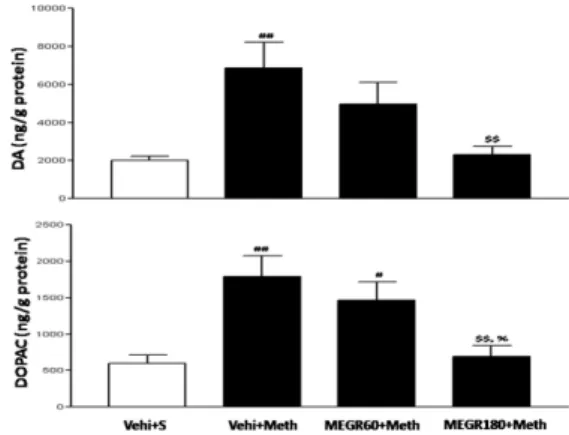
with MEGR dose dependently inhibited the hyperlocomotion. In parallel, there were markedly increased levels of dopamine and its metabolite 3, 4-dihydroxyphenylacetic acid in the nucleus accumbens tissues in Meth-treated rats, which were also almost completely reversed by 180 mg/kg MEGR.
Conclusions: These results showed that radix of Glycyrrhizae uralensis attenuates Meth-induced hyperlocomotion by inhibiting dopamine synthesis and utilization, suggesting that radix of Glycyrrhizae uralensis might be effective in blocking the rewarding effect of Meth.
Keyword : Methamphetamine, Glycyrrhizae uralensis, Locomotion, Dopamine, Nucleus accumbens
Ⅰ. Introduction
23)
Although methamphetamine (Meth) dependence is a serious worldwide public health problem with major medical, psychiatric, socioeconomic and legal consequences [1], there is currently no FDA approved pharmacotherapy for this addictive disorder. The rewarding effects of Meth including euphoria and many other psychostimulant effects are the mainstay to sustain the continued use of Meth, ultimately lead to Meth addition [2]. Therefore, nullifying the rewarding effects of Meth is a promising approach to treat Meth addition.
It is well documented that the commonly abused psychostimulants including Meth increase locomotor activity in rodents, and the biological mechanisms of this stimulant action are homologous with their rewarding effects [3]. Convincing evidence shows that the rewarding effects of Meth mainly come from enhanced dopaminergic neurostransmissions in the mesolimbic and mesocortical brain regions such as the ventral tegmental area, the nucleus accumbens (Nacc) and the medial prefrontal cortex, which also mediate Meth-induced hyperlocomotion [4]. Therefore, inhibition on the increased dopaminergic
activities in the mesolimbic brain regions biochemically prevents the formation of the biological bases for Meth addiction and behaviorally blocks hyperlocomotion induced by Meth. For example, a selective dopamine (DA) 1 receptor antagonist SCH23390 injection into the Nacc attenuates Meth-induced hyperlocomotion in rats [5], Limonene inhibits Meth-induced increased locomotor activity via regulation of enhanced accumbal DA release [6].
Radix of Glycyrrhizae uralensis (G. radix), one of the oldest and most popularly used botanicals in traditional Oriental medicine, contains flavonoids and pentacyclic triterpene saponin as major constituents, which include liquiritigenin, isoliquiritigenin, liquiritin, liquiritin apioside, and glycyrrihizin [7]. Being an important tonic, G. radix is generally used to replenish and invigorate deficient Qi and blood, and widely recommended for life-enhancing properties and cure of injury or swelling and for detoxification [8]. Historically, most experimental studies on pharmacological values of G. radix were devoted to its anti-inflammatory and anti-oxidative properties [9, 10], however, over the last decade, numerous reports from various laboratories have emphasized the neuropharmacological properties of G. radix, and these reports point to the
# Authors contributed equally.
* Corresponding author : Sang Chan Kim, Daegu Haany University, #1, Hanuidae-ro, Gyeongsan-si, Gyeongsangbuk-do, 712-715, South Korea.
․ Tel : 82-53-819-1863
․ E-mail: [email protected]
RongJie Zhao, Mudanjiang Medical University, #3 Tongxiang Street, Mudanjiang, China,
․ Tel : 86-453-2984682
․ E-mail: [email protected]
∙ Received : May 8, 2014 / Revised : May 30, 2014 / Accepted : June 7, 2014
Figure 1. The HPLC profiles of MEGR at 254 nm (liquiritigenin), 276 nm (glycyrrhizic acid), and 380 nm (isoliquiritigenin).
Flow rate: 1.0 mL/min, column: Waters XTerratTMRP18(150
× 4.6 mm, 5 μm)
neuroprotective effects of G. radix and its
bioactive ingredients against various noxious stimuli [11]. Most notably, studies done in our lab have shown that methanol extracts of G. radix (MEGR) and isoliquiritigenin suppress cocaine-induced extracellular dopamine release in rat Nacc through GABAb receptors [12], and isoliquiritigenin also prevents Meth-induced neurotoxicity in mouse brain by inhibiting NF-kB activation [13]. These observations conjunction with its neuroprotective effects reported by others lead to a hypothesis that G. radix have preventive effects against Meth-induced psychostimulant effects.
To test this hypothesis, in the present study we evaluate the effects of MEGR on locomotor activities and accumbal DA and 3, 4- dihydroxyphenylacetic acid (DOPAC) levels in rats following an acute Meth administration.
Ⅱ. Materials and methods
1. Preparation of MEGR
The air-dried G. radix was purchased from Daewon Pharmacy (Daegu, Republic of Korea), and its identity and composition was confirmed by Professor Sang Chan Kim of the College of Oriental Medicine at Daegu Haany University in Korea. The G. radix was crushed into powder and extracted with methanol three times for 12 h each at room temperature. The extract was filtered through a 0.2 μm filter (Nalgene, NY, USA), evaporated under reduced pressure at temperature of 37 °C and lyophilized. The amount of extracts was estimated from the dry weight of lyophilized powder, and the yield was 15.39%, and the high performance liquid chromatography (HPLC) fingerprint of MEGR is shown in Fig.
1. The MEGR was dissolved in 5% Tween-80 and orally administered.
2. Reagents
Sodium octanesulfonic acid acetonitrile, tetrahydrofurane, DA and DOPAC were purchased from Sigma Co. (St Louis, MO, USA). All other drugs were of analytical or HPLC grade.
3. Animals and experimental design
Adult male Sprague-Dawley rats (250-270 g) were obtained from Hyochang Science (Hyuchang, Pusan, South Korea), and housed under a controlled environment with food and water ad libitum throughout the course of the study. All animal procedures were approved by the Institutional Animal Care and Use Committee for the National Institutes of Health guidelines concerning the Care and Use of Laboratory Animals.
Meth (purity > 98%, obtained from Korean Food and Drug Administration) was dissolved in saline and administered intraperitoneally (i.p.) to rats in a single dose of 1.0 mg/kg. MEGR (60 and 180 mg/kg) or the vehicle was acutely applied to rats 60 min prior to Meth administration.
After MEGR (or vehicle) administration, each rat
was allowed to adapt the locomotor testing box
for 60 min followed by Meth administration,
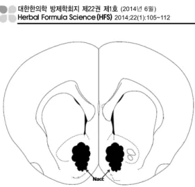
Figure 2. Diagrammatic representation of a coronal section showing the Nacc.
and immediately after the Meth the locomotor activity of each rat was measured in the box for 60 min. The box used to measure locomotor activity is a rectangular container (40 x 40 x 45 cm) equipped with a video camera above the center of the floor. The walls and floor were made of a clear plexiglas and painted black.
The ambulatory distance was recorded by a videotracking system using the Ethovision program (NoldusInformation Technology BV, Wageningen, Netherlands).
4. DA and DOPAC analysis
Immediately after the behavioral test, each rat was decapitated and the entire brain was moved and stored at -80℃. To determine the effects of MEGR on the Meth-induced changes of mesolimbic dopaminergic activities, the accumbal DA and DOPAC concentrations were measured using HPLC analysis. The stored brain was dissected using the coordinates of Paxinos and Watson [14] to obtain Nacc tissues(Fig. 2). The tissues were sonicated in 1 ml of 0.1 M HClO
4for 30 s, and centrifuged for 15 min at 26,000 g, 4ºC. Then, a 20-µl supernatant aliquot was injected directly into the HPLC with a coulmoetric detector (Coulochem II; ESA, Bedford, MA, USA). The
HPLC system consists of a C18 reverse-phase column(5 u ODS; Altex, Ann Arbor, MI, USA) and an electrochemical transducer with a glassy carbon electrode set at 350 mV. The mobile phase was 0.16 M citric acid, pH 3.0, containing 0.02 mM EDTA with 0.69 mM sodium octanesulfonic acid as an ion-pairing reagent, 4%(v/v) acetonitrile and 1.7% (v/v) tetrahydrofurane. Peaks and values of DA and DOPAC in samples were identified and calculated by comparing their retention times and peak heights with those of standards. Results were reported as ng/g protein. The protein concentration in brain homogenate was determined by the BCA protein assay.
5. Statistical analysis
All data were expressed as means ± standard error of the mean (SEM) and statistically analyzed by one-way analysis of variance (ANOVA) followed by the Newman-Keuls multiple comparison test using the commercially available GraphPad Prism 5.0 software (GraphPad Software, San Diego, CA, USA). A P value < 0.05 was considered to indicate statistical significance.
Ⅲ. Results
1. Effects of MEGR on Meth-induced locomotor activity
Figure. 3 shows the effect of MEGR on
locomotor activities. ANOVA on data from
MEGR treatment revealed a significant effect
[F (3, 20) = 17.8, p < 0.001]. Post hoc
comparisons revealed acute Meth significantly
increased locomotor activities in rats (vehicle+saline
vs. vehicle+Meth, p < 0.001), while pretreatment
with both doses of MEGR significantly inhibited
the Meth-induced hyperlocomotion (vehicle+Meth
Figure 3. Effects of MEGR on acute Meth-induced hyperlocomotion. The traveled distances were measured for 60 min immediately after Meth.
Data represent mean ± SEM (n = 6/group). Vehi: vehicle, S: saline, ###p < 0.001, ##p < 0.01, compared with vehicle-treated control group; $$$p < 0.001, $$p < 0.01, compared with Meth-treated control group; %p < 0.05, compared with MEGR60+Meth group by post hoc test.
Figure 4. Effects of MEGR on the locomotor activities of saline-treated rats.
Rats were treated with a single dose of saline 60 min after an acute 60 mg/kg or 180 mg/kg MEGR, and then the traveled distance of each rat was measured for 60 min immediately after the saline. Data represent mean ± SEM (n = 6/group). Vehi: vehicle, S: saline. Statistical analysis showed no difference among the groups.
Figure 5. Effects of MEGR on acute Meth-induced changes of accumbal DA and DOPAC concentrations.
The concentrations were measured by HPLC analysis. Data represent mean ± SEM (n = 6/group). Vehi: vehicle, S:
saline, ##p < 0.01, #p < 0.05, compared with vehicle- treated control group; $$p < 0.01, compared with Meth- treated control group; %p < 0.05, compared with MEGR60+
Meth group by post hoc test.