Ⅰ. 서 론
골 결손 부위의 수복에는 탈회 동결건조골이나 hydrox- yapatite 등의 기질을 이용하는 방법과 자가골 등을 사용하
는 기질과 세포를 동시에 이용하는 방법이 있는데, 최근에 는 골수 줄기세포를 이용한 분자 생물학적 기법에 대한 연 구가 활발히 진행되고 있다
1,2). 골수 줄기세포는 골수에서 생성되며 연골이나 지방, 근육, 골 조직 등의 다양한 조직으 윤정호∙윤정주∙장현석∙임재석∙이의석∙김대성∙권종진
고려대학교 의과대학 구강악안면외과학교실
Pleiotrophin이 골수 줄기 세포의 부착 및 골형성에 미치는 효과에 대한 연구
PLEIOTROPHIN EFFECTS ON BINDING AND SUBSEQUENT OSTEOGENESIS OF HUMAN MESENCHYMAL STEM CELLS
Jung-Ho Yoon, Jung-Ju Eune, Hyon-Seok Jang, Jae-Suk Rim, Eui-Seok Lee, Dae-Sung Kim, Jong-Jin Kwon Department of Oral and Maxillofacial Surgery, College of Medicine, Korea University
An area of current research is investigating the app1ication of human mesenchymal stem cells or hMSCs as a cell-based regenerative therapy. In order to achieve effective bone regeneration, appropriate matrices functioning as cell-carriers must be identified and optimized in terms of function, efficacy and biocompati- bility. Two methods of approaching optimization of matrices are to facilitate adhesion of the donor hMSCs and furthermore to facilitate recruitment of host progenitor cells to osteoblastic differentiation. Pleiotrophin is an extracellular matrix protein that was first identified in developing rat brains and believed to be asso- ciated with developing neuronal pathways. A recent publication by Imai and colleagues demonstrated that transgenic mice with upregulated pleiotrophin expression developed a greater volume of cortical as well as cancellous bone. The proposed mechanism of action of pleiotrophin is demonstrated here. Through either environmental stresses and/or intracellular regulation, there is an increase in pleiotrophin production. The pleiotrophin is released extracellularly into areas requiring bone deposition. A receptor-mediated process recruits host osteoprogenitor cells into these areas.
Therefore, the aim of our study was to investigate the osteoconductive properties of pleiotrophin. We wanted to determine if pleiotrophin coating facilitates cellular adhesion and furthermore if this has any effect on hMSCs derived bone formation in an animal model.
The results showed a dose dependent response of cellular adhesion in fibronectin samples, and cellular adhesion was facilitated with increasing pleiotrophin concentrations. Histologic findings taken after 5 weeks implantation in SCID mouse showed no presence of bone formation with only a dense fibrous con- nective tissue. Possible explanations for the results of the osteogenesis assay include inappropriate cell loading.
Key words : Pleiotrophin, Human mesenchymal stem cell, Matrix carrier, Osteogenesis Abstract
※ 본 연구는 보건복지부 보건의료기술진흥사업의 지원에 의하여 이루어진 것임(과제고유번호: A050801).
관여한다. 골수 줄기세포를 이식부위에 적용하기 위해서는 carrier가 필요하며, 현재 carrier로 사용될 수 있는 기질로 는 탈회 동결건조골이나 Bio-Oss
�, hydroxyapatite, tri- calcium phosphate 등을 들 수 있다. 기질 carrier는 분자 생물학적 처리과정을 통해서 그 기능을 증진시킬 수 있으며 현재 줄기세포의 효과적 이용을 위한 기능적이고 효율적이 며 생체 적합성이 양호한 cell-carrier에 대한 연구가 활발 히 진행되고 있어 기질의 표면을 거칠게 하거나 소공을 부 여하여 표면 특성을 변화 시키거나, 기질에 fibronectin이 나 pleiotrophin 등의 접착성 단백질을 표면에 피복해서 세 포의 부착 능력을 증진 시키고 bone morphogenic protein (BMP)이나 pleiotrophin, chitosan 등의 골전도성 물질을 피복해서 숙주세포의 조골세포로의 분화를 유도하는 등의 방법이 연구되고 있다
3).
Pleiotrophin은 heparin-binding growth associated molecule(HB-GAM), 또는 osteoblast stimulating fac- tor-1(OSF-1)으로도 알려진 세포외 기질단백질로 성장 중 인 쥐의 뇌 조직에서 최초로 발견 되었으며
4), 신경 조직의 기저막에서 발견되나 배아 형성이 일어나는 동안은 신경조 직이 아닌 부위에서도 발견된다
5). Pleiotrophin은 골 형성 과 밀접한 관련이 있는 단백질로 스트레스나 세포내 조절 기전으로 골 형성에 관여 하는 것으로 알려져 있다. Imai 등
6)은 pleiotrophin이 외상을 입은 골표면이나 골생성이 필 요한 부위에 급속히 분비되고 골전구세포나 조골세포가 pleiotrophin이 침착 된 부위로 이동되어 골 형성을 유도하 는 것을 보고하고 있다.
본 연구는 골수 줄기세포를 이용한 골 결손부 수복에 대한 사용 가능성을 확인하기 위하여 pleiotrophin의 세포 부착 및 골 형성능을 조사하여 보고 이러한 결과가 줄기세포에 의한 골 형성을 증진할 수 있는지 여부를 파악해 보고자 시 행하였다.
Ⅱ. 연구 재료 및 방법
1. 연구 재료
Pleiotrophin의 세포 부착능과, 골 형성능을 평가하기 위 하여 in vitro로 Bio-Oss
�(Osteohealth, Shirley, USA) 를 기질 carrier로 하여 pleiotrophin을 피복한 실험군과 fibronectin을 피복한 양성 대조군으로 분류하여 골수줄기 세포의 부착을 평가하였고 골형성 능력에 대한 실험은 in vivo로 pleiotrophin과 fibronectin을 피복한 Bio-Oss
�를 SCID mice에 이식하여 5주 후의 조직학적 소견을 관찰하
(1) Cell binding assay
Pleiotrophin을 실험군으로, 세포의 부착에 관여 한다고 알려진 fibronectin을 양성 대조군으로, 피복하지 않은 것 을 음성 대조군으로 하여 21개의 표본을 실험 대상으로 하 였다. 표본당 6.5 mg의 Bio-Oss
�를 4℃ PBS(phosphate buffered saline)에서 0.1~10 μg/ml 농도로 희석한 pleiotrophin과 fibronectin을 혼합하여 12시간 피복한 후, 피복된 Bio-Oss
�를 150,000개의 인간 골수줄기세포 혼합 액에서 세포부착을 위해 37℃의 110 μl DMEM (Dulbecco’s modified eagles medium) 배지에서 5시간 배양하였다. 각 표본은 4% paraformaldehyde에 포매하여 osmium coating, 탈수과정을 거쳐, gold/palladium sputter coating 한 후 JEOL SEM으로 관찰 하였다. 임의 의 구역을 설정하여 200배 확대하고 세포 부착의 양을 정량 화하기 위해 스테롤 격자를 이용하여 0.071 mm
2에 부착되 어 있는 세포의 수를 계산 하였고 Student’s t-test 이용하 여 통계학적 분석을 시행하였다.
(2) Osteogenesis assay
10mg의 Bio-Oss
�에 10 μg/ml의 pleiotrophin과 fibronectin을 피복하고, 피복된 Bio-Oss
�를 85,000개의 인간 골수줄기세포가 포함된 혼합액에 담가서 37℃의 110 μl DMEM에서 5시간 배양하였다. 피복된 Bio-Oss
�표본은 이식 전에 SEM을 위한 절편을 제작하여 200배 확 대하여 관찰하였다. 표본 이식을 위하여 2마리의 SCID mice의 등에 mid-sagittal incision을 가하여 subdermal pouch를 만들고 표본의 조작성 및 이식효과를 증진시키기 위해서 murine fibrinogen과 thrombin으로 표본을 응고 시킨 뒤 mice 한 마리 당 6개의 pouch를 형성하고 피복된 Bio-Oss
�를 이식하고 5주 후 mice를 희생시켜 12개의 조 직 표본을 얻어 통상적 헤마톡시린-에오진 염색을 이용하여 광학 현미경 하에서 관찰하였다.
Ⅲ. 연구 결과
(1) Cell binding assay
Bio-Oss
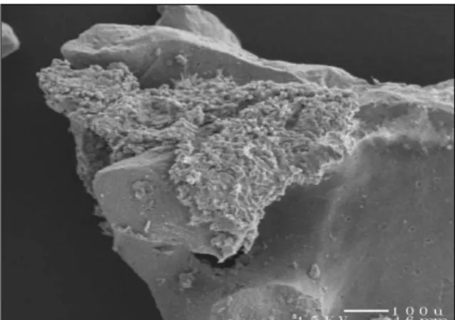
�는 약 0.5~1 mm 정도 크기의 불규칙한 입자
형태를 가지고 있었으며 적은 양의 소공이 존재하였다
(Fig. 1). 피복하지 않은 대조군에서는 Bio-Oss 표면에 소
수의 세포만이 부착되어 있음이 관찰되었고(Fig. 2), 양성
대조군인 fibronectin 피복 표본은 피복하지 않은 표본과
비교해서 부착세포의 수가 증가되어 있었으며(Fig. 3)
음성 대조군과 비교해서 증가 한 것을 관찰할 수 있었지만 (Fig. 4), fibronectin 피복 표본과 비교해 보면 부착량이 상대적으로 적게 관찰되었다. 세포의 부착량을 정량적으로 분석 해보면 pleiotrophin이 피복된 표본은 40.8(±
12.8)%, fibronectin이 피복된 표본은 50.6(±18.5)%, 대조군은 11.7(±6.6)%의 세포 부착을 나타내고 있어 pleiotrophin의 농도가 증가함에 따라 세포 부착량이 증가 했으며 fibronectin 피복 표본의 경우에도 유사한 결과를 보이고 있었다(Table 1). 통계학적 분석 결과, fibronectin 과 pleiotrophin 군은 대조군과 유의성 있는 차이를 보였으 나 pleiotrophin의 농도 사이에는 통계학적 유의성을 발견 할 수 없었다(Table 2). Pleiotrophin의 세포 부착에의 영 향은 대조군 보다는 현저히 높았지만 fibronectin 군 보다 는 적은 양상을 보이고 있었다.
(2) Osteogenesis Assay
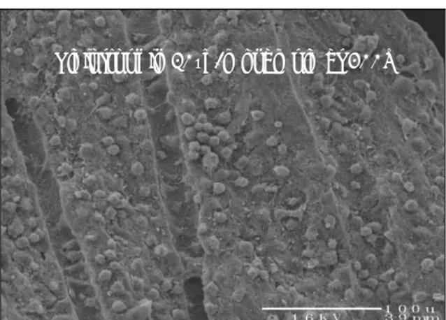
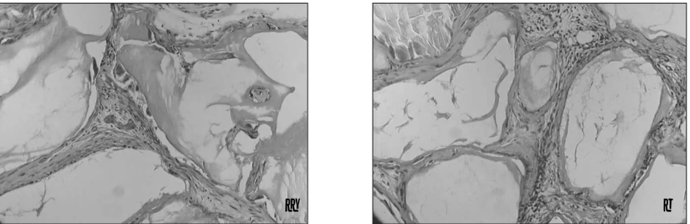
각 표본은 조직학적 분석을 시행하기 전에 세포부착 평가 를 위해 SEM으로 관찰하였는데 Bio-Oss의 소공에 세포들 이 밀집되어 뭉쳐있는 양상이 관찰되었으며(Fig. 5), 이식 5주 후 음성 대조군의 조직 표본에서는 골 형성은 보이지 않고 단지 치밀한 섬유성 결체조직 만이 관찰되고 있었으며 (Fig. 6a) 다핵성 파골세포가 기질 주변에 분포하고 있었다 (Fig. 6b). Fibronectin군의 5주 조직 표본에서도 역시 골 형성이 관찰되지 않았으며(Fig. 7a), 골양조직 (osteoid)의 형성을 보이지 않고 다핵성 파골세포 만이 관찰되고 있었다 (Fig. 7b). Pleiotrophin군의 5주 조직 표본에서도 골 형성 은 관찰되지 않았으며(Fig. 8a), 치밀한 섬유성 결체조직만 이 관찰되었다(Fig. 8b). Osteogenesis assay 결과, 실험 군과 대조군간에는 정성적 차이를 나타내지 않았다.
Fig. 1. Scanning electron microscopic finding of control Bio-Oss
�(X33).
Fig. 2. Cell binding assay image of control sample at 200X.
Fig. 3. Cell binding assay image of fibronectin 1.0μ g/ml sample at 200X.
Fig. 4. Cell binding assay image of pleiotrophin 10μ g/ml sample at 200X.
Control sample at 200X
Fibronectin 1.0μ g/ml sample at 200X Pleiotrophin 10μg/ml sample at 200X
Table 1. Bar diagram of Cell Binding assay results
Fig. 5. Scanning electron microscopic finding of osteo- genesis assay.
Table 2. Bar diagram of cell binding assay results with t-test comparison
Fig. 6a, b . Histologic finding of osteogenesis assay of control sample 5 weeks after implantation.
a b
Fig. 7a, b. Histologic finding of osteogenesis assay of Fibronectin sample 5 weeks after implantation.
Fig. 8a, b. Histologic finding of osteogenesis assay of Pleiotrophin sample 5 weeks after implantation.
Fig. 9. Histologic finding of hMSC-derived osteogenesis with HA/TCP as matrix.
a b
a b
외상이나 질병으로 인한 골 결손이 발생할 경우 결손부가 큰 경우 자연적인 골치유를 기대하기 힘들어 결손부에 대한 골 이식을 시행하여야 한다. 골 이식에는 골 대체물을 이식 하거나 자가골을 이식하는 방법 등이 있는데 골 대체물을 이식하는 경우 이식량에 한계가 있고 안정성에 대한 문제점 이 있으며 자가골 이식은 가장 안정적이고 믿을 만한 술식 으로 여겨 지기는 하지만 골 채취를 위한 부가적 수술을 해 야 하는 등의 여러 가지 문제점이 수반된다. 최근 분자 생물 학이 발전함에 따라 골 대체물의 효과를 증진시키기 위해 BMP 등의 성장 요소를 이용하거나 pleiotrophin이나 fibronectin같은 부착성 단백질을 골 대체물에 coating하는 방법에 대한 많은 연구가 이루어지고 있다.
Pleiotrophin은 HB-GAM, 또는 OSF-1으로도 알려진 세포외 기질 단백질로 신경 조직의 기저막에서 발견되는 단 백질이다
5). 배아성장이 일어나는 동안은 신경 연관 조직이 아닌 부위에서도 발견되며 murine sarcoma cell에서 이와 비슷한 염기 서열을 가진 분자가 발견되어 OSF-1이라는 명칭을 얻게 되었으며
7), 정상의 경우 골격 성장이 완료된 후에는 형성 및 분비가 감소되는 것으로 알려지고 있다.
N-syndecan은 pleiotrophin의 수용체인 당단백으로 신경 조직의 발생시에만 발현되고
8)HB-GAM과 N-syndecan에 의한 phospholyation이 세포의 이동을 유도하는 것으로 알 려지고 있다
9). Pleiotrophin이 골의 형성과 치유 과정에 미 치는 영향에 대한 기전은 Imai 등
6)에 의해 밝혀졌는데 pleiotrophin이 외상을 입은 골 표면이나 골 생성이 필요한 부위에 급속히 분비되어 침착되고, pleiotrophin/N-syn- decan에 의한 신호가 조골세포 등에 인지되며 조골세포가 pleiotrophin이 침착 된 부위로 이동되고 분화하여 궁극적 으로 골 형성이 일어 나게 된다.
Fibronectin은 교원질이나 다른 세포 외 기질 성분 그리 고 세포 표면의 integrin에 부착할 수 있는 수용기가 다양 하게 존재하는 다 부착성 기질단백(multiadhesive matrix protein)이다
10). 본 연구 결과에서 pleiotrophin과 fibronectin은 각각 40.8%, 50.6%의 부착량을 보여 대조 군의 11.7%에 비교하여 유의성 있는 세포부착의 증가를 나 타내었고(Table 1) pleiotrophin과 fibronectin의 농도가 증가함에 따라 세포 부착이 증가되었는데 이는 결국 골 대 체물에 pleiotrophin과 fibronectin을 피복하는 것은 골 대 체물의 안정성을 증가시켜 줄 수 있는 방법임을 나타낸다.
Pleiotrophin은 본 실험 결과가 나타내는 세포 부착을 증진 시키는 역할 이외에도 골 형성에 여러 가지 중요한 기능을 수행하는 것으로 알려지고 있다.
표본이나 대조군에서도 같은 결과를 보이고 있었다. 이는 이전의 hydroxyapatite와 tricalcium-phosphate를 matrix carrier로 이용한 실험 결과와 상이하였는데 hydroxyapatite와 tricalcium-phosphate matrix에 pleiotrophin을 같은 방법으로 피복 한 후 SCID mice에 이 식한 경우에는 6주 후 조직 표본에서 기질 경계부의 골수강 내에 골세포가 관찰되고 있었으며 광화된 골 주변으로 골기 질을 분비하는 세포가 관찰되고 있어(Fig. 9), 본 연구의 결 과와 커다란 차이가 있었다. 본 연구의 결과에서 골 형성이 일어나지 않은 원인에 대해서는 몇 가지 원인을 찾아볼 수 있는데 첫째로 부적절한 세포의 loading을 생각할 수 있다.
즉 표본 당 적용한 세포의 수가 너무 많아 Bio-Oss
�의 소공 부위에 세포가 밀집되어 뭉쳐있어 조골세포로의 분화를 유 발할 수 있는 signal transduction에 방해가 되었을 수 있 고 다른 원인으로는 Bio-Oss
�의 형태와 표면 성상이 부적 절한 것을 들 수 있다. Kuboki 등
11)은 고형성과 다공성 hydroxyapatite의 형태에 따른 골 형성의 차이를 비교한 연구에서 고형성의 hydroxyapatite granule은 다공성에 비하여 신생혈관의 생성이 저하되어 골형성이 감소되는 결 과를 보고하고 있다. 본 연구에 사용된 Bio-Oss
�는 gran- ule 형태의 기질로 소공의 크기가 비교적 작아 신생 혈관의 형성이 저하되고 결과적으로 골 형성이 되지 않았을 가능성 이 있으며 작은 소공 부위에 많은 수의 세포가 밀집되어 뭉 쳐있어 적절한 혈액공급이 이루어지지 않아 많은 양의 세포 가 죽음으로 해서 소공의 역할은 더욱 적어지지 않았나 추 측된다. 골수 줄기세포와 기질을 이용한 골 형성을 위한 조 직공학적 적용에는 부착 단백이나 cytokine의 피복 이외에 도 세포가 더 잘 부착될 수 있는 기질의 형태나 구조에 대한 연구를 통하여 성공적인 골재생을 얻을 수 있을 것으로 생 각된다.
Ⅴ. 결 론
Pleiotrophin의 세포 부착능과 골 형성능을 평가하기 위 하여 Bio-Oss
�를 기질로 이용하여 pleiotrophin을 피복한 실험군과 fibronectin을 피복한 양성 대조군으로 분류하여 골수 줄기세포의 부착을 평가하였고 pleiotrophin과 fibronectin을 피복한 Bio-Oss
�를 SCID mice에 이식하여 조직학적 소견을 통하여 골 형성능을 관찰하여 다음과 같은 결론을 얻었다.
1. Pleiotrophin과 fibronectin은 Bio-Oss
�에의 인간 골수 줄기세포의 세포 부착능력을 증진시켰다.
2. Pleiotrophin과 fibronectin의 피복은 세포부착을 증가
인간 골수줄기세포의 세포 부착능력을 증진 시켰지만 골수 줄기세포와 기질을 이용한 골 형성을 위한 조직공학적 적용 에는 부착 단백이나 cytokine의 피복 이외에도 기질의 형태 나 구조에 대한 연구가 필요하리라 사료되었으며 세포와 기 질 상호작용에 대한 더 많은 연구를 통하여 향후 임상적으 로 적용이 가능한 새로운 골형성 촉진법을 개발할 수 있을 것으로 사료된다.
참고문헌