Ⅰ. 서 론
기도 부위인 인두는 해부학적으로 크게 비인두, 구인두, 하인두인 세부분으로 나뉘며
1-3),악교정 수술과 관련한 기도
공간의 변화는 대부분 하악의 전,후방 이동술과 관련된 구 인두, 하인부 부위의 변화에 대한 보고이며 상악골의 이동 에 따른 기도공간의 변화에 대한 보고는 거의 없다.
주범기ㆍ김진태ㆍ조명철ㆍ허종기ㆍ김형곤ㆍ박광호 연세대학교 치과대학 구강악안면외과학교실 영동세브란스병원
골격성 제3급 부정교합자의 양악 수술 후 상기도 공간의 변화에 관한 두부 계측 방사선학적 연구
A RADIOGRAPHIC STUDY OF CHANGES OF UPPER RESPIRATORY AIRWAY SPACE AFTER ORTHOGNATHIC SURGERY OF BOTH JAWS IN
PATIENTS WITH SKELETAL CLASS III MALOCCLUSION
Bum-Ki Joo, Jin-Tae Kim, Myung-Chul Cho, Jong-Ki Huh, Hyung-Gon Kim, Kwang-Ho Park Department of Oral and Maxillofacial Surgery, College of Dentistry, Yonsei University
(Yongdong Severance Dental Hospital)
Purpose: The aim of this study is the changes of upper respiratory airway space in patients with mandibular prognathism after 2-jaw orthognathic surgery in patients with skeletal classs III malocclusion.
Method: We measured the lines between selected upper airway landmarks on lateral cephalometric x- ray films of skeletal class III 64 persons who had not been operated yet, were 6 months after operation.
The test subjects were divided into 3 groups according to maxillary movement, as follows; maxillary advancement (MA) group, maxillary posterior impaction (MPI) group, maxillary posterior impaction and superior repositioning (MPI+MSR) group.
Result: In this study, nasopharyngeal airway space in MPI+MSR group was significantly increased after operation (p<0.05).
Oropharygeal and hypopharyngeal airway space in MA group and MPI group were significantly decreased after operation (p<0.05).
From hyoid bone to anterior mandible point distance in MA group and MPI group were significantly decreased after operation (p<0.05).
Conclusion: Oropharygeal and hypopharyngeal airway space were influenced more by mandibular set- back than maxillary movement. Maxillary movement surgery as well as mandibular setback surgery should be taken into consideration in order to minimize symptoms related to obstructive sleep apnea syndrome after operation.
Key words: Orthognathic surgery, Upper respiratory airway space, Cephalometric study
Abstract
하악의 전,후방 이동술과 관련된 연구를 보면, 하악전돌증 환자의 하악골 후퇴 수술시 기도공간의 변화에 대해 악교정 수술 후 기도공간의 일시적인 감소가 일어나지만 시간경과 후 연조직의 생리적 적응을 통해 원래의 크기로 회복된다는 보고
4)와 기도공간의 감소가 수술 후 상당기간 경과 후에도 유지되었다는 보고
5)가 있으며 이러한 연구가 코골이와 수면 무호흡증의 발생과 더불어 중요하게 되었다
6,7).
본 교실에서도 폐쇄성 수면 무호흡증과 연관된 증상의 예 견 및 치료를 위한 기초 자료를 위해 골격성 제3급 부정교 합 환자의 악교정 수술 후 설골 위치와 상기도 크기의 변화 라는 연구를 통해 하악골 후퇴량이 클 경우 폐쇄성 수면 무 호흡증과 연관된 증상을 유발 할 수 있으리라 보고 한 바가 있다
8). 그러나 이러한 연구들이 대부분 하악에 중점을 두어 보고 되었으며 양악에 대한 보고는 많지가 않다. 이는 상악 만 단독으로 수술하는 경우가 드물며 대부분의 기도공간의 변화가 하악의 전,후방 이동술에 많은 영향을 받기 때문이 다. Hochban 등
9)은 하악골 후방이동술후 인두부 기도량은 확실히 줄어들지만 폐쇄성 무호흡증을 야기하지 않는다고 하였으며 후방인두부 기도량이 많이 협소화 된 경우 상악골 전방이동술을 포함한 양악 수술법을 고려해야 한다고 하였 다. 또한 DePonte 등
10)도 상악이 전상방으로 이동시 비인 후 기도공간이 증가한다고 보고한 바 있다. 이는 상악수술 을 포함한 양악수술이 기도공간에 영향을 끼친다고 볼 수 있다. 이에 본 교실에서는 이번 연구를 통해 골격성 제3급 부정교합자들을 대상으로 상악술식의 분류에 따른 양악 수 술 전후의 상기도 크기 변화를 알아보고 술 후에 야기될 수 있는 인후공간의 협소화와 향후 술 후에 발생 할 수 있는 폐
쇄성 수면 무호흡증과 관련된 증상에 대한 예측을 보다 용 이하게 하는데 그 목적이 있다.
Ⅱ. 연구대상 및 방법
1. 연구대상
본 연구의 대상은 2000년 7월부터 2005년 8월까지 연세 대학교 영동세브란스병원 구강악안면외과에서 악안면기형으 로 진단받고 수술을 시행받은 환자 154명 중, 골격성 제3급 부정교합으로 진단받고 상악은 Le Fort I 골절단술, 하악은 양측 수직골절단술이나 시상골절단술로 상하악 동시 이동 수술을 받은 환자 127명이 대상이었고, 이중 최소 6개월 이 상 추적이 가능했던 환자 64명(남자26명, 여자38명)을 선정 하였으며, 이들의 연령은 18세부터 31세까지로 평균연령 22.2세였다. 대상자는 술식 적용에 따라 3 군으로 분류하였 다.
1군(16명)은 상악을 전방 이동한 군(Maxillary adva- ncement group), 2군(33명)은 상악 후방부를 상방 이동 한 군(Maxillary posterior impaction group), 3군(15명) 은 상악 후방부 상방 이동 및 전체 상방 이동한 군(Maxi- llary posterior impaction and superior repositioning group)으로 분류하였다.
2. 연구방법
모든 방사선사진은 경조직과 연조직 구조를 0.1mm 두께
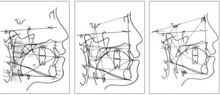
Fig. 1. Landmarks, Reference Lines. Fig. 2. Cephalometric Distances (mm). Fig. 3. Cephalometric Angles (� ).
Table 1. Cephalometric Landmarks & Reference Lines
S Sella Midpoint of fossa hypophysealis
N Nasion Anterior point at frontonasal suture
O Orbitale Most inferior point of the orbit
ANS Spina nasalis ant. Most anterior point of anterior nasal spine PNS Spina nasalis post. Most posterior point of hard palate
A A-point Deepest anterior point in concavity of anterior maxilla B B-point Deepest anterior point in concavity of anterior mandible Pg Pogonion Most anterior point of bony chin
Pm Pm-point Halfway B-point - Pogonion
Gn Gnathion Most antero-inferior point of bony chin
Me Menton Most inferior point of bony chin
Go Gonion A mid-plane point at the gonial angle located by bisecting the posterior and inferior mandibular borders
Ar Articulare A mid-plane point at the intersection of posterior ramus with inferior cranial base
C Condylion Most postero-superior point of mandibular condyle
Pt Pterygon Inferior border of foramen rotundum bisecting posterior border of pterygomaxillary fissure
Xi Xi-point Constructed point in the centre of the ramus
CF Pterygoid-Vertical Bisecting Frankfurt horizontal
DC Middle of condyle on plane Ba-N
Ba Basion Most inferior point on anterior foramen magnum Po Porion Most superior point of bony external auditory meatus
PhW1 Ba-PNS bisecting posterior pharyngeal wall
PhW2 ANS-PNS bisecting posterior pharyngeal wall
PhW3 Occlusal plane bisecting posterior pharyngeal wall PhW4 Mandibular plane bisecting posterior pharyngeal wall
PhW5 Me-H bisecting posterior pharyngeal wall
H Hyoid Most antero-superior point of hyoid
V Vallecula Most antero-inferior point of epiglottic fold T1 Tongue base ML-bisecting posterior margin of the tongue base T2 Back of tongue Most superior point of the back of the tongue to V-TT TT Tongue tip Most anterior point of the tip of the tongue
U1 Most superior point of soft palate distal to PNS
U2 Posterior margin of soft palate at its greatest thickness U3 Occlusal plane bisecting posterior margin of soft palate
UT Uvula tip Tip of uvula or soft palate
U4 Anterior margin of soft palate at its greatest thickness
U5 Most antero-superior margin of the soft palate
AA Ant. atlas Most anterior point of bony atlas
NSL Nasion-sella-line N-S
NL Nasal-line ANS-PNS
ML Mandibular-line Me-Go
FH Frankfurt horizontal O-Po
의 투사지상에 중첩시켜 2명의 구강악안면외과 의사에 의 해 0.3mm 굵기의 흑연필로 각각 2회씩 투사도를 완성하였 다. 모든 계측점을 컴퓨터 디지타이저(Summa-Sketch
�III, Summagraphics, USA)를 이용하여 입력시킨 후 계 측값을 컴퓨터 상에서 계산하였다.
이렇게 얻어진 자료는 각군 계측값의 유의성은 paired
t - test 로, 각군간의 차이의 유의성은 ANOVA test(mul-
tiple comparison: Tukey)이용하였으며 사용된 계측점과
계측항목은 Fig. 1, 2, 3과 Table 1, 2에 나타내고 있다.
Ⅲ. 연구 결과
측모두부규격 방사선사진상에서 인후 기도공간구조(pha- ryngeal dimension) 분석에 대한 각계측 6개, 선계측 29 개 각각 항목에 대하여 각 군마다의 술전(T0), 술후 6개월 (T1), 술후 변화량(T1-T0)의 평균치와 표준편차를 구하고 그룹간 비교를 하였다(Table 3, 4, 5, 6).
1. 비인두(nasopharynx) 부위의 기도공간의 변화
술후 비인두(nasopharynx) 부위의 기도공간(PhW1- PNS)은 모든 군에서 전반적으로 증가하는 양상을 보였으 며, 상악 후방부 상방 이동 및 전체 상방 이동한 군 (Group 3)에서는 술전에 평균 27.7mm 술후 6개월 평균 28.8mm 로 술후 변화량이 다른 군보다 비해 유의한 증가(p<0.05)를 보였다(Table 3, 4, 5, 6)
2. 구인두(oropharynx)부위의 기도공간의 변화
구인두(oropharynx)부위의 기도공간(PAS (UT))은 상 악을 전방 이동한 군(Group 1) 술후 변화량 -0.9±1.3과 상악 후방부를 상방 이동한 군(Group 2) 술후 변화량 -0.9
±2.2로 술후 유의한 감소를 보였으나(p<0.05), 3군은 별 차이가 없었다(Table 3, 4, 5, 6).
3. 하인두(hypopharynx) 부위의 기도공간의 변화
하인두(hypopharynx)부위의 기도공간(Vallecula- PhW)은 술후 6개월에도 협소화가 유지되었으며, 1군 술후 변화량 -1.3±1.6과 2군 술후 변화량 -1.0±2.2은 술전보 다 유의한 감소를 보였다(p<0.05) (Table 3, 4, 5, 6).
Table 2. Cephalometric Distances (mm)
S-Go Posterior facial height
N-Gn Anterior facial height
A/N-Pg Convexity
PAS (ML) Distance posterior pharyngeal wall-tongue base on ML
PAS (Occl.) Distance posterior pharyngeal wall-tongue base on occlusal plane PAS (NL) Distance posterior pharyngeal wall-tongue base on NL
PAS (UT) Distance posterior pharyngeal wall-uvula tip AA-PNS Distance anterior atlas-posterior nasal spine Ba-PNS Distance basion-posterior nasal spine
Ba-PhW1 Distance basion-posterior pharyngeal wall on Ba-PNS PhW1-PNS (PAS) Distance posterior pharyngeal wall-PNS on Ba-PNS
Go-PNS Posterior lower facial height
Ba-A Distance basion- point A
PNS-UT Length of the soft palate (uvula-length) U2-U4 Thickness of the soft palate (uvula-thickness)
V-Me Distance vallecula-menton
V-ANS Distance vallecula-anterior nasal spine
V-S Distance vallecula-sella
T1-ANS Distance tongue base-anterior nasal spine
T1-B Distance tongue base-point B
T1-PNS Distance tongue base-posterior nasal spine T1-TT Distance tongue base-tongue tip
T2/V-TT Tongue height
H-ML Shortest distance hyoid to mandibular plane
H-Me Distance hyoid-menton
H-B Distance hyoid-point B
H-PhW (Me-H) Distance hyoid-posterior pharyngeal wall on Me-H H-PhW Shortest distance hyoid to posterior pharyngeal wall
AA-H Distance hyoid-anterior Atlas
H-S Distance hyoid-sella
4. 설골과 기도공간의 변화
설골에서 하악의 전방 부위까지의 거리(H-Me, H-B)를 보면 3군 술후 변화량 (H-Me에서 -0.5±2.3, H-B에서 - 0.6±1.7)에 비해 1군 술후 변화량(H-Me에서 -3.8±3.3, H-B에서 -3.3±3.2)과 2군 술후 변화량(H-Me에서 -3.2
±3.3, H-B에서 -3.3±2.8)이 술후 감소 정도가 유의한 차 이를 보였다(P<0.05).
설골에서 후인두벽까지의 거리(H-PhW(Me-H), H-
PhW)의 감소는 세 군 사이에 유의한 차이가 없었다.
Ⅳ. 총괄 및 고찰
본 연구는 Hochban 등
11)이 1994년 제안한 분석법을 토 대로 하여, 상기도의 수평거리, 연구개, 혀, 설골 등과 관련 된 연조직 구조 분석을 시행하였다. 인두부 기도량을 측정 하기 위해 두부규격 방사선사진을 이용하는 방법은 상당히 실용성과 신뢰성이 있으며 무엇보다도 간편하고, 환자에게 Table 3. Analysis for Pharynx(Group 1, Maxillary advancement group)
T0 T1 T1-T0
Mean ±SD Mean ±SD Mean ±SD p-value
PAS (NL) 28.3±5.9 28.4±5.3 0.0±1.9 0.9578
PAS (OCCL) 26.3±8.7 24.8±6.3 -1.4±3.5 0.1193
PAS (UT) 13.3±4.0 12.4±3.3 -0.9±1.3 0.0158
PAS (ML) 15.5±4.9 14.4±4.1 -1.1±2.0 0.0402
AA-PNS 34.9±6.4 35.3±5.8 0.5±1.6 0.2456
Ba-PNS 47.7±8.1 48.3±7.5 0.6±1.3 0.0877
Ba-PhW1 20.5±4.2 20.8±4.1 0.3±1.2 0.3875
PhW1-PNS (PAS) 27.2±6.0 27.5±5.2 0.3±2.0 0.5391
Go-PNS 47.1±8.7 47.4±8.2 0.3±1.3 0.3455
Ba-A 96.5±12.7 98.0±12.9 1.4±0.8 0.0001
Uvula length 35.8±5.4 36.2±5.4 0.4±1.2 0.1662
Uvula thickness 9.6±1.7 8.8±1.4 -0.8±0.9 0.0040
Uvula angulation � 121.4±5.2 122.9±4.6 1.5±2.4 0.0240
Vallecula-Me 67.4±10.9 71.8±10.2 4.4±5.9 0.0097
Vallecula-ANS 100.4±15.8 94.8±18.3 -5.6±5.5 0.0010
Vallecula-S 123.1±22.3 121.7±22.5 -1.4±2.3 0.0244
Tongue base-ANS 81.9±10.7 83.2±11.3 1.3±2.1 0.0263
Tongue base-B 73.9±10.1 70.3±10.1 -3.6±2.8 0.0001
Tongue base-PNS 46.4±8.2 46.5±8.0 0.1±1.3 0.7118
Tongue base-TT 74.5±9.9 76.2±10.3 1.7±1.7 0.0012
Vallecula-PhW (PAS) 18.9±4.1 17.7±3.7 -1.3±1.6 0.0064
Vallecula-TT 82.5±13.0 84.0±13.4 1.5±2.3 0.0199
Tongue height 39.0±6.0 40.9±6.4 1.9±1.7 0.0012
V-TT/ML � 59.0±5.6 58.4±5.7 -0.6±2.2 0.3327
V-TT/FH � 25.3±5.8 24.0±5.9 -1.2±2.1 0.0366
H-ML 14.7±7.7 16.4±7.9 1.8±1.7 0.0010
H-Me 52.7±8.6 48.9±8.0 -3.8±3.4 0.0005
H-B 55.4±9.2 52.1±9.3 -3.3±3.2 0.0009
H-PhW (Me-H) 35.3±6.1 34.5±5.9 -0.8±1.4 0.0418
H-PhW 34.3±6.4 33.8±6.2 -0.4±1.2 0.1551
AA-H 73.7±17.3 74.4±17.0 -0.3±3.5 0.7633
H-S 122.0±23.6 121.5±23.6 -0.5±1.5 0.1816
N-S-H � 89.7±5.1 89.9±4.6 0.2±1.6 0.6163
NSL/Ar-H � 77.6±6.1 78.0±5.4 0.4±2.0 0.3745
ML/H � 16.6±7.8 20.3±8.8 3.6±2.5 0.0001
Angle : �, Another : mm
T0 : Pre-OP Data, T1 : Post-OP Data
불편을 덜 주며, 빠르게 비교 분석할 수 있다는 면에서 다른 방법보다 두부규격방사선사진을 이용한 설골과 혀 및 기도 량의 계측법이 널리 사용되고 있다
12).
측모두부규격 방사선사진에서 기도공간의 세부분을 보면 비인두는 인두정과 후비극을 잇는 선, 구개면과 인두후벽으 로 경계되는 부위로 나타나는데, 이 부위는 하악전돌증 환 자의 하악골 후퇴 수술시에 영향을 받지 않는다는 연구
13)와 측모 두부계측 방사선사진상에서 비인두부 기도공간이 감 소되었음을 보고한 연구
14)가 있다. 구인두는 비인두 하방경
계, 연구개와 혀의 후면을 지나며 구개면에 평행인 선으로 경계되는 부위로 하악골 후퇴 수술시 기도공간의 변화에 대 한 연구
4)가 주로 관찰되는 부위이다. 하인두는 구인두의 하 방경계, 후두개의 후면, 제4경추의 최전하방점을 통과하면 서 구개면에 수평인 선과 인두후벽에 의해 경계되며, 하악 골 후퇴 수술시 유의하게 영향 받지 않는 것으로 알려져 있 다. 이와 관련하여 골격성 제3급 부정교합 환자의 악교정 수술은 하악골을 후퇴하여 설골, 혀 및 기도공간의 변화를 가져온다. 이러한 변화가 하악골 후방이동술후 수면 무호흡 Table 4. Analysis for Pharynx(Group 2, Maxillary posterior impaction group)
T0 T1 T1-T0
Mean ±SD Mean ±SD Mean ±SD p-value
PAS (NL) 30.5±6.2 30.7±6.2 -0.1±2.3 0.7629
PAS (OCCL) 25.1±5.9 25.4±5.3 0.3±2.3 0.5037
PAS (UT) 13.7±3.7 12.7±3.3 -0.9±2.2 0.0313
PAS (ML) 15.4±4.3 14.4±3.8 -0.9±1.8 0.0067
AA-PNS 37.9±7.7 39.1±7.0 1.1±2.8 0.0242
Ba-PNS 51.7±9.7 51.8±9.5 0.1±2.1 0.7776
Ba-PhW1 22.0±5.3 21.5±4.7 -0.4±1.6 0.1399
PhW1-PNS (PAS) 29.4±4.6 29.9±4.4 0.4±1.9 0.1746
Go-PNS 52.6±9.0 52.2±9.4 -0.4±2.1 0.2818
Ba-A 103.7±13.9 103.0±13.7 -0.8±3.8 0.2298
Uvula length 37.6±5.8 38.5±5.7 0.8±2.1 0.0682
Uvula thickness 9.8±2.3 9.3±2.0 -0.5±1.4 0.0682
Uvula angulation � 123.5±5.5 123.7±5.5 0.2±2.5 0.6517
Vallecula-Me 68.9±9.5 67.3±10.7 -1.3±5.4 0.1835
Vallecula-ANS 103.1±16.1 102.7±16.2 -1.5±5.2 0.1144
Vallecula-S 124.4±21.7 122.7±21.5 -1.7±5.6 0.0931
Tongue base-ANS 88.3±13.0 80.0±13.1 -0.3±3.5 0.6014
Tongue base-B 75.5±10.9 72.1±10.7 -3.5±3.3 0.0001
Tongue base-PNS 50.8±8.6 50.5±9.0 -0.3±2.2 0.4070
Tongue base-TT 79.7±11.9 79.9±11.5 0.2±3.9 0.7209
Vallecula-PhW (PAS) 18.4±3.8 17.4±3.6 -1.0±2.2 0.0111
Vallecula-TT 85.7±12.9 85.7±13.0 -0.0±3.3 0.9712
Tongue height 41.2±7.2 42.3±6.4 1.1±2.7 0.7209
V-TT/ML � 53.6±4.6 54.1±4.3 0.5±2.8 0.3556
V-TT/FH � 24.0±5.1 22.8±3.8 -1.2±4.1 0.1120
H-ML 12.9±5.1 14.3±5.6 1.4±1.3 0.0001
H-Me 53.3±7.9 50.1±7.8 -3.2±3.3 0.0001
H-B 56.6±8.5 53.3±8.5 -3.3±2.8 0.0001
H-PhW (Me-H) 34.8±5.2 34.0±5.6 -0.8±1.6 0.0046
H-PhW 34.0±5.2 33.2±5.6 -0.8±1.5 0.0035
AA-H 75.4±13.0 74.5±12.5 -1.0±3.8 0.1472
H-S 124.1±21.1 122.5±21.1 -1.5±5.3 0.1010
N-S-H � 87.9±2.5 88.3±2.5 0.4±1.0 0.0469
NSL/Ar-H � 74.1±3.4 74.8±3.5 0.6±1.4 0.0179
ML/H � 14.2±5.2 17.2±6.1 3.0±1.7 0.0001
Angle : � , Another : mm
T0 : Pre-OP Data, T1 : Post-OP Data
증의 발생과 코골이 등과 같은 수면 무호흡증과 관련된 증 상을 나타낼 수 있다고 보고되고 있다
7). 또한 하악골 후방 이동술후 후방인두부 기도량이 하악평면 위치에서 10mm 이하이면 폐쇄성 수면무호흡증을 야기할 수 있으며 이런 경 우는 상악골 전방이동술을 포함한 양악수술법을 고려해야 한다는 보고가 있다
9). 이러한 점에서 본 연구는 골격성 제3 급 부정교합 환자의 악교정 수술시 하악골 후퇴에 따른 수 면 무호흡증과 관련된 증상을 고려하여 양악 수술시에 기도 공간의 변화를 관찰하였다.
비인두 부위에서 Holmberg 등
13)과 정 등
15)은 술후 기도 공간의 감소가 일어나지 않는다고 보고하였으나 본 연구에 서 측정한 술후 비인두(nasopharynx) 부위의 기도공간 (PhW1-PNS)은 모든 군에서 전반적으로 증가하는 양상을 보였으며, 상악 후방부 상방 이동 및 전체 상방 이동한 군 (Group 3)에서는 술후 변화량이 1.0±1.4로(p<0.05)로 다른 군에 비해 유의한 증가를 보였다. 이러한 3군의 유의 한 증가는 상악이 상방이동 및 전진됨으로써 연구개가 전진 하게 되어 비인두의 기도공간이 증가된 것으로 볼 수 있다.
Table 5. Analysis for Pharynx(Group 3, Maxillary posterior impaction and superior repositioning group)
T0 T1 T1-T0
Mean ±SD Mean ±SD Mean ±SD p-value
PAS (NL) 29.3±5.9 30.1±6.4 0.8±2.0 0.1418
PAS (OCCL) 22.5±7.5 23.1±6.4 0.6±2.4 0.3774
PAS (UT) 12.5±4.7 12.6±4.0 0.2±2.0 0.7113
PAS (ML) 15.4±6.3 14.4±5.3 -1.0±1.9 0.0664
AA-PNS 37.4±6.8 39.4±6.9 2.0±1.8 0.0001
Ba-PNS 47.8±7.4 49.2±7.9 1.5±1.7 0.0034
Ba-PhW1 19.5±3.5 19.8±3.8 0.3±1.2 0.2971
PhW1-PNS (PAS) 27.7±3.5 28.8±3.6 1.0±1.4 0.0066
Go-PNS 49.0±6.6 49.4±6.6 0.4±0.9 0.0967
Ba-A 95.7±9.0 97.0±9.4 1.3±0.9 0.0001
Uvula length 36.9±3.5 37.2±3.4 0.3±1.3 0.3670
Uvula thickness 9.1±1.5 8.6±1.4 -0.5±0.6 0.0068
Uvula angulation � 127.5±5.9 128.0±6.0 0.5±1.8 0.3435
Vallecula-Me 65.2±5.5 71.3±5.6 6.1±4.8 0.0002
Vallecula-ANS 97.4±9.5 93.0±10.7 -4.4±4.4 0.0016
Vallecula-S 110.9±16.6 110.9±15.7 0.0±1.3 0.8598
Tongue base-ANS 84.4±7.2 85.2±7.3 0.8±1.1 0.0210
Tongue base-B 71.0±5.4 70.0±5.8 -1.0±1.3 0.0115
Tongue base-PNS 47.4±6.3 47.5±6.2 0.1±1.0 0.7518
Tongue base-TT 77.7±6.1 78.4±6.3 0.8±1.2 0.0268
Vallecula-PhW (PAS) 17.8±3.8 17.0±3.1 -0.8±1.7 0.0892
Vallecula-TT 83.1±7.4 84.4±7.4 1.4±1.0 0.0001
Tongue height 37.9±4.4 38. 8±4.4 0.9±1.4 0.0273
V-TT/ML � 51.5±3.7 51.6±3.4 0.2±1.4 0.6453
V-TT/FH � 18.2±5.5 18.4±4.6 0.3±1.4 0.4597
H-ML 11.0±3.2 13.1±3.4 2.1±1.2 0.0001
H-Me 51.3±4.2 50.8±4.0 -0.5±2.3 0.3825
H-B 54.8±4.6 54.1±4.8 -0.6±1.7 0.1736
H-PhW (Me-H) 32.7±5.4 31.9±5.0 -0.8±0.8 0.0017
H-PhW 32.1±5.4 31.3±4.8 -0.9±1.0 0.0046
AA-H 64.3±13.2 63.8±13.2 -0.5±0.8 0.0470
H-S 110.7±16.8 110.4±16.8 -0.3±0.6 0.0695
N-S-H � 93.4±5.0 93.1±4.5 -0.2±0.8 0.2641
NSL/Ar-H � 80.2±6.1 80.2±5.7 0.0±0.9 0.8657
ML/H � 12.6±3.5 15.3±4.0 2.7±1.5 0.0001
Angle :� , Another : mm
T0 : Pre-OP Data, T1 : Post-OP Data
이는 상악이 전상방으로 이동시 비인두 기도공간이 증가한 다는 DePonte 등
10)의 보고와 일치한다고 볼 수 있다.
구인두 부위의 기도공간은 하악골 후퇴수술 직후 감소되 어 술후 약 1년 후에도 계속 감소된다는 보고가 있다
16). 본 연구에서 측정한 구인두(oropharynx)부위의 기도공간 (PAS (UT))은 상악을 전방 이동한 군(Group 1)과 상악 후방부를 상방 이동한 군(Group 2)에서는 술후 유의한 감 소를 보였으나(p<0.05), 3군은 별차이가 없었다. 3군의 유 의한 차이가 나지 않은 것은 상악의 상방이동으로 인한 연 구개를 둘러싸고 있는 근육의 긴장도가 늘어나서 생긴 결과
로 추정된다.
하인두 부위는 구인두부와 함께 기도공간의 협소화면에서 중요한 의미를 가진다. 하인두(hypopharynx)부위의 기도 공간(Vallecula-PhW)은 술후 6개월에도 협소화가 유지되 었으며, 1군, 2군은 수술전보다 유의한 감소를 보였다 (p<0.05). Greco 등
6)과 Enacar 등
16)은 수술 직후 줄어든 하인두부 기도량이 다시 술전의 상태로 회복되지 않고 영구 적으로 변화가 지속된다는 연구결과를 보고 하였으며 본 교 실의 이전 연구에서도 골격성 제3급 부정교합 환자에서 술 후 구인두와 하인두부의 협소화가 보고된 적이 있다. 이는 본 연구의 결과와 일치한다고 볼 수 있다.
설골과 혀 위치의 변화는 기도 크기의 유지와 상관관계가 있고 수술 결과의 안정성은 설골위치의 안정성과 관련이 있 다
17). 본 연구에서 측정한 설골에서 하악의 전방 부위까지의 거리(H-Me, H-B)를 보면 3군에 비해 1군과 2군이 술후 감소정도가 유의한 차이를 보였다(p<0.05). 또한 설골에서 후인두벽까지 거리(H-PhW(Me-H), H-PhW)의 감소는 세 군 사이에 유의한 차이가 없었다. 이러한 기도공간과 설 골의 이동량의 차이는 하악을 전방이동하여 설골을 잡아당 기는 힘에 의한 설골의 이동보다 하악을 후방이동하여 설골 을 밀어 이동시키는 힘에 의한 설골의 이동이 작기 때문이 라 사료된다. 또한 Takagi 등
18)은 하악골 후방이동술 직후 설골은 하방 이동되지만 경추에 대한 전후방 위치는 거의 변화가 없으며 이는 일정 기간이 지난 후 술전 상태로 복귀 한다고 하였으며 본 연구에서도 이를 관찰 할 수 있었다.
본 연구결과에서 상악을 전방 이동한 군과 상악 후방부를 상방 이동한 군은 상악이 전방 및 상방 이동 되었음에도 하 악골 후퇴가 영향을 미치는 구인두, 하인두에서는 유의한 감소를 보였다. 이는 상악골 이동과 관련된 비인두를 제외 한 구인두, 하인두에서는 상악의 이동보다는 하악의 후퇴정 도가 더 영향을 미치리라 사료된다. 따라서 하악의 후퇴량 이 많아 구인두, 하인두 기도공간의 협소화가 예상된다면 상악의 전, 상방을 이동하는 술식이 기도공간의 협소화를 줄여주는 한 방법이라고 본다.
Ⅴ. 결 론
골격성 제3급 부정교합 환자의 악교정 수술의 진단시 술 후 기도공간 변화량 예측은, 술후 하악골이 후방으로 이동 하면서 기도공간의 협소화에 따른 코골이나 폐쇄성 수면 무 호흡증과 같은 합병증이 예상되기 때문에 중요하다. 본 연 구는 상악술식의 분류에 따른 양악 수술 전후에 기도공간의 변화를, 술후 6개월간 추적 조사가 가능했던 골격성 제3급 부정교합 환자 64명을 대상으로 비교 분석을 통하여 다음 과 같은 결론을 얻었다.
1. 술후 비인두(nasopharynx)부위의 기도공간 후인두벽에 Table 6. Analysis for Pharynx(Multiple comparison
among 3 groups)
p-value Group
PAS (NL) 0.5476
PAS (OCCL) 0.0651
PAS (UT) 0.1978
PAS (ML) 0.9441
AA-PNS 0.1778
Ba-PNS 0.0633
Ba-PhW1 0.1407
PhW1-PNS (PAS) 0.5152
Go-PNS 0.2022
Ba-A 0.0109 1-2/2-3
Uvula length 0.5969 Uvula thickness 0.6498 Uvula angulation � 0.1792
Vallecula-Me 0.0001 1-2/2-3
Vallecula-ANS 0.0212 1-2
Vallecula-S 0.4609
Tongue base-ANS 0.1385
Tongue base-B 0.0143 1-3/2-3 Tongue base-PNS 0.6367
Tongue base-TT 0.2985 Vallecula-PhW (PAS) 0.7707 Vallecula-TT 0.1232 Tongue height 0.4408
V-TT/ML � 0.3866
V-TT/FH � 0.3281
H-ML 0.3029
H-Me 0.0099 1-3/2-3
H-B 0.0043 1-3/2-3
H-PhW (Me-H) 0.9868
H-PhW 0.5577
AA-H 0.7434
H-S 0.5095
N-S-H � 0.2226
NSL/Ar-H � 0.4861
ML/H � 0.3812
Angle :� , Another : mm
서 PNS까지 거리(PhW1-PNS)는 전반적으로 증가하는 양상을 보였으며, 상악 후방부 상방이동 및 전체 상방 이 동한 군(Group 3)에서는 유의한 증가를 보였다.
2. 술후 코골이를 유발할 수 있는 구인두(oropharynx)부위 의 기도공간 인후기도공간에서 uvula tip까지의 거리 (PAS (UT))는 상악을 전방 이동한 군(Group 1)과 상 악 후방부를 상방이동한 군(Group 2)에서 술후 유의한 감소를 보였으나, 3군은 별차이가 없었다.
3. 하인두(hypopharynx)부위의 기도공간 vallecula에서 후인두벽까지 거리(Vallecula-PhW)는 술후 6개월에도 협소화가 유지되었으며, 1군과 2군은 술 전보다 유의한 감소를 보였다.
4. 설골에서 하악의 전방 부위까지의 거리(H-Me, H-B)는 3군에 비해 1, 2군이 술후 감소 정도가 유의한 차이를 보였으나, 설골에서 후인두벽까지의 거리(H-PhW(Me- H), H-PhW)의 감소는 세 군 사이에 유의한 차이가 없 었다.
골격성 제3급 부정교합환자의 양악 수술시 비인두 부위는 상악골의 이동이 영향을 미치고 구인두, 하인두 부위는 상 악골 이동보다 하악골 후퇴량에 더 영향을 받는 것으로 보 인다. 따라서 수술 계획시 하악골 후퇴량과 함께 상악골 수 술 계획을 고려하는 것이 중요하다.
참고문헌