ISSN 0378-6471 (Print)⋅ISSN 2092-9374 (Online)
http://dx.doi.org/10.3341/jkos.2015.56.2.285
Case Report
필러 주입 후 발생한 신경학적 증상을 동반한 뇌경색과 안동맥폐쇄 1예
Multiple Cerebral Infarctions with Neurological Symptoms and Ophthalmic Artery Occlusion after Filler Injection
이원섭⋅윤원태⋅최연주⋅박성표
Won Sup Lee, MD, Won Tae Yoon, MD, Youn Joo Choi, MD, Sung Pyo Park, MD, PhD
한림대학교 의과대학 강동성심병원 안과학교실
Department of Ophthalmology, Kangdong Sacred Heart Hospital, Hallym University College of Medicine, Seoul, Korea
Purpose: To report a case of visual loss, side weakness and facial palsy due to ophthalmic artery occlusion with diffuse multiple cerebral infarctions after injection of hyaluronic acid.
Case summary: A 50-year-old female visited our clinic for visual loss in the left eye after filler injection in the glabella. Her best corrected visual acuity was 1.0 in the right eye and hand motion in the left eye. The intraocular pressure was 8 mm Hg in the right eye and 14 mm Hg in the left eye. In the left eye, there was abnormal pupillary light reflex and complete extra-ocular muscles pal- sy with blepharoptosis. A pale retina with a cherry-red-spot also appeared in the left fundus. A central retinal artery occlusion was observed on fluorescein angiography and brain magnetic resonance imaging showed multiple cerebral infarctions at the frontal, temporal, parietal and occipital lobes. Four days later, the motor weakness was aggravated and dysarthria and aphasia became worse. According to symptoms, a hemorrhagic transformation in subacute infarctions developed based on brain computed tomography. After 3 months of follow up, the visual acuity in the left eye was no light perception. However, the general conditions including ophthalmoplegia and motor weakness were improved.
J Korean Ophthalmol Soc 2015;56(2):285-290
Key Words: Central retinal artery occlusion, Cerebral infraction, Filler complication, Hemorrhagic transformation, Ophthalmic ar- tery occlusion
■Received: 2014. 8. 29. ■ Revised: 2014. 10. 11.
■Accepted: 2015. 1. 7.
■Address reprint requests to Sung Pyo Park, MD, PhD Department of Ophthalmology, Hallym University Kangdong Sacred Heart Hospital, #150 Seongan-ro, Gangdong-gu, Seoul 134-701, Korea
Tel: 82-2-2224-2274, Fax: 82-2-470-2088 E-mail: [email protected]
ⓒ2015 The Korean Ophthalmological Society
This is an Open Access article distributed under the terms of the Creative Commons Attribution Non-Commercial License (http://creativecommons.org/licenses/by-nc/3.0/) which permits unrestricted non-commercial use, distribution, and reproduction in any medium, provided the original work is properly cited.
최근 필러는 다양한 미용목적으로 사용되고 있다. 코끝, 콧대, 미간, 팔자 주름, 이마, 턱, 볼, 눈 밑, 입술까지 목적에 따라 다양한 부위 시행하고 있는데, 점차 시행빈도가 증가 함에 따라 합병증 발생도 증가하고 있으며, 부위별로 다양 한 합병증이 보고되고 있다.1
이러한 합병증은 대부분 경미하여 시간이 지나면 회복되 는 경우가 많으나 드물게 시력저하나 뇌경색과 같은 합병 증이 발생했을 경우 영구적인 후유증으로 남을 수 있기 때 문에 필러 주입술 시행에서 많은 주의가 필요하다.
본 증례의 경우 미간부위에 히알루론산 필러 주입 후 안 동맥폐쇄, 중심망막동맥폐쇄에 의한 시력장애와, 광범위한 국소 뇌경색으로 상지 근력저하와 구음장애, 실어증 및 뇌 경색의 출혈성 변화가 동반되어 나쁜 예후를 암시하였지만, 꾸준한 경과관찰과 재활치료로 실명을 제외한 다른 증상은 모두 회복된 사례이다, 기존에 필러 주입 후 시력을 소실한 증례들은 있어왔고,2-4 국내에서 Paik et al5은 국소 뇌경색 소견을 보인 증례를 보고한 바 있지만, 본 증례처럼 심각한 신경학적 증상을 동반한 광범위한 다발성 뇌경색이 발생한
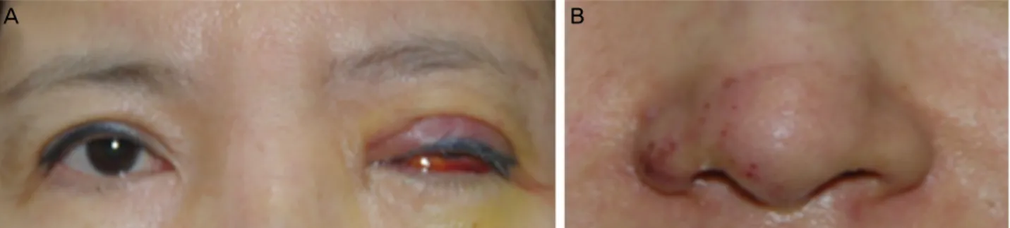
Figure 1. (A) Initial facial photographs show erythematous left eyelid swellings with blepharoptosis, severe conjunctival chemosis,
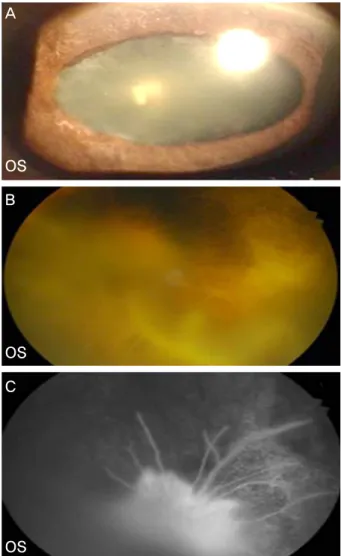
subconjunctival hemorrhage in the left eye and (B) purpura like rash at nasal ala and forehead.Figure 2. (A) Fundus pho-
tographs show normal in right eye, (B) but pale ret- ina and thin vessels with cherry red spot in the left eye. (C) Fundus fluorescein angiographs show normal in the right eye, (D) non- perfusion of retina and cho- roid in the left eye about 2 minute after dye injection.증례는 이전에 보고된 바 없어 이를 보고하고자 한다.
증례보고
기저질환 없는 50세 여자환자가 내원 4시간 전 미간에 히 알루론산 필러(Restylane; Q-Med AB, Uppsala, Sweden) 주 입 직후 발생한 좌안의 시력저하를 주소로 본원에 내원하 였다. 내원 시 최대교정시력은 우안 1.0, 좌안 안전수동이였 고, 안압은 우안 8 mmHg, 좌안 14 mmHg이었으며, 세극등 검사에서 경도의 각막부종과 데스메막 주름이 관찰되었다.
좌안의 동공은 직접대광반사는 보이지 않고, 간접대광반사 만 보였으며, 상대적 구심성 동공장애를 보였다. 좌안 안구 운동은 모든 방향에서 완전히 제한되어 전혀 움직이지 않
았으며, 좌안 상안검에 발적과 부종을 동반한 안검하수 소 견 관찰되었다. 콧대에도 황색 멍과 자반 양상의 피부병변 이 동반되었다(Fig. 1). 산동 후 시행한 안저검사에서 창백 한 망막과 망막부종, 앵두반점이 관찰되었고, 형광안저혈관 조영술 또한 조영이 매우 지연되는 중심망막동맥폐쇄에 해 당하는 소견을 보였다(Fig. 2). 좌안 안동맥폐쇄 진단하에 산소공급을 하며 30 gaze 바늘로 전방천자를 시행하였다.
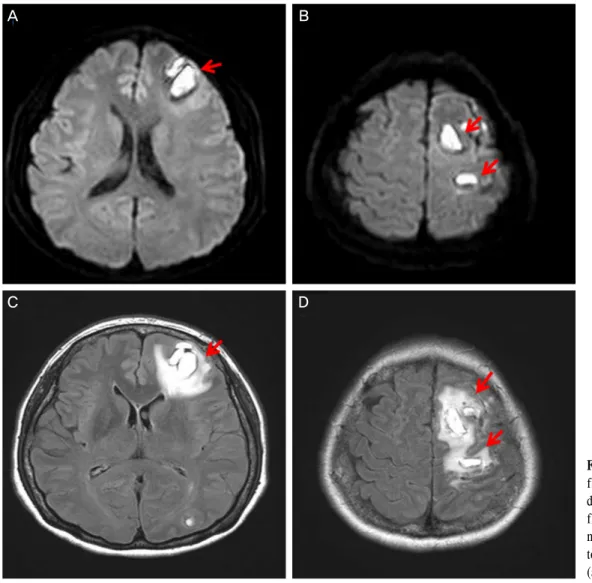
전신증상으로는 어지럼증과 의식 변화, 구음장애가 나타 났으며, 우측 상지는 3단계, 우측 하지는 4단계의 근력저하 가 나타났다. 바로 뇌 자기공명영상 촬영을 하였으며, 다발 성의 국소 고신호를 보이는 병변이 전두엽, 측두엽, 두정엽, 후두엽에서 발견되어, 광범위한 뇌경색이 동반되었음을 알 수 있었다(Fig. 3). 뇌경색에 대한 치료로 경구 아스피린 복
C D
A B
A B
OD OS
OD OS
Figure 3. (A, B) Brain diffuse MRI (axial view) shows multi-
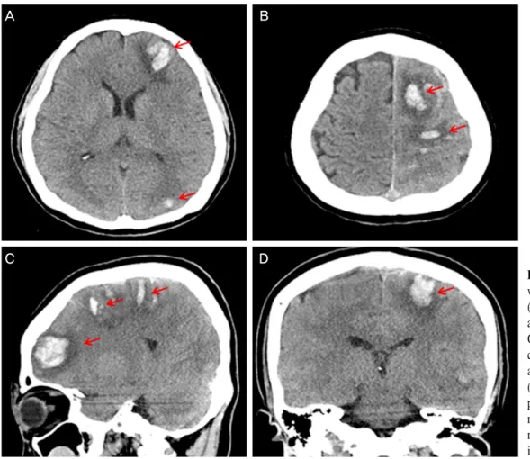
ple high signal intensity lesions on left cerebral hemisphere (frontal, temporal, occipital, parietal lobes) which were sus- pected recent infarctions of middle cerebral artery territory (arrows).Figure 4. (A, B: axial
view), (C: sagittal view), (D: coronal view) 4 days after filler injection, brain CT shows multiple hyper- dense hemorrhages (arrows) at left cerebral hemisphere (fro- ntal, temporal, occi- pital, parietal lobes) which mean hemorrhagic transfor- mation in subacute infarct- ions.용과 함께 다량의 수액공급을 시행하였다.
입원 4일째부터 좌안 시력은 광각무로 떨어졌으며, 안압 은 7 mmHg였다. 각막의 부종은 큰 변화 없이, 심한 결막부 종과 결막하 출혈이 나타났다. 외안근 마비는 호전되었지 만, 구음 장애와 실어증이 심해지고, 우측 상지 근력저하가 2단계로 악화되어 뇌 전산화 단층촬영을 재시행하였고, 경
색이 있던 부위에 출혈성 변화가 진행되었음을 관찰하였다 (Fig. 4).
약 3개월간의 언어 및 운동 재활치료 후 시력은 여전히 광각무였지만, 결막부종과 결막하 출혈, 외안근 마비는 호 전되었고, 구음장애, 실어증, 사지 근력저하 등의 전신증상 도 모두 소실되었다(Fig. 5). 하지만 안구 허혈의 후기 변화 로 좌안 동공은 빛에 대한 반응이 없고 홍채후 유착이 생겼 으며, 망막에는 섬유혈관조직이 관찰되었다. 뇌 자기공명영 상결과는 3개월 전과 큰 차이를 보이지 않았다(Fig. 6).
고 찰
미용에 대한 관심이 꾸준히 늘어가면서 성형수술보다 비 교적 비용도 저렴하고, 시술도 간단한 필러 주입이 성행하 고 있다. 필러의 성분으로는 히알루론산, 자가이식성 지방 (autologous fat)이 가장 흔히 사용되고 있고, 그 외에도 칼슘 수산화인회석(calcium hydroxyapatitie), 파라핀(paraffin), 실 리콘 오일(silicone oil), 부신피질호르몬(corticosteroids), 폴 리메틸 메타크릴산(polymethylmethacrylate) 등이 있다. 주사 부위도 다양한데, 목적에 따라 콧등, 코끝, 미간, 이마, 측두 부 등에 시술을 한다.1,4
A B
A B
C D
Figure 5. 3 months after filler injection (A) anterior segment
photograph shows posterior synechiae and oval-shape fixed pupil. (B) Fundus photograph shows fibrovasular membrane around the disc and posterior pole. (C) Fundus fluorescein an- giograph shows hyperfluorescence because of fibrovascular membrane about 1 minute after dye injection.본 증례에서 사용된 필러는 히알루론산 필러(Restylane;
Q-Med AB, Uppsala, Sweden)로 입자 크기는 약 400 마이크 로미터이며, 지속시간은 피부의 상태, 생활습관, 나이에 따 라 차이가 있으나 평균 6-12개월이다.
필러 주사 후 발생할 수 있는 합병증도 시술의 증가에 따 라 자연스럽게 증가하고 있다. 피부합병증으로는 발적, 부 종, 통증, 소양감, 주사물질에 대한 피부과민반응, 피부허혈, 2차적인 피부감염 등이 있으며, 드물게 피하 지방육아종, 피부괴사도 보고되고 있다. 안과적 합병증으로는, 외안근마 비, 망막동맥폐쇄(retinal artery occlusion)에 의안 실명 (blindness) 등이 있고, 그 외 전신합병증으로 필러 색전에 의한 뇌경색 등이 보고된 바 있다.1,4-8
본 증례는 미간에 필러 주사 후 심각한 합병증인 실명과,
다발성 국소 뇌경색에 의한 인지기능장애, 구음장애, 실어 증, 편측 사지 근력저하까지 발생한 경우로, 매우 광범위하 게 필러 색전이 발생했을 뿐만 아니라 경과 도중 뇌병변의 출혈성 변화로 신경학적 증상이 더욱 악화되었으나, 꾸준한 재활 치료로 시력을 제외한 임상경과가 호전된 사례이다.
이렇게 안와 주변의 필러 시술로 색전이 발생하는 이유 는 안주변의 혈관분포가 주요한 요인이라고 생각한다. 미간 의 혈액공급은 눈동맥(ophthalmic artery)에서 분지된, 도르 레위동맥(supratrochlear artery)과 안와위동맥(supraorbital ar- tery)이 주로 담당한다. 필러를 주입하는 과정에서 바늘 끝 이 이러한 동맥에 위치해 있다면, 필러 주입 시 압력에 의해 필러 물질이 동맥을 따라 역류하게 되고, 시술 종료 후에 혈 압에 의해 필러 물질은 다시 말단부로 향해 색전을 일으키 게 된다. 합병증의 발생 정도는 역류한 필러 물질의 양과, 어디까지 역류했느냐에 따라 크게 달라진다.9
예를 들어, 필러 물질의 역류가 중심망막동맥(central reti- nal artery)의 분지보다 원위부까지 발생했다면, 국소적인 피 부합병증만 나타나게 되고, 중심망막동맥 분지보다 근위부 까지 발생했다면, 중심망막동맥 폐쇄로 실명할 가능성이 매 우 높아진다. 역류가 좀 더 진행하여 속목동맥(internal car- otid artery)까지 진행한 경우에는 필러 물질 색전에 의한 뇌 경색까지 발생할 수 있다.
이렇게 필러 색전이 발생했을 때 시도할 수 있는 급성기 치료는 그 효과가 입증된 것이 없으나 우선 중심망막동맥 폐쇄에 준해, 전방천자, 산소공급, 안구지압 등을 시도할 수 있다. 필러 색전에 의한 뇌경색에 대해서는 다량의 수액공 급과 경구 아스피린을 투여를 해볼 수 있다. 경우에 따라서 는 조직 플라스미노겐 활성인자(tissue plasminogen activa- tor) 정맥내 주사도 시도해 볼 수 있지만, 그 효과는 입증되 지 않았다.10 그렇다면 색전이 발생하지 않도록 예방하는 것 이 중요한데, 필러 주입 전 주사기를 역류시켜 주사바늘 끝 이 혈관 내에 있지 않는 것을 확인하는 것이 가장 중요하다.
그리고 필러를 주입할 때 낮은 압력으로 천천히, 최소한의 양만 주입하여야 한다. 또한 외상의 기왕력이 있는 조직으 로의 주입은 피해야 하며, 같은 자리에 주입하는 것보다 바 늘 끝을 조금씩 움직여 주입하는 것이 좋다. 시술 전 혈관수 축제가 포함된 마취제로 마취하는 것도 혈관손상을 줄여 필러 색전의 예방에 도움이 된다.5,8
본 증례에서 또 한 가지 눈여겨 볼 만한 점은 급성 뇌경 색의 출혈성 변화(hemorrhagic transformation)이다. 한 연구 에 따르면, 혈전용해 치료를 시행하지 않은 급성 뇌경색 환 자에서 12.3% 에서 자발적인 출혈성 변화가 나타났다고 보 고되었으며, 그중에 36%에서는 신경학적 증상의 악화도 동 반됐다고 한다.11-13 또한 심방세동의 유무, 뇌경색 부위의
A
OS B
OS C
OS
Figure 6. 3 months after
filler injection, (A, B:difuseion MRI), (C, D:
flare MRI) there’s no sig- nificant change compare to earlier image (Fig. 5) (arrows).
면적이 통계적으로 유의한 출혈성 변화의 위험인자이다. 이 러한 출혈성 변화의 원인은 뇌조직에 허혈이 발생하면 산 소와 당이 부족해지고 ATPase의 기능이 떨어지는데, 이는 세포의 물질대사에 이상을 일으키며, 뇌 혈관 장벽의 손상 을 유발하기 때문이다.11-13
앞에서 말했다시피 필러는 사용이 간편하다는 장점으로 사용 빈도가 점점 늘고 있는 추세이고, 그에 대한 합병증 역 시 따라 증가하고 있다. 대부분의 합병증은 일시적인 것들 로 완전 회복이 가능하지만, 실명이나 뇌경색 같은 심각하 고 후유증이 남을 수 있는 합병증도 발생할 수 있다는 것을 항상 염두에 두어야 하며, 환자에게도 이러한 위험성을 고 지하고, 시술한 의사도 경각심을 가져야 한다. 특히 신경학 적 증상이 동반되는 뇌경색이 발생했을 경우, 즉시 전신적 인 평가가 이뤄져야 하며, 필요에 따라서 신경과전문의와 상의하여 적절하고 신속한 조치가 이루어져야 함은 물론, 급성기를 지나서도 꾸준한 재활치료를 병행하여 신경학적 증상을 호전시킬 수 있도록 해야 한다.
REFERENCES
1) Edwards PC, Fantasia JE. Review of long-term adverse effects as- sociated with the use of chemically-modified animal and non- animal source hyaluronic acid dermal fillers. Clin Interv Aging 2007;2:509-19.
2) Lee CM, Hong IH, Park SP. Ophthalmic artery obstruction and cer- ebral infarction following periocular injection of autologous fat.
Korean J Ophthalmol 2011;25:358-61.
3) Park KH, Kim YK, Woo SJ, et al. Iatrogenic occlusion of the oph- thalmic artery after cosmetic facial filler injections: a national sur- vey by the Korean Retina Society. JAMA Ophthalmol 2014;132:
714-23.
4) Sahin I, Isik S. Blindness following cosmetic injections of the face.
Plast Reconstr Surg 2012;130:738e; author reply 738e-740e.
5) Paik DW, Jang IB, Kim JS, et al. A case of visual loss and oph- thalmoplegia following injection of hyaluronic acid into the glabella. J Korean Ophthalmol Soc 2013;54:971-6.
6) Eun YS, Cho SH, Lee JD, Kim HS. Periorbital lipogranuloma re- lated to filler migration: a rare complication of facial fillers. J Cosmet Laser Ther 2014;16:149-50.
7) Banh K. Facial ischemia after hyaluronic acid injection. J Emerg
A B
C D
= 국문초록 =
필러 주입 후 발생한 신경학적 증상을 동반한 뇌경색과 안동맥폐쇄 1예
목적: 필러 주입 후 외안근 마비 및 안동맥폐쇄로 인한 시력소실 등의 안과적 합병증뿐만 아니라 다발성의 국소 뇌경색으로 인한 근력저하, 구음장애, 실어증 등의 전신 신경학적 증상이 함께 발생한 증례를 경험하여 이를 보고하고자 한다.
증례요약: 기저질환 없는 50세 여자가 필러 주입 직후 발생한 좌안의 시력저하를 주소로 내원하였다. 최대교정시력 우안 1.0, 좌안 안전수동, 안압은 우안 8 mmHg, 좌안 14 mmHg로 측정되었다. 좌안에는 동공반사 이상과 함께 안검하수를 동반한 전체 외안근 마비 가 있었다. 좌안 안와와 콧대 주위로 피부 병변과 눈꺼풀의 부종 및 홍반이 관찰되었다. 산동 후 안저 검사 결과 창백한 망막과 중심와 에는 앵두반점이 관찰되었으며, 중심망막동맥폐쇄에 대항하는 소견을 보였다. 뇌 자기공명영상검사에서 좌측 다발성의 국소 뇌경색 소견이 보였다. 며칠 후 환자는 우측 상지의 근럭저하가 악화되었고, 구음장애와 실어증도 심해졌으며, 뇌 전산화단층촬영에서 뇌경색 의 출혈성 변화가 관찰되었다. 3개월간 경과 관찰하였을 때 시력은 무광각으로, 호전되지 않았고, 홍채 유착이 관찰되었으나, 외안근 마비와 상지 마비를 포함한 신경학적 증상은 호전을 보였다.
<대한안과학회지 2015;56(2):285-290>
Med 2013;44:169-70.
8) Tracy L, Ridgway J, Nelson JS, et al. Calcium hydroxylapatite as- sociated soft tissue necrosis: a case report and treatment guideline.
J Plast Reconstr Aesthet Surg 2014;67:564-8.
9) Park SW, Woo SJ, Park KH, et al. Iatrogenic retinal artery occlu- sion caused by cosmetic facial filler injections. Am J Ophthalmol 2012;154:653-62.e1.
10) Park SJ, Woo SJ, Park KH, et al. Partial recovery after intraarterial pharmacomechanical thrombolysis in ophthalmic artery occlusion following nasal autologous fat injection. J Vasc Interv Radiol 2011;
22:251-4.
11) Sussman ES, Connolly ES Jr. Hemorrhagic transformation: a re- view of the rate of hemorrhage in the major clinical trials of acute ischemic stroke. Front Neurol 2013;4:69.
12) Khatri R, McKinney AM, Swenson B, Janardhan V. Blood-brain barrier, reperfusion injury, and hemorrhagic transformation in acute ischemic stroke. Neurology 2012;79(13 Suppl 1):S52-7.
13) Yassi N, Parsons MW, Christensen S, et al. Prediction of poststroke hemorrhagic transformation using computed tomography perfusion.
Stroke 2013;44:3039-43.