• 검색 결과가 없습니다.

The causes of LCN palsy are genetic, vascular, traumatic, iatrogenic, infectious, immunologic, metabolic, nutritional, degenerative, or neoplastic diseases.

N/A
N/A
Protected

Academic year: 2021

Share " The causes of LCN palsy are genetic, vascular, traumatic, iatrogenic, infectious, immunologic, metabolic, nutritional, degenerative, or neoplastic diseases."

Copied!
5
0
0

로드 중.... (전체 텍스트 보기)

전체 글

(1)

Introduction

Dysphagia and hoarseness are frequently observed in central nervous system disorders, neurodegenerative diseases, peripheral nervous system disorders, neuro- muscular junction disorders, myopathies, local ana-

tomical lesions, and psychogenic disorders.

1

However, the cranial neuropathy induced dysphagia with voice change is rarely reported. Lower cranial nerves (LCN) associated with these symptoms include glossopha- ryngeal (CN IX), vagal (CN X), accessory (CN XI) and hypoglossal (CN XII) nerves.

2

The causes of LCN palsy are genetic, vascular, traumatic, iatrogenic, infectious, immunologic, metabolic, nutritional, degenerative, or neoplastic diseases.

2

It is not common to develop dys- phagia by affecting LCN after infection.

Because there are diverse causes of dysphagia and voice changes, the differential diagnosis is important for the proper treatment and rehabilitation. The pur- pose of this case is to emphasize the accurate diagnosis

ISSN 1229-6066 https://doi.org/10.18214/jkaem.2018.20.1.33 J Korean Assoc EMG Electrodiagn Med 20(1):33-37, 2018

J Korean Assoc

Electrodiagn Med EMG

Copyright © by Korean Association of EMG Electrodiagnostic Medicine

This is an Open Ac cess article distributed under the terms of the Creative Commons Attribution Non-Commercial License (http://creativecommons.org/licenses/by-nc/4.0) which permits unrestricted non-commercial use, distribution, and reproduction in any medium, provided the original work is properly cited.

Received November 21, 2017

Revised (1st) March 19, 2018, (2nd) May 4, 2018 Accepted May 25, 2018

Corresponding Author: Chang Hwan Kim

Department of Physical & Rehabilitation Medicine, Inha University School of Medicine, 27 Inhang-ro, Jung-gu, Incheon 22332, Korea

Tel: 82-32-890-2480, Fax: 82-32-890-2486, E-mail: [email protected] This research was supported by Basic Science Research Program through the National Research Foundation of Korea (NRF) funded by the Ministry of Education (2010-0020163).

염증성 하부 뇌신경 마비 후 연하 장애: 증례보고

박찬혁

1

, 김창환

1

, 좌경림

1

, 정한영

1

, 김명옥

1

, 김지원

2

인하대학교 의과대학 1재활의학교실, 2이비인후과학교실

Dysphagia after Multiple Lower Cranial Nerves Palsy Induced by Inflammatory Lesion: A Case Report

Chan Hyuk Park

1

, Chang Hwan Kim

1

, Kyung Lim Joa

1

, Han Young Jung

1

, Myeong Ok Kim

1

, Ji Won Kim

2

Departments of

1

Physical & Rehabilitation Medicine and

2

Otolaryngology, Inha University School of Medicine, Incheon, Korea

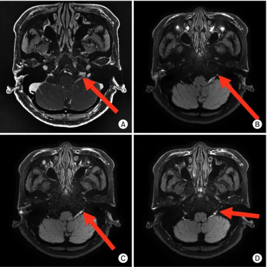
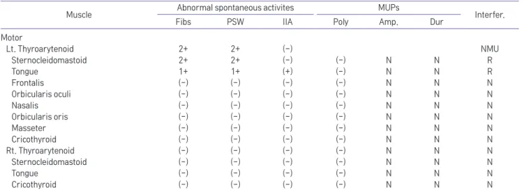
Speech and swallowing disorder is frequently observed in stroke, brain injury, tumors, and radiation therapy. A 65-year- old woman was admitted to otolaryngology department due to dysphagia and hoarseness. These symptoms occurred two weeks after upper respiratory infection (URI). She was diagnosed with inflammatory multiple lower cranial nerves by Videofluroscopic Swallowing Study (VFSS), MRI, and neurophysiologic examinations. Shaker’s exercise for dysphagia has been performed, and the dysphagia has improved after two months. However the hoarseness persisted, and this has been improved with thyroplasty. Dysphagia by unilateral inflammatory multiple lower cranial nerve palsy would be a rare complication of URI, and we experienced a case of multiple lower cranial neuropathy with successful management.

Key Words: lower cranial nerves, dysphagia

(2)

using electromyography of unilateral LCN palsy after

upper respiratory infection (URI) which could be easily neglected as a cause of these disorders.

Case Report

A 65-year-old woman was admitted to otolaryngol- ogy department due to dysphagia and hoarseness. Her medical history included hypertension, diabetes mel- litus, and rheumatoid arthritis without history of ge- netic disease or toxic exposure. Two weeks after URI, dysphagia and voice change were occurred. She was admitted because she complained of dysphagia.

On physical examination, muscle power and reflexes of all extremities were preserved, there were no sen- sory symptoms. Also there was no shingles on her skin.

The gag reflex was preserved, but vocal change and wet voice were noted after swallowing screening test.

Soft palate was bilaterally elevated (CN IX) and tongue deviation (CN XII) was not observed. Shoulder shrug- ging shows the difference between left and right (CN XI) and hoarseness (CN X) was noted.

In laboratory tests, CBC was normal but CRP (2.4 mg/dl) and ESR (52 mm/hr) were increased.

On laryngoscopy, 2 weeks after dysphagia, left vocal cord paralysis with intact airway patency and salivary pooling was shown, and there was no ulceration or ve- sicular lesion (Fig. 1).

Videofluoroscopic Swallowing Study (VFSS), 2 weeks

after dysphagia, revealed delayed aspiration due to residue in vallecular space and pyriform sinus with cough in 9 cc liquid while there was penetration with- out cough in 3 cc and 6 cc liquid. We also observed markedly decreased pharyngeal peristalsis and velo- pharyngeal insufficiency in pharyngeal phase (Fig. 1B).

We recommended Shaker’s exercise for dysphagia.

In cranial nerve Magnetic Resonance Image (MRI), performed at the same time as other evaluations, there were asymmetric mild thickening in cisternal segment of left CN X/XI, glossopharyngeal sulcus area and pars nervosa of jugular foramen on Fluid Attenuated In- version Recovery (FLAIR) and enhanced Fast Spoiled Gradient Echo (FSPGR) image and prominent venous plexus in root exit zone area of CN IX, X, XII. These findings were correlated with inflammatory condition such as neuritis (Fig. 2).

Both upper and lower extremities nerve conduc- tion studies (NCS) including F-waves were normal. In cranial NCS, although including facial, trigeminal, and blink reflex studies were within normal limits, acces- sory nerves were abnormal in left side (Table 1). The electromyography of left thyroarytenoid showed 2+

fibrillations and positive sharp waves with no motor units. The left sternocleidomastoid and tongue muscles (CN IX) showed abnormal spontaneous activities due to denervation. Unlike the left side, no specific physical findings were observed on the right side (Table 2).

We finally diagnosed this dysphagia caused by unilat-

A B C

Rt Lt

Fig. 1. (A) Laryngoscopy observed left vocal cord palsy with intact airway patency (there was no ulceration or vesicular lesion), and video-

fluoroscopic swallowing study (VFSS) showed residue in vallecular space, pyriform sinus in AP (B) and lateral view (C).

(3)

eral LCN palsy involving CN IX, X, XI, XII.

Two months after onset, there was a few residues in vallecular space and pyriform sinuses in VFSS, and in electromyography, a few motor units were noted in

the left thyroarytenoid and no abnormal findings were observed in the other muscles. As a result, she removed Levin-tube. However, because of voice change without improvement for 5 months onset, tyroplasty was per-

Table 1. Motor and Sensory Nerve Conduction Studies

Left side Right side

Lat. (ms) Amp. (mV) CV (m/s) Lat. (ms) Amp. (mV) CV (m/s)

Motor

Faical nerve (Nasialis) 4 1.5 4.0 1.3

Accessory 2.5* 2.9* 2.1 3.2

Median 3.1 5 59.3 3.2 5.1 58.5

Ulnar 2.6 6.7 59.7 2.5 5.8 62.2

Tibial 4.9 6.1 52.9

Peroneal (TA) 3.2 4.5

Sensory Lat. (ms) Amp. (mV) CV (m/s) Lat. (ms) Amp. (mV) CV (m/s)

Median 3.2 25.2 43.5 3.3 23.6 43.4

Ulnar 3.1 35.6 45.9 2.9 40.9 47.6

Peroneal 2.8 9.3 49.6

Sural 3.1 10.7 44.9

*: abnormal, Lat.: latency, Amp.: amplitude, CV: conduction velocity, TA: Tibialis anterior

A B

C D

Fig. 2. Asymmetric mild nerve thick-

ening in cisternal segment of left CN

X/XI, glossopharyngeal sulcus area

and pars nervosa of jugular foramen

on enhanced Fast Spoiled Gradient

Echo (FSPGR) (A) and FLAIR image

(B), (C), (D) and enhanced FSPGR im-

age and prominent venous plexus in

root exit zone area of CN IX,X,XII.

(4)

formed, and the hoarseness improved.

Discussion

Dysphagia and hoarseness are commonly caused by various diseases,

1

and cranial neuropathy is rarely seen in patients presenting with swallowing impairment.

3

The most common cause of multiple cranial neuropa- thy is tumor, followed by vascular disease, trauma and infections.

4

The cranial nerve associated with dysphagia is cranial nerve V, VII, IX, X, XII. It is a symptom seen in LCN palsy when appealing hoarseness and dysphagia like this. LCN injury could lead to dysphagia, coughing, speech disturbance, taste or autonomic function, neu- ralgia, swallowing disorder, head, pharyngeal or neck pain. In tumorous conditions, the complex anatomy of the vital structures at the skull base makes surgical resection or biopsy of lesions involving this area ex- tremely difficult.

5

LCN palsy has genetic, vascular, traumatic, iatrogenic, infectious, immunologic, metabolic, nutritional, degen- erative, or neoplastic casues.

2

According to LCN lesions various syndromes are also called Vernet-syndrome, Jugular-foramen-syndrome, Garcin-syndrome, and Vil- laret’s-syndrome associated with varicella-zoster virus.

2

The CN palsy by general infection is different from

this. The difference from Villaret’s syndrome, with a complete unilateral IX-XII cranial nerve palsy with Horner’s sign, is no involvement of cranial sympathetic nerve.

6

Our patient is distinguished from Ramsey- Hunt-syndrome because there is no facial nerve palsy, otic pain, and herpetic vesicles.

2

It is different from jugular foramen syndrome because there is no cranial, cervical pain and no herpetic eruption of skin. It is also one of the other features that is not enhanced around the jugular foramen.

3

It differs from Garcin’s syndrome because there is no proptosis.

7

No abnormality in up- per and lower NCV study and unilateral LCN palsy was also different from Guillain-Barré Syndrome.

8

Especially, dysphagia can lead to pneumonia, malnu- trition, dehydration, and increased mortality.

3

Since the initial diagnosis, this has slowed down and permanent impairment occurs, when symptoms occur, accurate diagnosis and evaluation.

9

In general, MRI, cerebro- spinal fluid (CSF) study, NCS/electromyography and VFSS are performed for diagnosis. Treatment is usually conservative measures such as shaker’s exercise for dysphagia

9

and anti-inflammatory, antiviral agents.

10

We diagnosed our patient CN IX (glossopharyngeal nerve), CN X (vagus nerve), CN XI (accessory nerve), and CN XII (hypoglossal nerve) palsy with physical ex- amination, VFSS, laryngoscopy, electromyography, and

Table 2. Initial Needle Electromyographic Examinations

Muscle Abnormal spontaneous activites MUPs

Interfer.

Fibs PSW IIA Poly Amp. Dur

Motor

Lt. Thyroarytenoid 2+ 2+ (-) NMU

Sternocleidomastoid 2+ 2+ (-) (-) N N R

Tongue 1+ 1+ (+) (-) N N R

Frontalis (-) (-) (-) (-) N N N

Orbicularis oculi (-) (-) (-) (-) N N N

Nasalis (-) (-) (-) (-) N N N

Orbicularis oris (-) (-) (-) (-) N N N

Masseter (-) (-) (-) (-) N N N

Cricothyroid (-) (-) (-) (-) N N N

Rt. Thyroarytenoid (-) (-) (-) (-) N N N

Sternocleidomastoid (-) (-) (-) (-) N N N

Tongue (-) (-) (-) (-) N N N

Cricothyroid (-) (-) (-) (-) N N N

MUPs: motor unit potentials, Interfer.: interference pattern, Fibs: fibrillation potentials, PSW: positive sharp waves, IIA: Increased insertion

activity, Poly: polyphasia, Dur: duration, N: normal, NMU: no motor unit, R: reduced recruitment

(5)

MRI for differential diagnosis.

In our case, we considered that intracranial edema due to inflammatory or immune process would be a cause of LCN.

3

The exact cause of viral etiology or au- toimmune attack cannot be revealed, the primary no- tice of LCN with clinical neurophysiologic examination followed by cranial nerve MRI has revealed the exact location and extent of disorders.

And the normal nerve conduction study with non- uniform denervation processes in cranial nerve in- nervated muscles would be associated with relatively mild cranial nerve injury except for the no motor unit observed in muscles innervated by recurrent laryngeal nerve. The limitation of this case is that there are no virology tests including CSF study. If these were per- formed, and antiviral agents would be applied accord- ing to virus etiologies, symptoms could be expected to improve earlier.

In conclusion, we have experienced LCN palsy due to intracranial lesion which might be associated with in- flammatory or immune attack. And this case dysphagia was successfully managed by conservative measures, and voice changes corrected with surgery.

References

1. Cook IJ: Diagnostic evaluation of dysphagia. Nat Clin Pract Gastroenterol Hepatol 2008: 5: 393-403

2. Finsterer J, Grisold W: Disorders of the lower cranial nerves.

J Neurosci Rural Pract 2015: 6: 377-391

3. Yeom J, Song YS, Lee WK, Oh BM, Han TR, Seo HG: Diagno- sis and clinical course of unexplained dysphagia. Ann Reha- bil Med 2016: 40: 95-101

4. Keane JR: Multiple cranial nerve palsies: Analysis of 979 cases. Arch Neurol 2005: 62: 1714-1717

5. Patmore H, Jebreel A, Uppal S, Raine CH, McWhinney P:

Skull base infection presenting with multiple lower cranial nerve palsies. Am J Otolaryngol 2010: 31: 376-380

6. Tiliket C, Petiot P, Arpin D, Mornex JF, Mornex F, Tournut P, et al: Clinical and radiological aspects of villaret’s syndrome.

Clin Neurol Neurosurg 1996: 98: 194-196

7. Alapatt JP, Premkumar S, Vasudevan RC: Garcin’s syndrome- a case report. Surg Neurol 2007: 67: 184-185

8. Mengi T, Secil Y, Incesu TK, Arici S, Akkiraz ZO, Gurgor N, et al: Guillain-barre syndrome and swallowing dysfunction.

J Clin Neurophysiol 2017: 34: 393-399

9. Langmore SE, Pisegna JM: Efficacy of exercises to rehabili- tate dysphagia: A critique of the literature. Int J Speech Lang Pathol 2015: 17: 222-229

10. Gilden D, Cohrs RJ, Mahalingam R, Nagel MA: Neurological

disease produced by varicella zoster virus reactivation with-

out rash. Curr Top Microbiol Immunol 2010: 342: 243-253

수치

Table 1.	Motor	and	Sensory	Nerve	Conduction	Studies
Table 2.	Initial	Needle	Electromyographic	Examinations

참조

관련 문서

Dependence of the chemical potentials of solid, liquid and gas phases on temperature at constant pressure.. The dashed lines are for

A goal can be broadened to include creative solutions by breaking it up into multiple specific goals and ranking the multiple goals according to their importance to you.

Space-time is the same in all the equal-size reactors of volume V i Let’s compare multiple MFR system with PFR system.. Volume of multiple mixed flow reactor system required

DNA-based testing includes pre- and postnatal genetic testing for the diagnosis of genetic diseases, carrier testing for genetic diseases, susceptibility genetic testing

It considers the energy use of the different components that are involved in the distribution and viewing of video content: data centres and content delivery networks

After first field tests, we expect electric passenger drones or eVTOL aircraft (short for electric vertical take-off and landing) to start providing commercial mobility

1 John Owen, Justification by Faith Alone, in The Works of John Owen, ed. John Bolt, trans. Scott Clark, "Do This and Live: Christ's Active Obedience as the

- distributes indexes across multiple computers and/or multiple sites - essential for fast query processing with large numbers of documents - many variations: