Can Suboccipital Release Followed by Cranio-Cervical Flexion Exercise Improve Shoulder Range of Motion, Pain, and Muscle Activity of Scapular
Upward Rotators in Subjects With Forward Head Posture?
Bo-been Kim1, BHSc, PT, Ji-hyun Lee1, MSc, PT, Hyo-jung Jeong2, MSc, PT, Heon-seock Cynn3,4, PhD, PT
1Dept. of Physical Therapy, The Graduate School, Yonsei University
2Dept. of Physical Therapy, College of Health Science, Sangji University
3Dept. of Physical Therapy, College of Health Science, Yonsei University
4Dept. of Ergonomic Therapy, The Graduate School of Health Science, Yonsei University
Abstract
Background: For the treatment of forward head posture (FHP) and forward shoulder posture, methods for strengthening scapular retractors and deep cervical flexors and stretching pectoralis and upper cervical extensors are generally used. No study has yet assessed whether suboccipital release (SR) followed by cranio-cervical flexion exercise (CCFE) (SR-CCFE) will result in a positive change in the shoulders and neck, showing a “downstream” effect.
Objects: The purpose of this study was to investigate the immediate effects of SR-CCFE on craniovertebral angle (CVA), shoulder abduction range of motion (ROM), shoulder pain, and muscle activities of upper trapezius (UT), lower trapezius (LT), and serratus anterior (SA) and LT/UT and SA/UT muscle activity ratios during maximal shoulder abduction in subjects with FHP.
Methods: In total, 19 subjects (7 males, 12 females) with FHP were recruited. The subject performed the fifth phase of CCFE immediately after receiving SR. CVA, shoulder abduction ROM, shoulder pain, muscle activities of UT, LT, and SA, and LT/UT and SA/UT muscle activity ratios during maximal shoulder abduction were measured immediately after SR-CCFE. A paired t-test and Wilcoxon signed-rank test were used to determine the significance of differences in scores between pre- and post-intervention in the same group.
Results: The CVA (p<.001) and shoulder abduction ROM (p<.001) were increased significantly post- versus pre-intervention. Shoulder pain was decreased significantly (p<.001), and LT (p<.05) and SA (p<.05) muscle activities were increased significantly post- versus pre-intervention. The LT/UT muscle activity ratio was increased significantly post- versus pre-intervention (p<.05). However, there was no significant change in UT muscle activity and SA/UT muscle activity ratio between pre- and post-intervention (p˃.05).
Conclusion: SR-CCFE was an effective intervention to improve FHP and induce downstream effect from the neck to the trunk and shoulders in subjects with FHP.
Key Words: Cranio-cervical flexion exercise; Forward head posture; Forward shoulder posture;
Suboccipital release.
Introduction
Forward head posture (FHP) is excessive anterior positioning of the head relative to a vertical reference line, coupled with increased lower cervical spine lor-
dosis (Harman et al, 2005). As cervical spine lordosis increases, the posture becomes characterized by a forward head, extended middle cervical spine, and flexed lower cervical spine (Harman et al, 2005).
FHP is related to lengthening and weakness in the Corresponding author: Heon-seock Cynn [email protected]
deep cervical flexor, lower trapezius (LT), and serratus anterior (SA), and tightness or shortening in the upper cervical extensors (Harman et al, 2005). Consequently, FHP leads to musculoskeletal dysfunction and neck and shoulder pain (Raine and Twomey, 1997).
An extensive literature has shown that the spine and shoulder complex have strong postural relation- ships and clinical correlations (Raine and Twomey, 1997; Roddey et al, 2002). FHP can change the scap- ular posture and decrease the mobility of scapular upward rotation, a common characteristic found in subjects with forward shoulder posture (FSP) (Lynch et al, 2010; Roddey et al, 2002; Yoo et al, 2008). FSP is related to LT weakness and shortening of the up- per trapezius (UT) (Peterson et al, 1997). The muscle imbalance and altered scapular mechanics decrease muscle activities in the LT and SA during arm ele- vation (Lynch et al, 2010).
For the treatment of FHP and FSP, methods for strengthening the scapular retractors and deep cer- vical flexors and stretching the pectoralis and upper cervical extensors are generally used (Roddey et al, 2002). In a previous study, activation of the deep cervical flexor was increased and muscle activity of the superficial cervical flexors was decreased with significant reduction in neck pain after cranio-cer- vical flexion exercises (CCFE) in subjects with chronic neck pain (Jull et al, 2009). Additionally, Rizo et al (2012) reported that suboccipital release (SR) immediately improved FHP in asymptomatic subjects, showing a significant increase in the craniovertebral angle (CVA) in both sitting and standing positions.
These previous studies showed a direct effect of the cervical approach to cervical problems (Jull et al, 2009; Rizo et al, 2012). However, interestingly, Lluch et al (2014) reported an indirect effect of an active scapular correction exercise on neck pain in subjects with chronic neck pain and scapular dysfunction. The results revealed an immediate reduction in neck pain and pressure-pain sensitivity.
Although there is evidence to suggest that improved FSP may lead to improved clinical outcomes in pa-
tients with neck pain (Lluch et al, 2014), little is known about how treatment for FHP may improve FSP. One previous study showed that greater thora- cic kyphosis was significantly associated with re- duced CVA (Quek et al, 2013). Furthermore, Quek et al (2013) suggested that addressing FHP improved cervical impairments and that addressing thoracic kyphosis impairments could constitute an “upstream”
approach. However, Quek et al (2013) failed to show that improving thoracic kyphosis resulted in im- proved FHP in subjects with neck dysfunction.
No study has yet assessed whether SR followed by CCFE (SR-CCFE) results in a positive change in shoulder as well as neck posture, representing a
“downstream” effect. Thus, this is the first reported study to investigate the immediate effects of SR-CCFE on (1) CVA, (2) shoulder abduction range of motion (ROM) and shoulder pain, and (3) muscle activities of the UT, LT, and SA and LT/UT and SA/UT muscle activity ratios during maximal shoulder ab- duction in subjects with FHP. We hypothesized that SR-CCFE would (1) increase CVA, (2) increase shoulder abduction ROM and decrease shoulder pain, and (3) increase LT and SA muscle activities and LT/UT and SA/UT muscle activity ratios, and de- crease UT muscle activity in subjects with FHP.
Methods
Subjects
A power analysis was performed using results from a pilot study with five subjects. A total sample size of 17 subjects was required to satisfy a significance level of .05, power of .80, and effect size of .64 (G-power software, ver. 3.1.2; Franz Faul, University of Kiel, Kiel, Germany). In total, 19 subjects (7 males, 12 females) with FHP were recruited [age=22.21±1.93 years, height=167.60±8.40 ㎝, weight=62.69±8.97 ㎏, body mass index=22.22±1.68 ㎏/㎡, FHP (pre-inter- ventions)=44.43±4.66°]. When assessed clinically, FHP is determined by the CVA, with a smaller CVA in-
dicating greater FHP (Quek et al, 2013). Inclusion criteria were (1) CVA<53° (Kim et al, 2015), (2) shoulder pain, and (3) limitation of abduction at the shoulder joint. The more painful side (greater visual analogue scale; greater VAS) was chosen to collect the data (18 right and 1 left side). Exclusion criteria were (1) medical/health care for low back pain over the past year (Harman et al, 2005), (2) dysfunction of the spine, (3) cervical, thoracic, or shoulder girdle fractures or anomalies (Peterson et al, 1997), and (4) obesity, as determined by body mass index>30 ㎏/㎡
(Hallman et al, 2011). Prior to the beginning of data collection, the experimental protocol was explained to all subjects by the principal investigator (PI), and partic- ipants signed an informed consent form approved by the Yonsei University Wonju Institutional Review Board (approval number: 1041849-201512-BM-085-02).
Surface electromyography recording and data processing
Electromyography (EMG) data were collected us- ing a Tele-Myo DTS EMG instrument with a wire- less telemetry system (Noraxon Inc., Scottsdale, AZ, USA) and analyzed using the Noraxon MyoResearch 1.06 software (Noraxon Inc., Scottsdale, AZ, USA).
Data were collected from the UT, LT, and SA on the subject’s tested side. The data were recorded at a 1000 ㎐ sampling rate. A digital band-pass filter (Lancosh FIR) was used between 20 and 450 ㎐ to filter the raw signals. A common mode rejection ra- tio was 92 ㏈ at 60 ㎐. Root-mean-square values with a moving window of 50 ㎳ were calculated.
The PI prepared the electrode sites to diminish impedance to the EMG signal by shaving the sub- jects’ hair, and cleaning the skin with rubbing alco- hol using a sterile gauze pad. Then, bipolar electrodes (Ag/AgCl) were attached in the direction of the mus- cle fibers, 2 ㎝ apart (Cram et al, 1998). Electrodes were placed as follows: UT: one half the distance between the mastoid process and the root of the scapular spine at approximately the angle of the neck and shoulder; LT: two finger widths medial
from the inferior angle of the scapula at a 45° angle toward the T10 spinous process; SA: below the axil- lary area, anterior to the latissimus dorsi, over the 4th through 6th ribs at an angle of 30° above the nipple line (Thigpen et al, 2010).
The maximal voluntary isometric contraction (MVIC) was used to normalize the EMG data of UT, LT, and SA. To collect MVIC data, we used stand- ard manual muscle-testing positions as follows. UT:
sitting with the neck extended posterolateral and 90°
abduction of the arm against resistance from should- er depression; LT: prone, 150° abduction of the arm, raising the arm upward against downward resistance applied at the elbow; SA: sitting, 90° flexion of the arm, protraction of the scapula against backward re- sistance applied to the hand (Kendall et al, 2005).
The mean value of two trials was used for data analysis, and the middle 3 seconds of each trial was used. Subjects performed the movement and held the position for 5 seconds, with a 3 minutes rest between muscle contractions. The intra-class correlation co- efficients (ICC) for the MVIC of UT, LT, and SA were .98 (95% confidence interval; 95% CI=.94∼.99), .98 (95% CI=.96∼.99), and .98 (95% CI=.95∼.99), respectively. EMG activity for the UT, LT, and SA during maximal arm abduction was expressed as a percentage of the mean MVIC (%MVIC).
CVA
The CVA was recorded with a digital camera (PL150, Samsung, Seoul, Korea), and the ImageJ image analysis software (U.S. National Institutes of Health, Maryland, USA) was used to assess the kinematic data. The dig- ital camera was placed perpendicular to the ground, with its lens 80 ㎝ from the lateral aspect of the subject and pointing directly at the subject’s shoulder to minimize parallax error (Yoo et al, 2008). The subject sat on the stool placed in the reference area, assuming a natural and relaxed position. The subject was asked to put both feet on the ground and to place the hands on the thighs while relaxing the back. Next, the PI instructed the subject to fix their
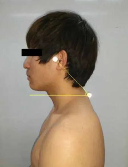
Figure 1. Measurement of craniovertebral angle (CVA).
gaze on the point marked on the wall directly ahead.
The PI attached two body markers on the external auditory meatus, and the spinous process of C7 (Lewis et al, 2005; Yoo et al, 2008). Two photographs were taken of the tested side of the subject’s upper body in the lateral aspect with a flash. After the first pho- tograph was made, the subject was asked to stand and then sit again. The CVA was determined as the angle between the line from the external auditory meatus to C7 and the line parallel to the spine at C7 (Lewis et al, 2005; Yip et al, 2008; Yoo et al, 2008) (Figure 1).
Shoulder abduction ROM
The shoulder abduction ROM was measured by two investigators using a universal goniometer (Baseline, Fabrication Enterprises, White Plains, New York, USA). While the PI measured the ROM, the other investigator prevented all compensatory movements. A reference line for standing was marked on the floor using tape. A plastic pole was set as a guide to maintain the frontal plane during maximal shoulder abduction. The measurement of shoulder abduction ROM was performed with the subject standing at the reference line. The subject held the extension of the elbow joint with the thumb directed
up toward the ceiling to create the external rotation necessary to prevent shoulder impingement (Kolber and Hanney, 2012). The subject was instructed to maximally abduct the shoulder in the frontal plane with chin tucked for stabilization and to prevent later- al flexion of the neck. Once active end-range was ac- complished, the angle of maximum shoulder abduction ROM was measured. The axis of a goniometer was set on the posterior part of the middle of the gleno- humeral joint, the fixed arm was parallel to the trunk, and the moveable arm was parallel to the longitudinal axis of the humerus (Kolber and Hanney, 2012).
Shoulder pain
Shoulder pain was measured using a VAS. The VAS entails a 10 ㎝ line and a scale completed by the subject. The subject was asked to check on the VAS line to identify the present intensity of shoulder pain on the tested side. The scores, which begin from the zero point indicating “no pain” are marked by the sub- ject using a ruler. The distance (㎜) indicates the pain score, which ranges from 0 to 100, providing 101 lev- els of pain intensity at intervals of 1 ㎜. The cut-off points for VAS scores were as follows: 0∼4 ㎜ (no pain), 5∼44 ㎜ (mild pain), 45∼74 ㎜ (moderate pain), and 75∼100 ㎜ (severe pain) (Hawker et al, 2011).
Procedure
Prior to measurement, the subject was asked to identify their painful side for the shoulder and to show their upper body by taking off their top if male or wearing a tank top if female. The subject completed a familiarization session for the CCFE at each phase of the exercise (up to the fifth phase) to ensure optimal performance capability. Then, the subject performed the fifth phase of CCFE immedi- ately after receiving the SR. All variables were measured twice by the PI before and after the SR-CCFE. The mean of the two measurements was used for statistical analysis. The other investigator was blinded to the experimental condition being test- ed during analysis of the variables.
CCFE
CCFE is a low-load exercise of the cranio-cervical flexors that involves contracting the deep cervical flex- ors of the upper cervical part (longus capitis and lon- gus colli) without recruitment of the superficial flexors (sternocleidomastoid and anterior scalene). The protocol for CCFE was established in the previous studies (Jull et al, 2002; Jull et al, 2009). In the first phase of the exercise, the PI taught the subject to perform con- trolled CCFE slowly in a supine position. The subject focused on sagittal rotation movement of the head slide in caudad and cephalad directions on the bed rather than a retraction movement. Once the first CCFE phase was achieved correctly, the subject performed the second phase using an air-filled pressure sensor (Stabilizer, Chattanooga Group Inc., Hixson, USA) placed between the back of the head and the bed. The subject was asked to perform progressive CCFE by increasing the amount of pressure, as shown by the feedback dial, thereby flattening cervical lordosis. Gradually, the sub- ject performed CCFE, increasing the pressure in 2 ㎜Hg increments, to reach the fifth phase, with a target pressure level of 20∼30 ㎜Hg. The PI confirmed that the subject could hold the target level consistently for 10 seconds without depending on retraction, dominant contraction of superficial neck flexor muscles, or a quick CCFE movement. Recruitment of the superficial muscles was monitored by the PI using palpation. At each tar- get level, the contraction time was 10 seconds with 10 repetitions; the subject was given a 3∼5 seconds rest period between contractions (Jull et al, 2009).
SR
SR is a suboccipital muscle inhibition technique that influences the craniocervical region and reduces ten- sion in the deep upper cervical tissues. First, the PI sat at the head of the bed and placed the palms un- der the head of the subject, who was in a supine position. Next, the PI placed the middle and ring fin- gers of both hands under the space between the oc- cipital condyles and the spinal process of C2. Then, the PI rested the base of the skull with 90° flexion of
the metacarpophalangeal joints and maintained the technique until the PI perceived relaxation of subject’s suboccipital muscle. It was important that pressure was applied ventrally without pain, using the ex- tended index, middle, and ring fingers of both hands. A slight traction force was allowed cranially to focus on the suboccipital zone. After tissue relaxation was ach- ieved, the pressure was smoothly released, leaving the subject’s head on the bed. During the SR, the subject was asked to keep their eyes closed to prevent eye movements that might influence suboccipital muscle tone.
The intervention time was 4 minutes (Rizo et al, 2012).
Statistical analysis
The test-retest reliability of EMG data was as- sessed by calculating the ICC and 95% CI, which was interpreted based on the following criteria:
<.69=poor, .70∼.79=moderate, .80∼.89=good, and .90
∼.99=excellent (T’Jonck et al, 1996). A one-sample Kolmogorov-Smirnov test was conducted to confirm the assumption of a normal distribution. Paired t-tests were conducted to assess the significance of differ- ences in the CVA, shoulder abduction ROM, muscle activities of the UT, LT, and SA, and LT/UT and SA/UT muscle activity ratios during maximal should- er abduction in subjects with FHP between pre- and post-intervention. A Wilcoxon signed-rank test was used to assess the significance of differences in shoulder pain between pre- and post-intervention. The level of significance was set at .05. The effect size (ES) is generally suggested to identify meaningful changes by accounting for group variability. The ES was cal- culated for the difference between measures pre- and post-intervention (Portney and Watkins, 2009). Data are presented as mean±standard deviation. All statistical analyses were performed using the SPSS ver. 21.0 (SPSS Inc., Chicago, IL, USA).
Results
The test-retest reliabilities of EMG data in max-
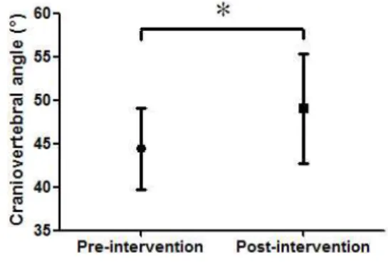
Figure 2. Craniovertebral angle (CVA) was increased significantly post-intervention versus pre-intervention (*p<.001).
Figure 4. The mean visual analog scale (VAS) score for shoulder pain was decreased significantly post-intervention versus pre-intervention (*p<.001).
Figure 3. Shoulder abduction range of motion (ROM) was increased significantly post-intervention versus pre-intervention (*p<.001).
imal shoulder abduction were excellent, except for UT post-intervention (good): UT [pre-intervention:
.95 (.86∼.98), post-intervention: .88 (.72∼.95)]; LT [pre-intervention: .96 (.91∼.99), post-intervention: .97 (.92∼.99)], and SA [pre-intervention: .91 (.79∼.97), post-intervention: .97 (.92∼.99)].
CVA
CVA was increased significantly, from 44.43±4.66°
pre-intervention to 49.03±6.32° post-intervention (p<.001, ES=1.21) (Figure 2).
Shoulder abduction ROM and shoulder pain Shoulder abduction ROM was increased significantly, from 126.03±5.70° pre-intervention to 133.87±7.64°
post-intervention (p<.001, ES=1.70) (Figure 3). The VAS score for shoulder pain was decreased significantly from 13.68±9.55 ㎜ pre-intervention to 1.58±6.88 ㎜ post-intervention (p<.001, ES=1.48) (Figure 4).
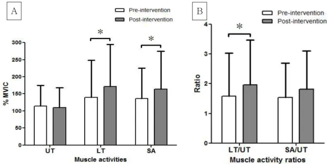
Muscle activities and muscle activity ratios LT and SA muscle activities were increased sig- nificantly, from 139.73±108.94 and 135.79±89.62 pre-in- tervention to 170.92±123.43 and 163.27±111.78 post-in- tervention (p<.05, ES=.78 and p<.05, ES=.75), respectively.
However, there was no significant change in UT muscle activity between pre- and post-intervention (p>.05, ES=.11). The LT/UT muscle activity ratio was increased significantly, from 1.58±1.45 pre-inter-
vention to 1.96±1.51 post-intervention (p<.05, ES=.88).
However, there was no significant change in the SA/UT muscle activity ratio between pre- and post-intervention (p>.05, ES=.43) (Figure 5).
Discussion
The purpose of this study was to investigate the effects of SR-CCFE on (1) CVA, (2) shoulder ab- duction ROM and shoulder pain, and (3) muscle ac- tivities of the UT, LT, and SA and LT/UT and SA/UT muscle activity ratios during maximal should- er abduction in subjects with FHP. The results of the kinematic data partially supported our research hypothesis. Comparisons of pre- and post-inter- vention measures showed that the CVA and shoulder
A B
Figure 5. Muscle activities during maximal shoulder abduction of pre- and post- intervention (A), muscle activity ratios during maximal shoulder abduction of pre- and post- intervention (B) (MVIC: maximal voluntary isometric contraction, UT: upper trapezius, LT: lower trapezius, SA:
serratus anterior, *p<.05).
abduction ROM were increased significantly and shoulder pain was decreased significantly post-inter- vention compared with pre-intervention. Additionally, LT and SA muscle activities during maximal shoulder abduction were increased significantly post-intervention compared with pre-intervention.
Also, the LT/UT muscle activity ratio during max- imal shoulder abduction was increased significantly post-intervention compared with pre-intervention.
The CVA increased significantly, by 10.35%, post-intervention versus pre-intervention. The CVA indicates the degree of FHP, and an increased CVA means improved FHP. This finding is similar to those reported by Rizo et al (2012) and Falla et al (2007). The SR treatment significantly increased CVA in both the sitting (about 14%) and the stand- ing (about 7%) positions in subjects with a history of orthodontics use (Rizo et al, 2012). Additionally, CCFE was associated with a significant reduction in the increase of CVA during a computer task in sub- jects with neck pain (Falla et al, 2007). Furthermore, Falla et al (2007) suggested that CCFE improved the ability to maintain a neutral cervical posture during
prolonged sitting. In this study, we used SR-CCFE as an intervention in FHP, and our results indicate that SR-CCFE was effective in reducing FHP through increasing CVA immediately.
The shoulder abduction ROM was increased significantly, by 6.22%, post-intervention versus pre-intervention.
We expected that if SR-CCFE improved FHP, then trunk and shoulder dysfunction would also be im- proved along with the positive change in the neck.
The increase in shoulder abduction ROM could be considered improvement of thoracic hyperkyphosis and FSP. The mechanism of these changes in the shoulder is likely craniocervicothoracic stabilization resulting from SR-CCFE. SR-CCFE may activate the deep segmental muscles of the craniocervicothoracic spine, building a stable base during shoulder movement.
A previous study reported that postural changes in- tegrated scapular orientation with spinal posture cor- rection, especially in patients who have FHP with FSP and thoracic hyperkyphosis (Cools et al, 2014).
Kebaetse et al (1999) found that a slumped posture was associated with reduced active shoulder abduc- tion, by about 23.6°, versus an upright posture. In
addition, Lewis and Valentine (2010) reported that thoracic hyperkyphosis reduced shoulder abduction ROM. Along with the evidences from the previous studies showing that CCFE improved the ability to maintain an upright trunk posture in subjects with neck pain (Falla et al, 2007), the increase in shoulder abduction ROM in this study reflects a downstream effect of SR-CCFE. Thus, SR-CCFE may be pre- ferred to enhance shoulder abduction ROM.
Shoulder pain was decreased significantly, by 88%, post-intervention versus pre-intervention. In a pre- vious study, SR decreased the tightness and hyper- activation of deep neck extensors resulting from FHP (Wang et al, 2003). The mechanism behind this change is believed that the SR technique could help to reduce central sensitization through relaxation of upper cervical tissues (Rizo et al, 2012). CCFE also significantly decreased the average intensity of neck pain (numerical rating scale) (Falla et al, 2007; Jull et al, 2009). Heintz and Hegedus (2008) matched in- tervention to an individual’s signs and symptoms in a single subject with neck pain, FHP, and FSP. The intervention, which included SR and deep neck flexor strengthening, obtained positive results with pain reduction. In this study, the resultant decrease in shoulder pain was considered that SR-CCFE lead to a downstream effect from neck to shoulders.
LT and SA muscle activities during maximal shoulder abduction were significantly greater, by 22.32% and 20.24%, respectively, post-intervention versus pre-intervention. The UT, LT, and SA per- form important roles in upward rotation of the scap- ula during shoulder abduction (Selkowitz et al, 2007).
When the subject performs an overhead activity, the UT and SA muscles work together for upward rota- tion of the scapula (Decker et al, 1999; Ekstrom et al, 2003). In addition, LT and SA serve as upward rotators of the scapula because of their origins and insertions (Kelly and Thomas, 2011). However, vari- ous conditions, such as postural changes, influence the scapular position and scapulohumeral rhythm (Ekstrom et al, 2003; Kebaetse et al, 1999). Shoulder
derangements may inhibit movement of the LT and SA while activating the UT. Athletes with scapular dyskinesis often have dominance of the UT and per- form a shoulder shrug motion during retraction, which is counterproductive (Kelly and Thomas, 2011). In this study, the LT/UT muscle activity ratio during maximal shoulder abduction was increased significantly, by 24.05%, post-intervention versus pre-intervention. The reduction in UT dominance relative to LT indicated improvement in the shoulder muscle imbalance during overhead activity. No pre- vious study has examined changes in scapular up- ward rotation activities during maximal shoulder ab- duction after SR-CCFE; thus, it is not possible to compare our results with previous work. Our results suggest that SR-CCFE could improve shoulder de- rangements via a downstream effect.
This study has some limitations. First, because it was performed to investigate the immediate effects of SR-CCFE using a cross-sectional design, the long-term effects of the intervention could not be determined. Second, the findings of this study have limited generalizability to other patient populations with shoulder and trunk pathologies because only subjects who had FHP with shoulder pain and limi- tation of abduction were recruited. Future studies should investigate the long-term effects of SR-CCFE in subjects with other medical histories, such as thoracic hyperkyphosis, FSP, winged scapula, or shoulder impingement syndrome. Third, we did not consider or exclude rhomboid muscle activity, which has a role in scapular downward rotation and could be variable among subjects. Further studies are needed to exclude subjects with a dominant rhomboid. Fourth, because we used combination of SR and CCFE as an intervention, it was uncertain that the effects resulted from whether combined in- tervention or not. Finally, because our study was not a randomized controlled trial, the results could be in- fluenced by other unrecognized factors. As a result, accurate evaluation of SR-CCFE from a large randomized controlled trial is needed.
Conclusion
SR-CCFE can increase the CVA and shoulder ab- duction ROM and decrease shoulder pain. The present findings suggest that SR-CCFE is an effective inter- vention to improve FHP and bring about downstream effect from the neck to the trunk and shoulders in subjects with FHP. Based on our EMG data, SR-CCFE can be effective in activating the LT and SA muscles during maximal shoulder abduction. In addition, if a subject has a relatively dominant UT among the scap- ular upward rotators, SR-CCFE can be recommended to elicit LT rather than UT muscle activity.
References
Cools AM, Struyf F, De Mey K, et al. Rehabilitation of scapular dyskinesis: From the office worker to the elite overhead athlete. Br J Sports Med. 2014;
48(8):692-697. http://dx.doi.org/10.1136/bjsports-2013- 092148
Cram JR, Kasman GS, Holtz J. Introduction to Surface Electromyography. 1st ed. Gaithersburg, MD, Aspen Publishers, 1998:65-74.
Decker MJ, Hintermeister RA, Faber KJ, et al.
Serratus anterior muscle activity during selected rehabilitation exercises. Am J Sports Med. 1999;
27(6):784-791.
Ekstrom RA, Donatelli RA, Soderberg GL. Surface electromyographic analysis of exercises for the trapezius and serratus anterior muscles. J Orthop Sports Phys Ther. 2003;33(5):247-258.
Falla D, Jull G, Russell T, et al. Effect of neck ex- ercise on sitting posture in patients with chronic neck pain. Phys Ther. 2007;87(4):408-417.
Hallman DM, Olsson EM, von Schéele B, et al. Effects of heart rate variability biofeedback in subjects with stress-related chronic neck pain: A pilot study. Appl Psychophysiol Biofeedback. 2011;36(2):
71-80. http://dx.doi.org/10.1007/s10484-011-9147-0 Harman K, Hubley-Kozey CL, Butler H. Effectiveness
of an exercise program to improve forward head posture in normal adults: A randomized, con- trolled 10-week trial. J Man Manip Ther. 2005;
13(3):163-176.
Hawker GA, Mian S, Kendzerska T, et al. Measures of adult pain: Visual Analog Scale for Pain (VAS Pain), Numeric Rating Scale for Pain (NRS Pain), McGill Pain Questionnaire (MPQ), Short-Form McGill Pain Questionnaire (SF-MPQ), Chronic Pain Grade Scale (CPGS), Short Form-36 Bodily Pain Scale (SF-36 BPS), and Measure of Intermittent and Constant Osteoarthritis Pain (ICOAP). Arthritis Care Res (Hoboken). 2011;63(Suppl 11):S240-S252.
http://dx.doi.org/10.1002/acr.20543
Heintz MM, Hegedus EJ. Multimodal management of mechanical neck pain using a treatment based classification system. J Man Manip Ther. 2008;
16(4):217-224.
Heredia Rizo AM, Pascual-Vaca ÁO, Cabello MA, et al.
Immediate effects of the suboccipital muscle in- hibition technique in craniocervical posture and greater occipital nerve mechanosensitivity in sub- jects with a history of orthodontia use: A random- ized trial. J Manipulative Physiol Ther. 2012;35(6):
446-453. http://dx.doi.org/10.1016/j.jmpt.2012.06.006 Jull GA, Falla D, Vicenzino B, et al. The effect of therapeutic exercise on activation of the deep cer- vical flexor muscles in people with chronic neck pain. Man Ther. 2009;14(6):696-701. http://dx.doi.org/
10.1016/j.math.2009.05.004
Jull G, Trott P, Potter H, et al. A randomized con- trolled trial of exercise and manipulative therapy for cervicogenic headache. Spine (Phila Pa 1976).
2002;27(17):1835-1843.
Kebaetse M, McClure P, Pratt NA. Thoracic position effect on shoulder range of motion, strength, and three-dimensional scapular kinematics. Arch Phys Med Rehabil. 1999;80(8):945-950.
Kelly JD, Thomas SJ. Identifying and managing scap- ular problems in overhead athletes. J Musculoskel Med. 2011;28:465-471.
Kendall F, McCreary E, Provance P, et al. Muscles:
This article was received January 27, 2016, was reviewed January 27, 2016, and was accepted March 9, 2016.
Testing and function with posture and pain. 5th ed. Baltimore, Williams & Wilkins, 2005:330-337.
Kim KH, Kim SG, Hwangbo G. The effects of horse-riding simulator exercise and Kendall exercise on the forward head posture. J Phys Ther Sci. 2015;
27(4):1125-1127. http://dx.doi.org/10.1589/jpts.27.1125 Kolber MJ, Hanney WJ. The reliability and con-
current validity of shoulder mobility measure- ments using a digital inclinometer and goni- ometer: A technical report. Int J Sports Phys Ther. 2012;7(3):306-313.
Lewis JS, Valentine RE. Clinical measurement of the thoracic kyphosis. A study of the intra-rater re- liability in subjects with and without shoulder pain. BMC Musculoskelet Disord. 2010;11:39.
http://dx.doi.org/10.1186/1471-2474-11-39
Lewis JS, Wright C, Green A. Subacromial impinge- ment syndrome: The effect of changing posture on shoulder range of movement. J Orthop Sports Phys Ther. 2005;35(2):72-87.
Lluch E, Arguisuelas MD, Calvente Quesada O, et al.
Immediate Effects of Active Versus Passive Scapular Correction on Pain and Pressure Pain Threshold in Patients With Chronic Neck Pain.
J Manipulative Physiol Ther. 2014;37(9):660-666.
http://dx.doi.org/10.1016/j.jmpt.2014.08.007
Lynch SS, Thigpen CA, Mihalik JP, et al. The effects of an exercise intervention on forward head and rounded shoulder postures in elite swimmers. Br J Sports Med. 2010;44(5):376-381. http://dx.doi.org/
10.1136/bjsm.2009.066837
Peterson DE, Blankenship KR, Robb JB, et al.
Investigation of the validity and reliability of four objective techniques for measuring forward shoulder posture. J Orthop Sports Phys Ther.
1997;25(1):34-42.
Portney L, Watkins M. Foundations of Clinical Research:
Applications to practice. 3rd ed. New Jersey, Pearson Prentice Hall, 2009:373-374, 423, 648-649.
Quek J, Pua YH, Clark RA, et al. Effects of thoracic kyphosis and forward head posture on cervical range of motion in older adults. Man Ther. 2013;
18(1):65-71. http://dx.doi.org/10.1016/j.math.2012.07.005 Raine S, Twomey LT. Head and shoulder posture
variations in 160 asymptomatic women and men.
Arch Phys Med Rehabil. 1997;78(11):1215-1223.
Roddey TS, Olson SL, Grant SE. The effect of pec- toralis muscle stretching on the resting position of the scapula in persons with varying degrees of forward head/rounded shoulder posture. J Man Manip Ther. 2002;10(3):124-128.
Selkowitz DM, Chaney C, Stuckey SJ, et al. The ef- fects of scapular taping on the surface electro- myographic signal amplitude of shoulder girdle muscles during upper extremity elevation in in- dividuals with suspected shoulder impingement syndrome. J Orthop Sports Phys Ther. 2007;37(11):
694-702.
Thigpen CA, Padua DA, Michener LA, et al. Head and shoulder posture affect scapular mechanics and muscle activity in overhead tasks. J Electromyogr Kinesiol. 2010;20(4):701-709. http://dx.doi.org/10.1016/
j.jelekin.2009.12.003
T’Jonck L, Lysens R, Grasse G. Measurements of scapular position and rotation: A reliability study. Physiother Res Int. 1996;1(3):148-158.
Wang WT, Olson SL, Campbell AH, et al.
Effectiveness of physical therapy for patients with neck pain: An individualized approach us- ing a clinical decision-making algorithm. Am J Phys Med Rehabil. 2003;82(3):203-218.
Yip CH, Chiu TT, Poon AT. The relationship be- tween head posture and severity and disability of patients with neck pain. Man Ther. 2008;
13(2):148-154.
Yoo WG, Yi CH, Cho SH, et al. Effects of the height of ball-backrest on head and shoulder posture and trunk muscle activity in VDT workers. Ind Health. 2008;46(3):289-297.