Introduction
Shoulder pain is the most common musculoskeletal symptom encountered in the primary clinical care set- ting, which represents a significant health problem as- sociated with functional impairments (Walther et al, 2004). The prevalence of shoulder pain throughout the whole lifetime is estimated to be approximately 35%
(Guerra de Hoyos et al, 2004). Shoulder problems were
believed to be connected with abnormal muscle ten- sion, spasms, and inflammation in the shoulder region like the rotator cuff syndrome as well as associated joints such as glenohumeral, scapulothoracic, sternocla- vicular and acromioclavicular (Ratcliffe et al, 2014).
A variety of shoulder stabilization exercise includ- ing glenohumeral stabilization exercise (GSE) and scapular stabilization exercise (SSE) are used, al- though outcome studies have failed to provide clinical Corresponding author: Seung-chul Chon [email protected]
Comparison of Glenohumeral Stabilization Exercise and Scapular Stabilization Exercise on Upper Extremity Stability, Alignment,
Pain, Muscle Power and Range of Motion in Patients With Nonspecific Shoulder Pain
Na-young Jeon1, MSc, PT, Seung-chul Chon2, PhD, PT
1Dept. of Rehabilitation Medicine, Konyang University Hospital
2Dept. of Physical Therapy, College of Medical Science, Konyang University
Abstract
1)Background: Shoulder stabilization commonly involves two components: the glenohumeral stabilization exercise (GSE) and scapular stabilization exercise (SSE). Despite the fact that the shoulder stabilization has advantageous merit, to our knowledge, only a few studies have compared the superior of the GSE and the SSE.
Objects: The purpose of this study was to assess the effects of GSE in patients with nonspecific shoulder pain.
Methods: Thirty subjects with nonspecific shoulder pain were randomly divided into an experimental group and control group, each with 15 patients. The experimental group used an GSE, whereas the control group did SSE. All subjects were measured in shoulder stability, scapular symmetric alignment, pain, muscle power, and range of motion before and after the intervention.
Results: GSE resulted in significantly better shoulder stability (p=.046, from 8.67±7.54 score to 13.93±9.40) in the experimental group compared with SSE in the control group. However, no significant effects were observed for scapular symmetric alignment including the angles of inferior scapular distance (p=.829) and inferior scapular height difference (p=.735), pain (p=.113), muscle power including shoulder flexion (p=.723) and abduction (p=.897) and range of motion including shoulder flexion (p=.853) and abduction (p=.472).
Conclusion: These findings suggest that GSE may be more effective in increasing the shoulder stability than the SSE in patients with nonspecific shoulder pain, probably through a centralization effect on the shoulder mechanism.
Key Words : Glenohumeral stabilization; Scapular stabilization; Shoulder pain; Shoulder stabilization;
Upper extremity.
evidence for the superiority of any particular technique. In addition, despite the fact that all of these stabilization exercises have been used in the management of individuals with shoulder pain, it is difficult to reach a clinical decision in adopting any one of them because their therapeutic efficacy has yet to be demonstrated.
Many clinical trials investigating the different ef- fects of management on shoulder problems including anti-inflammatory drugs, injection, laser therapy, ul- trasound therapy, electric stimulation and therapeutic exercise have been widely used, but showed variable results (Sangwan et al, 2015; Voight and Thomson, 2000). For example, previous research examining the therapeutic effect of painless range of motion, strength and endurance showed enhanced returning to normal daily living activities, whereas other stud- ies demonstrated little change in these outcome measures (Camargo et al, 2009; Faber et al, 2006).
Furthermore, the underlying mechanism of ther- apeutic effect remains unclear because of a lack of quantitative measurement.
Recently, there has been growing interest in the application of shoulder stabilization techniques in the rehabilitation field for reducing pain and improving function in patients with shoulder pain. Shoulder sta- bility is defined as the condition where in the hum- eral head remains in place or promptly returns to proper alignment within the glenoid fossa. Shoulder stabilization involves two components such as gleno- humeral joint and scapula. With regard to GSE, the humeral head remains in the same location and promptly returns to the proper position within the glenoid fossa via a coupling force (Myers et al, 2006). Glenohumeral instability may result in dys- function or pain due to repetitive microtrauma.
Furthermore, this instability can contribute to the development of secondary rotator cuff disease or im- pingement (Sangwan et al, 2015), which has an ad- verse effect on shoulder’s neuromuscular performance, resulting in subsequent shoulder injury (Voight and Thomson, 2000).
SSE focuses on balancing the trapezius, rhomboid, and serratus anterior muscles (Ludewig and Reynolds, 2009). Biomechanically, the scapular provides a stable base while the rotator cuff muscles provide dynamic force in order to maintain the humeral head in the glenoid fossa as glenohumeral movement occurs.
Consequently, these occurs arise the dynamic stability by the scapulothoracic joint and associated with sur- rounding musculature (Ludewig and Reynolds, 2009).
Therefore, the role of the scapular should be managed in the upper extremity, integrated scapular stabiliza- tion in order to maintain the scapular in the proper position with the length-tension relationships of the surrounding musculature (Grant et al, 2004).
To quantitatively investigate the effect of GSE on the scapular symmetric alignment using three dimen- sional spinal diagnostic imaging, this study focused on muscle power using hand-held dynamometer, range of motion using inclinometer and associated clinical outcome measures including shoulder stability and pain in patients with nonspecific shoulder pain.
Our basic premise was that our GSE would sig- nificantly reduce the pain in patients with nonspecific shoulder pain, thereby improving shoulder stability, scapular symmetric alignment, muscle power and range of motion during daily activities.
Methods
Subjects
This study recruited 35 patients with nonspecific shoulder pain who visited a local hospital. Subjects were excluded if they had been advised by their physician to abstain from exercise, had chest pain, dizziness and hypertension. Five subjects were ex- cluded because they declined to take part in this study. A total of 30 patients with nonspecific shoulder pain were recruited, and then they were randomly allocated into the experimental (n1=15) or control group (n2=15). Randomization was performed using sealed envelopes. A piece of paper in the
sealed envelope was given to the subjects for group allocation. Allocation was conducted before the initial assessment. All the procedures were explained to the subjects, and each subject signed an informed con- sent form. General characteristics of the subjects are presented (Table 1).
Instruments
Upper extremity stability
We used the closed kinetic chain upper extremity stability test (Tucci et al, 2014). The subjects as- sumed a push-up position with each hand placed on a piece of tape on the floor while keeping the body as straight and as parallel to the ground as possible.
The subjects’ shoulders were positioned directly over his/her hands. When the examiner said “go”, the subjects removed only one hand from the floor, touched the opposite line, and then returned the same hand to its original position on the line. A single test was comprised of continuing this alternating activity for 15 seconds. The test was performed twice for each subject in random order with a resting time of 1 minute. The subjects attempted as many touches as possible in the allotted time. The data of the two tests were averaged and recorded as the test scores.
The closed kinetic chain upper extremity stability test has a reliability of .93 and a validity of .64 (Negrete et al, 2010; Tucci et al, 2014).
Three-dimensional spinal diagnostic imaging
Scapular symmetric alignment was determined us- ing a three dimensional spinal diagnostic imaging system (Backmapper, ABW, Frickenhausen, Germany).
This equipment precisely measured the form, location, and distortion of the spine structure from the for- ward, backward, upward, and downward directions.
This device was used to analyze the position such as left and right heights of the scapula, which also ana- lyzed the distribution of the musculoskeletal structure.
Markers were attached on a level with the spinous process of the seventh cervical vertebra, both scap- ular inferior angle, both posterior superior iliac spine and sacrum. This three dimensional spinal diagnostic imaging system has a reliability of .93 and a validity of .64 (Golpayegani et al, 2013).
Pain intensity
Pain intensity was measured using a numeric rat- ing scale (NRS). The NRS is a clinically standard instrument used to assess in patients with chronic pain. The NRS involved asking the patients to rate their pain from 0 (best) to 10 (worst), with 0 repre- senting one end of the pain intensity. It has a reli- ability of .95 and a validity ranging from .86 to .95 (Hawker et al, 2011).
Muscle power
The strength of the shoulder girdle muscles was evaluated using a hand-held dynamometer (Microfet2, Hoggan Health Industries, West Jordan, USA).
Isometric strength was assessed by maintaining at shoulder 90° abduction and flexion in a sitting position. Each muscle test was performed three times with a resting time of 30 seconds between each test, and the three tests were averaged. The hand-held dynamometer has a high intra-reliability and coefficient correlation of .78 for the validity (Jubany et al, 2015).
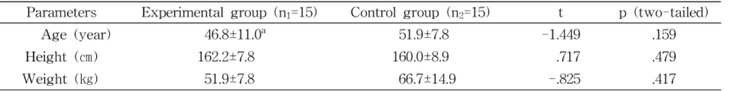
Parameters Experimental group (n1=15) Control group (n2=15) t p (two-tailed)
Age (year) 46.8±11.0a 51.9±7.8 -1.449 .159
Height (㎝) 162.2±7.8 160.0±8.9 .717 .479
Weight (㎏) 51.9±7.8 66.7±14.9 -.825 .417
amean±standard deviation.
Table 1. General characteristics of subjects (N=30)
Range of motion (ROM)
The ROM was actively measured using an elec- tronic inclinometer (Dualer IQ the smarter in- clinometer, JTECH medical, Salt lake, USA) during shoulder flexion and abduction in sitting positions.
The ROM test was performed three times consec- utively without pain and the average of the tests was calculated. This device has a reliability of .95 and a validity of .85 (Kolber and Hanney, 2012).
Procedures
The subjects were engaged in either the ex- perimental group (conventional physical therapy for 20 minutes followed by the GSE for 30 minutes) or con- trol group (conventional physical therapy for 20 mi- nutes followed by the SSE for 30 minutes). All sub- jects received conventional physical therapy for 20 mi- nutes regardless of treatment allocation. Two physi- otherapists were instructed in this study by the inves- tigator (a physiotherapist with 20 years’ experience).
The GSE and SSE were administered one-on-one by one of the trained physiotherapists. The outcome as- sessments were measured before and after interventions. The physiotherapists undertaking the outcome assessment were blinded to group allocation.
Glenohumeral stabilization exercise The GSE is a form of centralization of the gleno- humeral joint. All movements consisted of 3 sets of 15 repetitions in four different directions such as right, left, anterior, and posterior in the glenohumeral
joint with keeping the stability. The GSE involved 5 stages: (1) education how to maintain the humeral head in a neutral position in the glenohumeral fossa (2) grasping on the bar-stool with the shoulder 90°
flexion and 90° abduction (3) placed subject’s hand on the cushion ball in a sitting position (4) placed subject’s hands on the cushion ball with should- er-width apart at prone position on the bed (5) hold- ing a .5 ㎏ weight in sitting position (Jaggi and Lambert, 2010; Moghadam et al, 2011; Sangwan et al, 2015) (Figure 1).
Scapular stabilization exercise
All movements consisted of 3 sets of 15 repeti- tions at a feeling without any pain or fatigue. The SSE consisted of 5 stages: (1) education for exact movements of the scapular in 4 different directions (2) shoulder external and internal rotation with the elbow 90° flexion in sidelying position (3) shoulder horizontal abduction at the prone position (4) should- er 125° abduction combined with scapular protraction (5) placed subject’s hands on the ground with shoulder-width apart at prone position on the bed, and then extended subject’s elbows to push-up posi- tion for the protracting the scapular (De Mey et al, 2009; Hess et al, 2005) (Figure 2).
Statistical analysis
The data were analyzed using SPSS ver. 18.0 (SPSS Inc., Chicago, IL, USA). All variables were tested using the Kolmogorov-Smirnov test, which
A B C D E
Figure 1. Glenohumeral stabilization exercise (A: education how to maintain the humeral head, B:
grasping on the bar-stool, C: cushion ball in a sitting position, D: prone position on the bed, E:
holding a weight in sitting position).
showed a normal distribution of the data. The in- dependent t-test was used to determine significant differences in the general characteristics of the sub- jects between the groups. Two-way analysis of var- iance (ANOVA) with repeated measures was used to assess the main effects (group and time effects) and interaction effects of upper extremity stability, scap- ular symmetric alignment, pain, muscle power, and ROM between the groups. The significance level was set at a p value of <.05.
Results
Two-way ANOVA with repeated measures showed
a significant group×time interaction effect for upper extremity stability (F=4.370, p=.046), suggesting that the GSE showed remarkable improvement compared with the SSE. Group effect was not significant ob- served for upper extremity stability (F=.345, p=.562).
Time effect was significant observed for upper ex- tremity stability (F=67.763, p<.001) (Table 2).
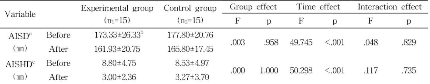
However, no significant group×time interaction effect was observed for angle of inferior scapular distance (F=.048, p=.829), angle of inferior scapular height dif- ference (F=.117, p=.735). Group effect was not sig- nificant observed for angle of inferior scapular distance (F=.003, p=.958), angle of inferior scapular height dif- ference (F=.000, p=1.000) (Table 3). Time effect was significant observed for angle of inferior scapular dis-
A B C D E
Figure 2. Scapular stabilization exercise (A: education for movements of the scapular, B: shoulder external and internal rotation, C: shoulder horizontal abduction, D: shoulder abduction combined with scapular protraction, E: push-up position for the protracting the scapular).
Variable Experimental group (n1=15)
Control group (n2=15)
Group effect Time effect Interaction effect
F p F p F p
UESa (score)
Before 8.67±7.54b 8.13±5.51
.345 .562 67.763 <.001 4.370 .046
After 13.93±9.40 11.27±7.41
aupper extremity stability,bmean±standard deviation.
Table 2. The upper extremity stability before and after treatment in the experimental and control group
Variable Experimental group (n1=15)
Control group (n2=15)
Group effect Time effect Interaction effect
F p F p F p
AISDa (㎜)
Before 173.33±26.33b 177.80±20.76
.003 .958 49.745 <.001 .048 .829 After 161.93±20.75 165.80±17.45
AISHDc (㎜)
Before 8.80±4.75 8.53±4.97
.000 1.000 50.298 <.001 .117 .735
After 3.00±2.36 3.27±3.70
aangle of inferior scapular distance, bmean±standard deviation, cangle of inferior scapular height difference.
Table 3. The scapular symmetric alignment before and after treatment in the experimental and control group
tance (F=49.745, p<.001), angle of inferior scapular height difference (F=50.298, p<.001) (Table 3).
There is no significant group×time interaction ef- fect was observed for pain (F=2.680, p=.113). Group effect was not significant observed for pain (F=.073, p=.789). Time effect was significant observed for pain (F=59.555, p<.001) (Table 4).
There is no significant group×time interaction effect was observed for shoulder flexion strength (F=.128, p=.723), shoulder abduction strength (F=.017, p=.897).
Group effect was not significant observed for shoulder flexion muscle strength (F=.042, p=.838), shoulder ab- duction muscle strength (F=.010, p=.923). Time effect was significant observed for shoulder flexion muscle strength (F=58.310, p<.001), shoulder abduction muscle strength (F=65.600, p<.001) (Table 5).
There is no significant group×time interaction ef-
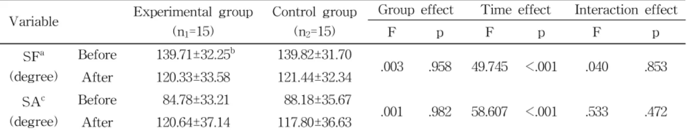
fect was observed for shoulder flexion ROM (F=.040, p=.853), shoulder abduction ROM (F=.533, p=.472).
Group effect was not significant observed for should- er flexion ROM (F=.003, p=.958), shoulder abduction ROM (F=.001, p=.982). Time effect was significant observed for shoulder flexion ROM (F=49.745, p<.001), shoulder abduction ROM (F=58.607, p<.001) (Table 6).
Discussion
This study demonstrated whether shoulder stabiliza- tion exercise could improve the shoulder stability, scapular symmetric alignment, pain, muscle power, and range of motion in patients with nonspecific shoulder pain. The upper extremity stability was significantly Variable Experimental group
(n1=15)
Control group (n2=15)
Group effect Time effect Interaction effect
F p F p F p
SFa (degree)
Before 139.71±32.25b 139.82±31.70
.003 .958 49.745 <.001 .040 .853 After 120.33±33.58 121.44±32.34
SAc (degree)
Before 84.78±33.21 88.18±35.67
.001 .982 58.607 <.001 .533 .472 After 120.64±37.14 117.80±36.63
ashoulder flexion,bmean±standard deviation,cshoulder abduction.
Table 6. The ROM before and after treatment in the experimental and control group Variable Experimental group
(n1=15)
Control group (n2=15)
Group effect Time effect Interaction effect
F p F p F p
NRSa (score)
Before 6.87±2.61b 6.20±2.11
.073 .789 59.555 <.001 2.680 .113
After 4.20±1.93 4.46±1.96
anumerical rating scale, bmean±standard deviation.
Table 4. The numeric rating scale before and after treatment in the experimental and control group
Variable Experimental group (n1=15)
Control group (n2=15)
Group effect Time effect Interaction effect
F p F p F p
SFSa (N) Before 25.42±14.60b 26.89±13.00
.042 .838 58.310 <.001 .128 .723 After 34.76±14.89 35.39±14.70
SASc (N) Before 25.47±15.49 24.79±16.23
.010 .923 65.600 <.001 .017 .897 After 34.04±14.00 33.64±15.76
ashoulder flexion strength, bmean±standard deviation, cshoulder abduction strength.
Table 5. The muscle power before and after treatment in the experimental and control group
improved the experimental group (GSE) compared to the control group (SSE) as a centralization effect of the shoulder mechanism. Intervention-related changes in al- tered functions such as scapular symmetric alignment were successfully quantified by 3 dimensional spinal diagnostic imaging. These alignment changes in scapular symmetric measure were paralleled with in- creased stability of the upper extremity in GSE. The functional improvements were corroborated by the centralization effect instigated by the stability of the humeral head and glenoid fossa in the shoulder joint.
Previous studies have reported that the shoulder stabilization exercise had positive effects on the pain, muscle strength, and ROM in individuals with vari- ous shoulder problems (Moghadam et al, 2011; Roy et al, 2009). Yet, most of these studies relied on a few clinical measurements such as the NRS, manual muscle testing and ROM of the upper extremity without considering the need for more scientific and quantitative measurements. Although Roy et al (2009) recently demonstrated that the shoulder stabilization exercise is safe and has positive effects on stability and mobility in patients with nonspecific shoulder pain, there still remains a lack of a standardized method on shoulder function.
Various parameters such as upper extremity sta- bility, pain intensity, muscle power and ROM of the shoulder part have been measured in shoulder stabi- lization exercise; however, in contrast to most varia- bles examined in shoulder stabilization studies, shoulder function in patients with shoulder pain has only been measured using limited clinical measure- ments (Sangwan et al, 2015). Kinematic data ob- tained from 3 dimensional spinal diagnostic imaging could provide more valid information on shoulder function when combined with a quantitative dyna- mometer and electronic inclinometer data on muscle power and ROM, specifically indicating increased shoulder function. However, to our knowledge, no study has provided valid and reliable measurements, such as data obtained from 3 dimensional spinal di- agnostic imaging analysis, on the effects of GSE
compared with SSE in patients with nonspecific shoulder pain.
To improve the efficacy of the GSE and SSE, we used the exercise methods reported by Park et al (2013) for the shoulder pain patients. Several studies have reported the exercise methods for the applica- tion of the shoulder stabilization exercise (Cricchio and Frazer, 2011); however, specific exercise guide- lines for shoulder pain are lacking and only short term effects have been reported. The effects of the shoulder stabilization exercise on hand grip muscle strength, non-electric manual ROM, and clinical measures of the upper extremity function in various patients have been examined (Jaggi and Lambert, 2010). In this current study, the subjects performed exercises consisted of specific 5 stages while ex- posed to the GSE and the SSE. In addition, exposure to GSE and SSE was limited to 30 minutes per ses- sion and 3 sets of 15 repetitions at a feeling without any pain or fatigue.
In this study, improved upper extremity stability was observed to a greater extent in the experimental group after the GSE. In fact, the present stability score among GSE group was increased by approx- imately 63% from 8 to 13 score after the intervention compared with that of the SSE alone, 37% from 8 to 11 score. Importantly, upper extremity stability is in line with the findings by a few previous studies (Jaggi and Lambert, 2010; Sangwan et al, 2015). In general, upper extremity stability and shoulder func- tion in patients with shoulder pain is known to be strongly correlated (Moghadam et al, 2011; Myers et al, 2006). Although the interaction effects failed to achieve statistical significance, scapular symmetric alignment, pain intensity, muscle strength, and ROM in both groups showed a tendency to improve after interventions through the time effects, suggesting that patients with nonspecific shoulder pain showed improvement with all measures.
Notwithstanding its significant results, this research has a few limitations that should be addressed in a more robust and large-scale study. First, the num-
bers of subjects were very small. Second, this re- search represents immediate effects of the GSE in patients with nonspecific shoulder pain. Lastly, we didn’t have a control group provided a baseline.
Therefore, our results cannot be generalized because of limited sample size. A larger clinical controlled trial involving patients with shoulder pain is needed to investigate the therapeutic effects of GSE to aug- ment upper extremity stability, scapular symmetric alignment, pain, muscle power and ROM in clinical practice.
Conclusion
This study suggests that the GSE is more benefi- cial than the SSE for improving upper extremity stability in patients with nonspecific shoulder pain.
Certainly, both the GSE and the SSE are effective to the scapular symmetric alignment, pain, muscle pow- er and ROM through the significant time effects of each measurement. These results offer clinical insight into the additive effect of GSE in upper extremity stability, and suggest that it may be used as an ad- ditional shoulder intervention technique for the man- agement of nonspecific shoulder pain.
References
Camargo PR, Haik MN, Ludewig PM, et al. Effects of strengthening and stretching exercises applied during working hours on pain and physical im- pairment in workers with subacromial impingement syndrome. Physiother Theory Pract. 2009;25(7):
463-475. https://doi.org/10.3109/09593980802662145 Cricchio M, Frazer C. Scapulothoracic and scap-
ulohumeral exercises: A narrative review of electromyographic studies. J Hand Ther. 2011;
24(4):322-333. https://doi.org/10.1016/j.jht.2011.06.001 De Mey K, Cagnie B, Danneels LA, et al. Trapezius muscle timing during selected shoulder re-
habilitation exercises. J Orthop Sports Phys Ther. 2009;39(10):743-752. https://doi.org/10.2519/
jospt.2009.3089
Faber E, Kuiper JI, Burdorf A, et al. Treatment of impingement syndrome: A systematic review of the effects on functional limitations and return to work. J Occup Rehabil. 2006;16(1):7-25.
Golpayegani M, Mahtabi S, Shahjerdi S, et al. The study of validity and reliability of formetric 4D system in measuring of deformites in kyphosis and lordosis in women. Shahrekord University of Medical Sciences Journal. 2013;15(3):74-81.
Grant HJ, Arthur A, Pichora DR. Evaluation of in- terventions for rotator cuff pathology: A sys- tematic review. J Hand Ther. 2004;17(2):274-299.
Guerra de Hoyos JA, Andrés Martín Mdel C, Bassas y Baena de Leon E, et al. Randomised trial of long term effect of acupuncture for shoulder pain. Pain. 2004;112(3):289-298.
Hawker GA, Mian S, Kendzerska T, et al. Measures of adult pain: Visual Analog Scale for Pain (VAS Pain), Numeric Rating Scale for Pain (NRS Pain), McGill Pain Questionnaire (MPQ), Short-Form McGill Pain Questionnaire (SF-MPQ), Chronic Pain Grade Scale (CPGS), Short Form-36 Bodily Pain Scale (SF-36 BPS), and Measure of Intermittent and Constant Osteoarthritis Pain (ICOAP). Arthritis Care Res (Hoboken). 2011;
63 Suppl 11:S240-S252. https://doi.org/10.1002/acr.20543 Hess SA, Richardson C, Darnell R, et al. Timing of rotator cuff activation during shoulder external rotation in throwers with and without symptoms of pain. J Orthop Sports Phys Ther. 2005;
35(12):812-820.
Jaggi A, Lambert S. Rehabilitation for shoulder instability. Br J Sports Med. 2010;44(5):333-340.
https://doi.org/10.1136/bjsm.2009.059311
Jubany J, Busquets A, Marina M, et al. Reliability and validity of a custom-made instrument including a hand-held dynamometer for measuring trunk muscle strength. J Back Musculoskelet Rehabil.
2015;28(2):317-326. https://doi.org/10.3233/BMR-140522
Kolber MJ, Hanney WJ. The reliability and con- current validity of shoulder mobility measure- ments using a digital inclinometer and goni- ometer: A technical report. Int J Sports Phys Ther. 2012;7(3):306-313.
Ludewig PM, Reynolds JF. The association of scapular kinematics and glenohumeral joint pathologies. J Orthop Sports Phys Ther. 2009;39(2):90-104.
https://doi.org/10.2519/jospt.2009.2808
Moghadam AN, Mohammadi R, Arab AM, et al. The effect of shoulder core exercises on isometric torque of glenohumeral joint movements in healthy young females. J Res Med Sci. 2011;
16(12):1555-1563.
Myers JB, Wassinger CA, Lephart SM. Sensorimotor contribution to shoulder stability: Effect of in- jury and rehabilitation. Man Ther. 2006;11(3):
197-201.
Negrete RJ, Hanney WJ, Kolber MJ, et al. Reliability, minimal detectable change, and normative values for tests of upper extremity function and power.
J Strength Cond Res. 2010;24(12):3318-3325.
https://doi.org/10.1519/JSC.0b013e3181e7259c Park SI, Choi YK, Lee JH, et al. Effects of shoulder
stabilization exercise on pain and functional re- covery of shoulder impingement syndrome patients. J Phys Ther Sci. 2013;25(11):1359-1362.
Ratcliffe E, Pickering S, McLean S, et al. Is there a relationship between subacromial impingement syndrome and scapular orientation? A systematic
review. Br J Sports Med. 2014;48(16):1251-1256.
https://doi.org/10.1136/bjsports-2013-092389 Roy JS, Moffet H, Hébert LJ, et al. Effect of motor
control and strengthening exercises on shoulder function in persons with impingement syndrome: A single-subject study design. Man Ther. 2009;14(2):
180-188. https://doi.org/10.1016/j.math.2008.01.010 Sangwan S, Green RA, Taylor NF. Stabilizing char-
acteristics of rotator cuff muscles: A systematic review. Disabil Rehabil. 2015;37(12):1033-1043.
https://doi.org/10.3109/09638288.2014.949357 Tucci HT, Martins J, Sposito Gde C, et al. Closed
Kinetic Chain Upper Extremity Stability test (CKCUES test): A reliability study in persons with and without shoulder impingement syndrome.
BMC Musculoskelet Disord. 2014;15:1. https://doi.org/
10.1186/1471-2474-15-1
Voight ML, Thomson BC. The role of the scapula in the rehabilitation of shoulder injuries. J Athl Train. 2000;35(3):364-372.
Walther M, Werner A, Stahlschmidt T, et al. The sub- acromial impingement syndrome of the shoulder treated by conventional physiotherapy, self-train- ing, and a shoulder brace: Results of a prospective, randomized study. J Shoulder Elbow Surg. 2004;
13(4):417-423. https://doi.org/10.1016/S1058274604000485
This article was received October 7, 2016, was reviewed October 7, 2016, and was accepted November 7, 2016.