Introduction
Functioning of the serratus anterior (SA) muscle contributes to the ability to maintain a normal scap- ulothoracic position and motion, such as upward ro- tation, protraction, and posterior tilt during forward flexion (FF) of the shoulder (FFS) (Decker et al, 1999; Ludewig and Cook, 2000; Ludewig et al, 2004;
Marshall and Murphy, 2006; Park and Yoo, 2011;
Thigpen et al, 2010). SA muscle function is essential
to ensure normal scapulohumeral rhythm during FFS.
Scapular motion during FFS should be commenced at various shoulder flexion angles (Ludewig and Cook, 2000; Lukasiewicz et al, 1999). Scapular up- ward rotation and posterior tipping markedly increase at an angle of 120° during FFS (Ludewig and Cook, 2000; Lukasiewicz et al, 1999). Moseley et al (1992) reported that average SA muscle activity equated to 96% of maximum manual muscle strength at an an- gle between 120° and 150° during FFS. Impaired SA Corresponding author: Oh-yun Kwon [email protected]
The Effect of a Combination of Scapular Protraction With Resistance and Forward Flexion of the Shoulder on Serratus Anterior Muscle Activity
Sung-hoon Jung1,2, BPT, PT, In-cheol Jeon1,2, MSc, PT, Ui-jae Hwang1,2, BPT, PT Jun-hee Kim1,2, BPT, PT, Oh-yun Kwon1,3,4, PhD, PT
1Kinetic Ergocise Based on Movement Analysis Laboratory
2Dept. of Physical Therapy, The Graduate School, Yonsei University
3Dept. of Physical Therapy, College of Health Science, Yonsei University
4Dept. of Ergonomic Therapy, The Graduate School of Health and Environment, Yonsei University
Abstract
1)Background: The functioning of the serratus anterior (SA) muscle is essential to normal scapulohumeral rhythm during forward flexion (FF) of the shoulder. Also, SA weakness and overuse of the upper trapezius (UT) is observed in patients with shoulder dysfunction and trapezius myalgia. We designed a combination exercise involving FF and scapular protraction with resistance (CFFSP) to activate the SA muscle and to deactivate the UT muscle.
Objects: The purpose of this study was to determine whether or not CFFSP would be more effective in activating the SA muscle than FF alone and FF with scapular protraction (FFP).
Methods: Nineteen subjects (12 men and 7 women) participated in this study and performed FF, FFP, and CFFSP at 120°. Surface electromyography was applied to the SA, UT, and pectoralis major (PM) muscles, as was one-way analysis of variance (ANOVA) with repeated measures. Statistical significance was set at .05. Bonferroni adjustment was used to counteract the problem of multiple comparisons, with a statistical level of significance of .017 (.05/3).
Results: A statistically significant difference was found in relation to the three positions for the SA muscle (p<.001) and the SA/UT ratio (p=.005) using ANOVA. Significantly different results, depending on the position, were also demonstrated using the Bonferroni post-hoc test for the SA muscle (FF=28.27±16.20, FFP=45.66±15.81, and CFFSP=62.4±27.21) and for the SA/UT ratio (FF=3.04±2.14, FFP=3.61±2.38, and CFFSP=5.95±3.01). Significant differences between the three positions was not found regarding the average amplitude of SA/PM muscle ratio (SA/PM: p=.060).
Conclusion: We recommend the use of CFFSP to strengthen the SA muscle at 120°.
Key Words: Forward flexion, Protraction, Serratus anterior, Upper trapezius.
muscle function is associated with abnormal scap- ulohumeral rhythm during FFS (Ludewig and Cook, 2000; Ludewig et al, 2004).
Identification of the most effective exercise with which to activate the SA muscle during functional movement, including FFS, has been investigated in many studies (Cools et al, 2007; Decker et al, 1999;
Hardwick et al, 2006; Ludewig et al, 2004; Martins et al, 2008; McClure et al, 2004). Various SA muscle exercises, such as FF, humeral elevation push-up plus, weight-resisted elevation of the humerus and wall slide, have been introduced.
It was reported in previous studies that the push-up plus exercise was effective in SA muscle strengthening (Hardwick et al, 2006; Lehman et al, 2006; Ludewig et al, 2004; Sandhu et al, 2008).
However, Hardwick et al (2006) observed that the wall slide exercise was more effective in activating the SA muscle than the push-up plus exercise in a humeral position above 90° during FFS. It was also found that the push-up plus exercise was more effi- cient in ensuring SA muscle activation than the con- ventional push-up plus exercise in a humeral position of 120° in FFS (Hwang et al, 2016). Therefore, SA muscle exercises performed above 90° FFS are con- sidered more effective in activating the SA muscle than those at 90° FFS.
Weakness in the SA muscle contributes to ex- cessive contraction of the upper trapezius (UT) (Ludewig et al, 2004) and pectoralis major (PM) muscles (Kim et al, 2010; Park et al, 2013) during FFS. Excess activation of the UT muscle, combined with decreased control with the SA muscle, has been proposed as a contributing factor to abnormal scap- ular motion during FFS (Cools et al, 2007; McClure et al, 2004; Ludewig and Cook, 2000; Lukasiewicz et al, 1999; Peat and Grahame, 1977). In addition, SA muscle weakness and overuse of the UT muscle has been observed in patients with shoulder dysfunction (Martins et al, 2008) and trapezius myalgia (Vollenbroek-Hutten et al, 2006). Weakness of the SA muscle may cause shoulder pathologies, such as
subacromial impingement, bursitis, and rotator cuff injury. Therefore, strengthening the SA muscle and reducing the UT/SA muscle ratio during FFS is es- sential in preventing the incidence of shoulder path- ology (Ludewig et al, 2004).
We designed a combination exercise involving FFS and scapular protraction with resistance (CFFSP) to activate and deactivate the SA and UT muscles, respectively. This exercise combines FF and self-re- sistive scapular protraction, involving the use of the opposite hand in close kinetic chain exercise, i.e., pushing against a wall. The study objective was to determine whether or not CFFSP would activate the SA muscle more effectively than FF and FF with scapular protraction (FFP). We hypothesized that SA muscle activity would be greatest during CFFSP exercise.
Methods
Subjects
Nineteen subjects (12 men and 7 women) aged 21
∼32 years, with a mean age of 22.5±2.0 years, who were students at Yonsei University, Wonju Campus, South Korea, voluntarily participated in this study.
Inclusion criteria included being healthy, free of met- abolic, neuromuscular, and musculoskeletal disorders, no history of shoulder pain, and the absence of pain in any part of the body at the time of testing.
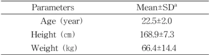
Exclusion criteria included the presence of pain in the shoulder region and a previous history of shoulder surgery. The mean height and weight of the subjects was 168.9±7.3 ㎝ and 66.4±14.4 ㎏, re- spectively (Table 1). The study was approved by the
Parameters Mean±SDa
Age (year) 22.5±2.0
Height (㎝) 168.9±7.3
Weight (㎏) 66.4±14.4
amean±standard deviation.
Table 1. General characteristics of the subjects (N=19)
Institutional Review Board of Yonsei University (1041849-201603-BM-008-01).
Instrumentation and data reduction Activation of the UT, SA, and PM muscles was measured by surface electromyography (EMG) (TeleMyo 2400T, Noraxon, Scottsdale, AZ, USA).
Two separate bipolar (20 ㎜ center-to-center) surface electrodes were placed two centimeters apart on the SA muscle in the dominant arm, just below the axilla at the level of the inferior angle of the scapula, ante- rior to the latissimus dorsi. UT muscle electrodes were attached at a point approximately halfway be- tween the acromion and the spinous processes of the seventh cervical vertebrae. The electrodes for the PM muscle (sternal fiber) were attached to the chest wall, horizontal to the arising muscle mass (2 ㎝ from the axillary fold) (Criswell, 2010).
The signals were collected using MyoResearch XP Master Edition (Noraxon Inc., Scottsdale, USA). A bandpass filter of between 20 ㎐ and 450 ㎐ was applied to the raw EMG signals and the root mean square values were calculated using a 50 ㎳ window.
The processed data were recorded at a sampling rate of 1000 ㎐. Maximum voluntary contraction (MVC) was performed to normalize the EMG signals.
Each subject was asked to position the shoulder flexed at 120° for the purpose of measuring MVC in the SA muscle. Maximum resistance against scap- ular protraction was applied to the elbow by the ex- aminer (Kendall et al, 2005). MVC in the PM mus- cle was determined while the subjects adducted their shoulders to a 90° flexion angle with their palms pressed together. Resistance was applied by the ex- aminer by pushing the palms of the participants to- gether, while in the supine position (Dark et al, 2007). MVC in the UT muscle was measured during resisted isometric scapular elevation. The partic- ipants were seated during scapular elevation (Kendall et al, 2005). Each MVC was collected for a period of five seconds and the mean values from the middle three seconds were averaged to calculate the
mean value. All data are expressed as a percentage of the MVC.
Procedures
Each exercise was performed three times, with a resting time of three minutes between them. Prior to the trials, the order of the exercises was assigned, based on a random number generated using Microsoft Excel ver. 2010 (Microsoft Corporation, Washington, US) to eliminate any order effects. The subjects sat with their backs placed flat against a chair in an upright sitting position with the hip and knee joints at 90° flexion. The participants positioned their feet shoulder width apart and in contact with the floor, with the two femurs in a parallel position. The tar- get bar was placed next to the subjects to control the plane and degree of flexion. An angle of 120°
shoulder flexion was measured in each subject using a smartphone inclinometer to determine the height of the horizontal bars.
Forward flexion
The subjects were instructed to lift the arm with the elbows extended and pronation of the forearm and wrist in a neutral position, while brushing against the vertical pole with the dorsal side of the forearm to maintain the shoulder in the sagittal plane. The subjects maintained the FF posture for five seconds, while slightly touching the horizontal bar which was placed at 120° shoulder flexion.
Forward flexion with scapular protraction The study participants were asked to lift their arm with the elbow extended and protract the scapular without trunk rotation (protraction). With pronation of the forearm and wrist in a neutral position, they simultaneously lifted the arm, while brushing the vertical pole with the dorsal side of the forearm to maintain the shoulders in the sagittal plane. They maintained FF with protraction for five seconds, while slightly touching the horizontal bar which was placed at 120° shoulder flexion.
Combination exercise involving forward flexion of the shoulder and scapular protraction with resistance
The subjects were instructed to grip the back of the dominant hand (the right side in every instance) with the palm of the non-dominant hand. They lifted the arm with the elbow extended and the wrist in a neutral position. The wrist brushed along the vertical pole during the lifting process, while the dominant hand simultaneously pushed down on the non-domi- nant hand. Thereafter, the non-dominant hand pulled up the dominant hand (resistance against pro- traction). The subjects maintained FF with resistance against protraction for five seconds while touching the horizontal bar slightly during 120° shoulder flexion. The subjects sat with their backs placed flat against a chair in an upright sitting position and wrist touch horizontal bar and vertical pole during maintaining FF with resistance against protraction in order to prevent compensative motion in scapular and trunk.
Statistical analysis
SPSS ver. 18.0 (SPSS Inc., Chicago, IL, USA) was used to analyze any differences between the SA/UT and the SA/PM ratio. The Kolmogorov-Smirnov test was conducted to ensure normal distribution of the var-
iables and this was confirmed. Thus, a parametric stat- istical test was conducted. One-way analysis of var- iance (ANOVA) with repeated measures was applied.
Statistical significance was set at .05. Bonferroni ad- justment was used to eliminate any differences and statistical significance was set at .017 (.05/3).
Results
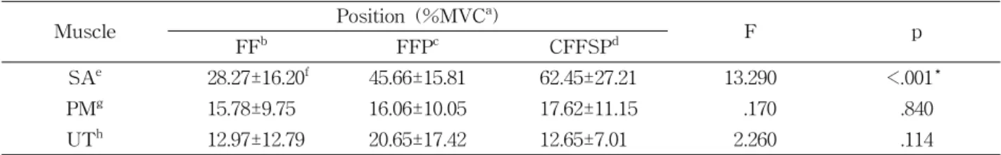
The average amplitude of SA, PM, UT muscle ac- tivity and standard deviation according to the posi- tion used are shown in Table 2. A statistically sig- nificant difference between the three positions with respect to average amplitude of the SA muscle and the SA/UT muscle ratio was demonstrated using ANOVA (p<.017). Significantly different results, again dependent on the position used, were observed using the Bonferroni post-hoc test for the average ampli- tude of the SA muscle (FF vs. FFP: p=.034 and FF vs. CFFSP: p<.001; Table 2; Figure 2), and for the SA/UT ratio (FF vs. FFP: p=.009 and FF vs.
CFFSP: p=.019; Table 3; Figure 3). Significant dif- ferences between the three positions was not found regarding the average amplitude of the PM, UT muscles and the SA/PM muscle ratio (PM: p=.840, UT: p=.114, SA/PM: p=.060; Table 3; Figure 4).
A B C
Figure 1. The serratus anterior muscle being exercised in three positions (A) forward flexion, (B) forward flexion with protraction, and (C) combination exercise involving forward flexion of the shoulder and scapular protraction with resistance.
Discussion
Activity in the SA muscle was shown to be greatest during CFFSP exercise in this study. To the best of our knowledge, this is the first study in which the activity of the SA, UT, and PM muscles
has been investigated and the ratio of the activity in the SA/UT muscles and that in the SA/PM muscles has been investigated during CFFSP compared FF and FFP. Activation of the SA muscle is essential for the rehabilitation of shoulder dysfunction caused by weakness in this muscle and by muscle strength
Figure 2. A comparison of SA muscle activity using the three different exercises (CFFSP: combination exercise involving forward flexion of the shoulder and scapular protraction with resistance, FF:
forward flexion, FFP: forward flexion with scapular protraction, SA: serratus anterior, MVC: maximal voluntary contraction).
Figure 3. A comparison of SA/UT muscle activity using the three different exercises (CFFSP: combination exercise involving forward flexion of the shoulder and scapular protraction with resistance, FF: forward flexion, FFP: forward flexion with scapular protraction, SA:
serratus anterior, UT: upper trapezius).
Muscle Position (%MVCa)
F p
FFb FFPc CFFSPd
SAe 28.27±16.20f 45.66±15.81 62.45±27.21 13.290 <.001*
PMg 15.78±9.75 16.06±10.05 17.62±11.15 .170 .840
UTh 12.97±12.79 20.65±17.42 12.65±7.01 2.260 .114
amaximal voluntary contraction, bforward flexion,cforward flexion with scapular protraction, dcombination exercise involving forward flexion of the shoulder and scapular protraction with resistance, eserratus anterior, fmean±standard deviation, gpectoralis major, hupper trapezius, *p<.017.
Table 2. A depiction of the average amplitude of serratus anterior, pectoralis major, and upper trapezius mus- cle activity and standard deviation according to the position used
Muscle Position
F p
FFa FFPb CFFSPc
SAd/UTe 3.04±2.14f 3.61±2.38 5.95±3.01 5.890 .005*
SA/PMg 2.53±2.19 3.88±2.75 4.97±4.02 2.974 .060
aforward flexion, bforward flexion with scapular protraction, ccombination exercise involving forward flexion of the shoulder and scapular protraction with resistance, dserratus anterior,eupper trapezius, fmean±standard deviation,gpectoralis major, *p<.017.
Table 3. The average and standard deviation of the serratus anterior/upper trapezius and the serratus ante- rior/pectoralis ratio, according to the position used
imbalance in the scapulothoracic and glenohumeral joints (Cools et al, 2004; Madeleine et al, 2008).
In previous studies, the push-up plus exercise was reported to be the effective exercise for the purposes of activating the SA muscle (Lehman et al, 2006;
Ludewig et al, 2004; Sandhu et al, 2008). In recent studies (Castelein et al, 2016; Ha et al, 2012; Hardwick et al, 2006; Hwang et al, 2016; Park et al, 2013; Park et al, 2014a), exercise to accompany the functional movement, such as shoulder flexion, was reported to be effective in activating the SA muscle. Choung et al (2013) reported that 130° shoulder flexion resulted in greater activation of the SA muscle than 90° shoulder flexion in the sagittal plane (MVC of 60.7% at 130°
and MVC of 51.3% at 90°). Hardwick et al (2006) ob- served that the wall slide exercise with shoulder flex- ion up to 120° was more effective in activating the SA muscle (MVC of 58.3%) than the push-up plus ex- ercise (MVC of 31.3%). And wall slide exercise more activated activity of middle SA than wall push-up (MVC of 63.5% and MVC of 43.1%) (Park et al, 2014b). The CFFSP exercise is similar to pushing against a wall (used in the wall slide exercise) be- cause one hand pushes forward (protraction) while the opposite hand pulls to provide resistance. Thus, our data reflect a high degree of activation of the SA muscle with the use of CFFSP (MVC of 62.5%).
Patients with shoulder dysfunction can more easily perform the wall slide exercise (leaning into the wall against partial gravity) than the push-up plus ex- ercise (leaning into the floor against full gravity) (Hardwick et al, 2006). CFFSP can easily be per- formed in any position anywhere, using resistance applied by the opposing hand. Therefore, patients can easily perform the exercises because additional tools, such as a wall, floor, or dumbbell, are not required.
The value of the SA/UT ratio during CFFSP was more significant than that recorded during FF and FFP. Activation of the UT muscle during CFFSP (MVC of 12.7%) was lower than that for the UT muscle during FFP (MVC of 20.7%). There are sev- eral reasons for this. Firstly, the focus is on scapular protraction as the opposite hand is pulling during CFFSP. This may decrease activation in the UT muscle because shoulder elevation is limited in fo- cused scapular protraction during CFFSP. Secondly, the opposite hand is involved in close kinetic chain exercises. CFFSP cause the scapular to be pushed during close kinetic chain exercises which have been shown to stimulate the mechanoreceptors, thus con- tributing to shoulder joint stabilization (De Mey et al, 2014). Improving the stability of the closed kinetic chain exercises (resistance against protraction during CFFSP) may contribute to increasing and decreasing SA and UT muscle activity, respectively.
There were several limitations to the current study.
Firstly, we only considered the SA, UT, and PM mus- cles, and did not investigate activity in other muscles which might also have affected scapular control, such as the lower trapezius, rhomboids, and levator scapulae. Secondly, we did not investigate scapula kin- ematics during the study. Future studies need to be conducted so that scapular kinematics can be evaluated in order to assess scapular movement during CFFSP in comparison with that recorded using other methods.
Thirdly, only healthy and relatively young subjects (21
∼32 years) were recruited into our study. Thus, our findings cannot be generalized to individuals with shoulder dysfunction or to other patient populations.
Figure 4. A comparison of SA/PM muscle activity using the three different exercises (CFFSP: combination exercise involving forward flexion of the shoulder and scapular protraction with resistance, FF: forward flexion, FFP: forward flexion with scapular protraction, PM:
pectoralis major, SA: serratus anterior).
Further research is required to investigate the effect of CFFSP on SA muscle activity in individuals with shoulder dysfunction. FF with resistance against pro- traction is selected by individuals duirng CCFSP.
Thus, this study cannot be generalized to each individual. Further research is required to control the pressure of FF with resistance during CFFSP.
Conclusion
This is the first study in which the activity of the SA muscle, the ratio of the activity in the SA/UT muscles and that in the SA/PM muscles has been investigated during CFFSP compared FF and FFP.
Activation of the SA muscle significantly increased in the CFFSP compared to that in the FF and FFP, and the activity was significantly greater in the CFFSP. The results suggest that CFFSP is effective exercise to strengthen the SA muscle at 120°.
References
Castelein B, Cagnie B, Parlevliet T, et al. Superficial and deep scapulothoracic muscle electromyo- graphic activity during elevation exercises in the scapular plane. J Orthop Sports Phys Ther. 2016;
46(3):184-193. https://doi.org/10.2519/jospt.2016.5927 Choung SD, Weon JH, Jung DY. Effect of movement
plane and shoulder flexion angle on scapular up- ward rotator during scapular protraction exercise.
J Korean Soc Phys Med. 2013;8(1):41-48.
Cools AM, Dewitte V, Lanszweert F, et al.
Rehabilitation of scapular muscle balance: Which exercises to prescribe? Am J Sports Med.
2007;35(10):1744-1751.
Cools AM, Witvrouw EE, Declercq GA, et al.
Evaluation of isokinetic force production and as- sociated muscle activity in the scapular rotators during a protraction-retraction movement in overhead athletes with impingement symptoms.
Br J Sports Med. 2004;38(1):64-68.
Criswell, E. Cram’s Introduction to Surface Electromyography. 2nd ed. Sudlbury, Jones &
Bartlett Learning, 2010:289, 297, 307.
Dark A, Ginn KA, Halaki M. Shoulder muscle re- cruitment patterns during commonly used rotator cuff exercises: An electromyographic study.
Phys Ther. 2007;87(8):1039-1046.
Decker MJ, Hintermeister RA, Faber KJ, et al.
Serratus anterior muscle activity during selected rehabilitation exercises. Am J Sports Med.
1999;27(6):784-791.
De Mey K, Danneels L, Cagnie B, et al. Shoulder muscle activation levels during four closed kinetic chain exercises with and without Redcord slings.
J Strength Cond Res. 2014;28(6):1626-1635.
https://doi.org/10.1519/JSC.0000000000000292 Ha SM, Kwon OY, Cynn HS, et al. Comparison of
electromyographic activity of the lower trapezius and serratus anterior muscle in different arm-lifting scapular posterior tilt exercises. Phys Ther Sport. 2012;13(4):227-232. https://doi.org/
10.1016/j.ptsp.2011.11.002
Hardwick DH, Beebe JA, McDonnell MK, et al. A comparison of serratus anterior muscle activa- tion during a wall slide exercise and other tra- ditional exercises. J Orthop Sports Phys Ther.
2006;36(12):903-910.
Hwang UJ, Kwon OY, Jeon IC, et al. Effect of hum- eral elevation angle on electromyographic activ- ity in the serratus anterior during the push-up plus exercise. J Sport Rehabil. 2016 (Article in Press).
Kendall FP, McCreary EK, Provance PG, et al.
Muscles: Testing and function. 5th ed. Baltimore, Lippincott Williams & Wilkins, 2005:333.
Kim BG, Gong WT, Lee SY. The effect of push-up plus exercise with visual biofeedback on the ac- tivity of shoulder stabilizer muscles for winged scapula. J Phys Ther Sci. 2010;22(4):355-358.
Lehman GJ, MacMillan B, MacIntyre I, et al.
Shoulder muscle EMG activity during push up
variations on and off a Swiss ball. Dyn Med.
2006;5:7.
Ludewig PM, Cook TM. Alterations in shoulder kin- ematics and associated muscle activity in people with symptoms of shoulder impingement. Phys Ther. 2000;80(3):276-291.
Ludewig PM, Hoff MS, Osowski EE, et al. Relative balance of serratus anterior and upper trapezius muscle activity during push-up exercises. Am J Sports Med. 2004;32(2):484-493.
Lukasiewicz AC, McClure P, Michener L, et al.
Comparison of 3-dimensional scapular position and orientation between subjects with and with- out shoulder impingement. J Orthop Sports Phys Ther. 1999;29(10):574-583.
Madeleine P, Mathiassen SE, Arendt-Nielsen L.
Changes in the degree of motor variability as- sociated with experimental and chronic neck-shoulder pain during a standardized repeti- tive arm movement. Exp Brain Res. 2008;185(4):
689-698.
Marshall P, Murphy B. Changes in muscle activity and perceived exertion during exercises per- formed on a Swiss ball. Appl Physiol Nutr Metab. 2006;31(4):376-383.
Martins J, Tucci HT, Andrade R, et al.
Electromyographic amplitude ratio of serratus anterior and upper trapezius muscles during modified push-ups and bench press exercises.
J Strength Cond Res. 2008;22(2):477-484.
https://doi.org/10.1519/JSC.0b013e3181660748 McClure PW, Bialker J, Neff N, et al. Shoulder func-
tion and 3-dimensional kinematics in people with shoulder impingement syndrome before and after a 6-week exercise program. Phys Ther.
2004;84(9):832-848.
Moseley JB Jr, Jobe FW, Pink M, et al. EMG analy- sis of the scapular muscles during a shoulder rehabilitation program. Am J Sports Med.
1992;20(2):128-134.
Park KM, Cynn HS, Kwon OY, et al. Comparison of pectoralis major and serratus anterior muscle
activities during different push-up plus exercises in subjects with and without scapular winging.
J Strength Cond Res. 2014a;28(9):2546-2551.
https://doi.org/10.1519/JSC.0000000000000443 Park KM, Cynn HS, Yi CH, et al. Effect of isometric
horizontal abduction on pectoralis major and serratus anterior EMG activity during three ex- ercises in subjects with scapular winging. J Electromyogr Kinesiol. 2013;23(2):462-468.
https://doi.org/10.1016/j.jelekin.2012.11.013
Park SY, Ahn TK, Eom JH, et al. Scapulothoracic muscle activity during use of a wall slide device (WSD), a comparison with the general wall push up plus. J Phys Ther Sci. 2014b;26(6):805-806.
https://doi.org/10.1589/jpts.26.805
Park SY, Yoo WG. Differential activation of parts of the serratus anterior muscle during push-up variations on stable and unstable bases of support. J Electromyogr Kinesiol. 2011;21(5):861-867.
https://doi.org/10.1016/j.jelekin.2011.07.001
Peat M, Grahame RE. Electromyographic analysis of soft tissue lesions affecting shoulder function.
Am J Phys Med. 1977;56(5):223-240.
Sandhu JS, Mahajan S, Shenoy S. An electromyo- graphic analysis of shoulder muscle activation during push-up variations on stable and labile surfaces. Int J Shoulder Surg. 2008;2(2):30-35.
https://doi.org/10.4103/0973-6042.40456
Thigpen CA, Padua DA, Michener LA, et al. Head and shoulder posture affect scapular mechan- ics and muscle activity in overhead tasks.
J Electromyogr Kinesiol. 2010;20(4):701-709.
https://doi.org/10.1016/j.jelekin.2009.12.003
Vollenbroek-Hutten M, Hermens H, Voerman G, et al. Are changes in pain induced by myofeedback training related to changes in muscle activation patterns in patients with work-related myalgia?.
Eur J Appl Physiol. 2006;96(2):209-215.
This article was received October 10, 2016, was reviewed October 10, 2016, and was accepted November 10, 2016.