J. of Korean Bone & Joint Tumor Soc.
Volume 15, Number 1, June, 2009
─ 52 ─
※통신저자: 박박 용용 구구
서울특별시 동대문구 회기동 1번지 경희의료원 병리과
Tel: 02) 958-8742, Fax: 02) 957-0489, E-mail: [email protected]
늑골에서 발생한 거대세포종: 1예 보고 및 문헌 고찰
경희의료원 병리과, 경희대학교 동서신의학병원 흉부외과*, 병리과� 김현수∙김대현*∙임성직�∙박용구
거대세포종은 늑골에서 드물게 발생할 수 있으며, 후중격에서 발생한 종괴로 나타난 늑골의 거대세포종은 지금까지 4 예가 보고되었다. 38세 남자의 늑골에서 발생하여 후중격 종괴로 보 인 거대세포종 1 예를 문헌 고찰과 함께 보고한다. 흉부 전산화 단층 촬영에서 우측 후상부 중격의 대부분을 차지하는, 경계가 명확한 다분엽성의 종괴가 우측 3번 늑골과 흉추를 침범하 고 있었다. 임상적으로는 후중격에서 발생한 신경절신경종 혹은 그와 동반된 악성 변화를 의 심하였다. 그러나 육안적으로 종괴는 우측 3번 늑골에서 발생하여 늑골 바깥쪽으로 성장하는 모습을 보였고, 현미경적으로 균일하게 산재된 다핵 거대 세포와 단핵 기질 세포로 구성되어 있어 늑골에서 발생한 거대세포종에 합당하였다. 거대세포종의 치료를 위해서는 재발과 전이 의 가능성을 고려하여 광범위한 수술적 절제와 술후 방사선 치료를 고려해야 한다. 후중격 신 경절신경종은 수술적 절제만으로 치료가 가능한 종양이므로, 거대세포종과 반드시 감별해야 한다.
색인 단어: 늑골, 거대세포종, 신경절신경종
INTRODUCTION
Giant cell tumor (GCT) represents 5% of all primary bone tumors and approximately 25% of benign bone tumors1). GCT usually involves the long bone epiphysis in skeletally mature patients and the metaphysis in skeletally immature patients2). GCT rarely occurs in the rib. Plain chest roentgenogra-
phy shows a well-defined, expansile, oste- olytic lesion, which is usually covered with a thin shell of reactive new bone. Computed tomography (CT) also shows a well-defined, expansile lesion of soft tissue density.
Internal calcified septae and punctuate calci- fication are frequently seen3). When GCT occurs in the posterior arc of the rib, it may form a large destructive mass that presents
as a posterior mediastinal mass3). We describe a case of GCT showing substantial growth out of the rib and mimicking a pos- terior mediastinal ganglioneuroma. We also review the relevant literature (GN).
CASE REPORT
A 38-year-old man presented with a 2-year history of pain in his right upper back and chest wall. He had recurrent episodes of this pain associated with decreased sensation in the right axilla. He had no remarkable past or family histories. On admission, the physi- cal examination revealed no abnormalities.
Routine laboratory tests, including serum calcium, phosphorus, alkaline phosphatase and tumor markers, were within normal range.
A plain chest roentgenography showed an ovoid, expansile upper paratracheal mass (Fig. 1). A chest CT showed a 5.5×4.2×4.3- cm well-defined, heterogeneous, multi-lobu- lated mass of soft tissue density with rela- tive enhancement in the right posterior mediastinum. The mass appeared to invade the medial end of the right third rib and caused a bulging appearance in the posterior
cortex (Fig. 2). Erosive change was also identified in the body of the third thoracic vertebra, but extensions into the spinal canal, esophagus or trachea were not identi- fied. Although most of the mass was of soft tissue density, it also showed subtle, irregu- lar calcified densities. The lung parenchyma was unremarkable.
A technetium-99m methylene diphospho- nate bone scan showed focally increased uptake of the radionuclide around the right third costovertebral joint, consistent with rib invasion. Based on the radiologic finding, it was thought to be a posterior mediastinal GN. Because tumor invasion into the rib and vertebra was strongly suspected, gan- glioneuroblastoma was included in the dif- ferential diagnosis. The mass, which occu- pied the mediastinum, the right third and fourth ribs and the right second to fourth intercostal muscles, was excised.
Grossly, there was a 2.2×0.7×0.7-cm red to brown firm tumor mass in the marrow cavity of the third rib, which was expand- ing and destroying the cortical bone. The third costovertebral joint was involved by
Fig. 1. Plain chest roentgenography shows an expansile upper paratracheal mass (arrowheads).
Fig. 2. Contrast-enhanced computed tomography shows a well-defined, lobulated, heterogeneous mass of soft tissue density associated with destruction of the right third rib and thoracic vertebra in the right posterior mediastinum.
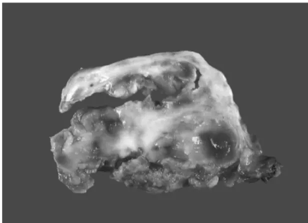
the tumor. Fragmented mediastinal masses were relatively well-circumscribed. The largest mass measured 5×4.5×3 cm and was partly enclosed by a yellow to pink calcified capsule. The cut surfaces of the mass showed beefy-red viable-appearing areas accompanied by multiple foci of hemorrhage (Fig. 3). Histologically, the tumor consisted of diffusely interspersed, multi-nucleated, osteoclast-type giant cells and oval to spin- dle-shaped stromal mononuclear cells with- out significant cytologic atypia (Fig. 4). The appearance of the giant cell nuclei was simi- lar to that of the stromal cells. Mitotic activity was apparent in the stromal cells but not in the giant cells; no atypical mito- sis was identified. Some areas of the tumor contained small, multi-focal, aneurismal bone cyst-like spaces. Reactive osteoid and woven bone were also found, especially at the periphery.
The pathologic diagnosis was a GCT of the rib involving the posterior mediastinum.
Twelve months after the surgery, the post- operative course has been uneventful, except for mild chest discomfort and anhidrosis of
the right side of the face and the right hand due to the sympathectomy.
DISCUSSION
Although GCT of the rib usually arises in the posterior arc, no reports have mentioned the differential diagnosis between GCT and GN, which is the most common benign tumor arising in the posterior mediastinum.
We found several radiologic findings in our case that may contribute to a misdiagnosis of GN. CT showed that the lesion formed a large paravertebral mass and that the medi- al end of the rib adjacent to the mass showed cortical thinning and expansion.
Based on these findings, the radiologist’s interpretation was that the paravertebral mass had invaded the rib, not that the pri- mary rib tumor extended into the medi- astinum, which was the case. In addition, internal calcification was punctuated and more subtle than that of typical GCT, and neither a sclerotic rim nor peripheral enhancement was identified. Based on these findings, we considered GN, which is the
─ 54 ─ Fig. 3. Grossly, the mediastinal mass is partly enclosed
by a calcified capsule and consists of beefy red to yellow, viable-appearing areas and multiple foci of hemorrhage. There are cleft-like empty spaces beneath the capsule.
Fig. 4. Microscopically, the tumor consists of diffusely interspersed multi-nucleated, osteoclast-like giant cells on a background of oval to spindle-shaped mononuclear stromal cells (Hematoxylin & eosin stain. Original magnification, ×400).
most common tumor of the posterior medi- astinum.
On a plain chest roentgenography, posteri- or mediastinal GN typically appears as a large, well-defined round or oval paraverte- bral mass4). By contrast-enhanced CT, it manifests as a well-defined, lobulated, homogenous or mildly heterogeneous par- avertebral mass with mild to moderate enhancement. Fine, speckled or coarse calci- fications may be also detected. We suggest that when there is a large, expansile soft tissue mass in the posterior mediastinum, GCT of the rib should be differentiated from posterior mediastinal GN. Because GCT is locally aggressive, with a high local recur- rence and low metastatic potential, radical surgical resection with postoperative radia- tion therapy is required to control local recurrence.
Neoplastic lesions that can present as pos- terior mediastinal masses include primary tumors of the chest wall, posterior medi- astinum or lung, as well as metastatic tumors. Among these, primary chest wall tumors are relatively uncommon, and prima- ry tumors involving the bony skeleton of the chest wall are even more rare. In particular, primary rib tumors comprise only 5 to 7% of all primary bone neoplasms5). About 0.5% of GCTs occur in the rib1). From 1991 to 2008, 19 cases of GCT were reported in the rib, including 16 in the English and 3 in the Korean literature. The clinicopathologic fea- tures of these reports and for our case are summarized in Table 1.
Of 17 cases that reported tumor locations, 10 cases arose in the anterior arc of the rib and 7 in the posterior arc. In 4 of 7 cases where GCT arose in the posterior arc, the tumor subsequently invaded the thoracic vertebra or posterior chest wall. In cases
with vertebral invasion, complete excision may be difficult due to anatomical and neu- rologic limitations. In 2 cases, the tumors could not be resected due to massive bleed- ing and extensive involvement for nearly half of the vertebral body6). These 2 patients received postoperative radiation therapy for residual tumors.
In fact, the use of postoperative radiation therapy for GCT has not been recommended due to the possibility of malignant transfor- mation. Campanacci et al. reported malignant transformations in 8 of 27 GCT patients (29%) following irradiation7). In a series by McGrath et al., 5 of 21 GCT patients devel- oped sarcomatous changes at the irradiation site8). In contrast, Nair et al. reported that details regarding energy and radiation sched- ules were lacking for most of the earlier stud- ies9). Recent reports suggest that mega-voltage radiotherapy is an effective, well-tolerated alternative to surgery without risk of malig- nant transformation9). Nonetheless, optimal management of GCT of the bone is complete tumor resection with wide margins, if possi- ble10). Postoperative radiation therapy may be necessary to treat patients with exten- sive, aggressive or incompletely resected GCT. In our case, although the tumor invaded the costovertebral joint, complete excision was possible. The patient received no postoperative radiation therapy.
In addition, 3 patients received preopera- tive radiation therapy for improving resec- tion outcomes or chemoradiation therapy due to tumor aggressiveness and pain. Additional studies on the therapeutic effects of preoper- ative radiation and chemoradiation should be performed. Although tumors did not recur in any of these patients, follow-up periods longer than 5 years after surgery were available for 2 cases only. Longer observa-
─ 56 ─
Table 1. Clinicopathologic features of giant cell tumor of the rib reported in the English and Korean literature NoAge/sexLocationSize (cm)TreatmentPresentationInvolvementRecurrenceFollow-upReference 138/MPosterior3rd5.5×4.2CRChest wall painCV jointNo12 monthsOur case 228/MEntire hemithorax25.0×17.0Preop. RT + CRAtelectasisNoNo24 monthsCordeiro et al. (2008) 328/FAnterior4th15.0×11.0CRBreast lumpNoN/AN/ARashid et al. (2007) 434/MPosterior10thN/ACRBack painN/AN/AN/AMaki et al. (2007) 530/FAnterior5thN/ACRBreast swellingNoNo4 yearsKumar et al. (2007) 646/FAnterior9th8.0×5.0CRPainless massNoNoN/AAl-Otaibi et al (2006) 736/MPosterior3rd11.5×10.0CR + Postop. RTMediastinal massChest wallNo14 monthsVolmar et al. (2004) 857/MPosterior5th7.0×6.5CR + Postop. RTBack painVertebraNo6 yearsSakao et al. (2003) 925/MAnterior8th8.0×5.0CRPainless massNoN/AN/AReddy et al. (2003) 1012/FPosterior9th8.0×8.0CRPainless massNoNo12 monthsAthanassiadou et al. (2003) 1127/FAnterior2nd12.0×8.0CRPainful massNoNo5 yearsBriccoli et al. (2003) 1240/MAnterior2nd8.0×6.5CRPainful massNoNo11 monthsShin et al. (2002) 1325/FPosterior4th6.0×3.5CRPainless massNoNo6 monthsNinomiya et al. (2002) 1447/MAnterior6th5.0×5.0CRPainful massNoNo9 monthsChang et al. (2002) 1520/MAnterior3rd6.0×6.0CRPainful swellingNoNoN/AGupta et al. (2000) 1651/MPosterior7th12.5×6.5Preop. CRT + CRChest wall painVertebraNoN/ATanaka et al. (1996) 1731/MN/A1st9.0×8.0CRChest wall massN/AN/AN/ASong et al. (1993) 1829/MAnterior9th8.0×6.0Preop. CRT + CRPainful massNoNo19 monthsHanna et al. (1992) 1950/MAnterior3rd7.0×6.0CRPainful massNoNo6 monthsHendra et al. (1991) 2024/MPosterior2ndN/AIR + Postop. RTPainful massVertebraNoN/AJu et al. (1991) CR indicates complete resection; CV, costovertebral; preop., preoperative; postop., postoperative; RT, radiation therapy; CRT, chemoradiation therapy; IR, incom- plete resection; N/A, not available.
tion periods and more cases are needed to clarify prognoses.
In summary, we described a GCT arising in the posterior arc of the rib. Because GCT has a high potential for local recurrence and metastasis, it must be differentiated from posterior mediastinal GN that can be treated with surgical excision alone. For GCT, a wide surgical excision with elective postoper- ative radiation therapy remains the main- stay for management. Appropriate clinical, radiologic and pathologic assessments may facilitate diagnosis and avoid delays in treatment.
REFERENCES
01) Volmar KE, Sporn TA, Toloza EM, Martinez S, Dodd LG, Xie HB: Giant cell tumor of rib mas- querading as thymoma: a diagnostic pitfall in nee- dle core biopsy of the mediastinum. Arch Pathol Lab Med. 128: 452-455, 2004.
02) Kumar A, Varshney MK, Trikha V, Rastogi S:
An unusual presentation of a rare chest wall tumour: giant cell tumour of bone. Joint Bone
Spine. 74: 100-102, 2007.
03) Unni KK, Inwards CY, Bridge JA, Kindblom L, Wold LE: Tumors of the Bones and Joints.
Washington, DC: American Registry of Pathology, 281-298, 2005.
04) Lonergan GJ, Schwab CM, Suarez ES, Carlson CL: Neuroblastoma, ganglioneuroblastoma, and ganglioneuroma: radiologic-pathologic correlation.
Radiographics. 22: 911-934, 2002.
05) Hughes EK, James SL, Butt S, Davies AM, Saifuddin A: Benign primary tumours of the ribs.
Clin Radiol. 61: 314-322, 2006.
06) Ju HD, Ju HD, Park KS, Park KS, Lim SP, Lim SP: Giant cell tumor arising from the rib: a case report. Korean J Thorac Cardiovasc Surg. 24: 1024- 1027, 1991.
07) Campanacci M, Baldini N, Boriani S, Sudanese A: Giant-cell tumor of bone. J Bone Joint Surg Am.
69: 106-114, 1987.
08) McGrath PJ: Giant-cell tumour of bone: an analy- sis of fifty-two cases. J Bone Joint Surg Br. 54:
216-229, 1972.
09) Nair MK, Jyothirmayi R: Radiation therapy in the treatment of giant cell tumor of bone. Int J Radiat Oncol Biol Phys. 43: 1065-1069, 1999.
10) Refai D, Dunn GP, Santiago P: Giant cell tumor of the thoracic spine: case report and review of the lit- erature. Surg Neurol. 2008.
─ 58 ─
Giant Cell Tumor of the Rib: A Case Report and Review of the Literature
Hyun-Soo Kim, M.D., Dae Hyun Kim, M.D.*, Sung-Jig Lim, M.D.�, Yong-Koo Park, M.D.
Department of Pathology, Kyung Hee Medical Center, Seoul, Korea, Departments of Thoracic and Cardiovascular Surgery* and Pathology
�, East-West Neo Medical Center,
School of Medicine, Kyung Hee University, Seoul, Korea
Giant cell tumor (GCT) of the rib may present as a posterior mediastinal mass when it involves the posterior arc. Only 4 cases of GCT of the rib presenting as a posterior mediastinal mass have been reported. We report a case of a 38-year-old man with GCT of the rib. Computed tomography revealed a well-defined, multi-lobulated, heterogeneous mass in the right supero- posterior mediastinum, which appeared to invade the right third rib and thoracic vertebra. It was thought to be a posterior mediastinal ganglioneuroma or its malignant transformation. Grossly, the tumor mass arose in the posterior arc and showed substantial growth out of the rib.
Microscopically, the tumor consisted of interspersed multi-nucleated giant cells and stromal mononuclear cells, compatible with GCT. For GCT, a wide excision with elective radiotherapy should be considered. GCT must be differentiated from posterior mediastinal ganglioneuroma that can be treated by surgical excision alone.
Key Words: Rib, Giant cell tumor, Ganglioneuroma
Address reprint requests to Yong-Koo Park, M.D.
Department of Pathology, Kyung Hee Medical Center, School of Medicine, Kyung Hee University, 1 Hoegi-dong, Dongdaemun-gu, 130-702 Seoul, Korea TEL: 82-2-958-8742, FAX: 82-2-957-0489, E-mail: [email protected]
Abstract