change, has been preferred in the treatment of discoid meniscus with tear. Many methods of partial meniscectomy have been introduced, such as open excision, piecemeal arthroscopic excision, morcellation excision and semiarthroscopic technique2,7-10). Current treatment commonly involves arthroscopic partial meniscectomy to reshape the meniscus, referred to as saucerization, in conjunct- ion with repair of any detached or unstable fragment. Hayashi et al.9) and Vandermeer and Cunningham10) suggested the one- piece technique, but did not present an appropriate surgical technique in detail. Kim et al.7) described an arthroscopic one- piece excision technique for 30 cases of treatment of symptomatic lateral discoid meniscus in 1996, and presented good clinical results. Ogata8) suggested an arthroscopic two-piece excision technique rather than Kim et al.’s7) one piece technique. However, these procedures are difficult to perform because of the confined working space within the compartment, and the difficulty in determining the width of the retained rim. In addition, these one- or two-piece techniques require enlargement of the portal to accommodate removal of a large meniscal piece. On the other
Results of Arthroscopic Partial Meniscectomy for Lateral Discoid Meniscus Tears Associated with New Technique
Chul-Hyung Lee, MD
1, In-Soo Song, MD
1, Sung-Won Jang, MD
1, and Hong-Eun Cha, MD
21Department of Orthopedic Surgery, Sun General Hospital, Daejeon; 2Department of Orthopedic Surgery, Daejeon St. Mary’s Hospital, The Catholic University College of Medicine, Daejeon, Korea
pISSN 2234-0726 · eISSN 2234-2451
Knee Surgery & Related Research
Purpose: To introduce and evaluate the clinical results of a new arthroscopic technique for partial meniscectomy of symptomatic lateral discoid meniscus using a knife.
Materials and Methods: From March 2005 to October 2010, 60 knees of 58 patients underwent arthroscopic partial meniscectomies for lateral discoid meniscus. The average age was 28.9 years (range, 12 to 63 years), and average follow-up was 26 months (range, 8 to 72 years). In this procedure, using a No. 11 knife holder inserted through the high far anteromedial portal, a stab incision on the anterior meniscal horn and following piecemeal meniscal excision were made. Clinical results were assessed using the scale of Ikeuchi and Lysholm score.
Results: Meniscus shape was complete in 32 knees (53.3%) and incomplete in 28 knees (46.6%). The shape of tears in complete type lesions was horizontal cleavage in 17 knees (53.1%), flap or complex degenerated tears in 10 knees (31.2%) and radial tears in 5 knees (15.6%). Clinical results assessed using the scale of Ikeuchi were excellent in 38 (63.3%), good in 13 (21.6%), fair in 8 (13.3%) and poor in 1 knee (1.6%). The average Lysholm score was improved from 82.8 preoperatively to 95.4 postoperatively.
Conclusions: Our new arthroscopic technique in lateral discoid partial meniscectomy suggests convenient methods and successful clinical results.
Keywords: Lateral meniscus, Discoid, Meniscectomy
Received October 17, 2011; Revised (1st) November 14, 2011;
(2nd) August 20, 2012; Accepted October 16, 2012 Correspondence to: In-Soo Song, MD
Department of Orthopedic Surgery, Sun General Hospital, 29 Mokjung- ro, Jung-gu, Daejeon 301-725, Korea
Tel: +82-42-220-8220, Fax: +82-42-254-4955 E-mail: [email protected]
Introduction
The discoid meniscus was first described by Young in 1889, and the prevalence has been reported to range from 0.4% to 20% depending on the method of investigation, selection criteria and the patient population1-6). Considering the important function of the meniscus, partial meniscectomy, rather than total meniscectomy leading to late radiographic degenerative
30
This is an Open Access article distributed under the terms of the Creative Commons Attribution Non-Commercial License (http://creativecommons.org/licenses/by-nc/3.0/) which permits unrestricted non-commercial use, distribution, and reproduction in any medium, provided the original work is properly cited.
Copyright © 2013. THE KOREAN KNEE SOCIETY www.jksrr.org
hand, an arthroscopic piecemeal excision is technically easy, but is a time-consuming procedure and poses the risk of damage to the articular surface due to frequent use of instruments8). The purpose of this study is to introduce a new surgical technique for arthroscopic partial meniscectomy of the symptomatic lateral discoid meniscus and investigate clinical results. Our hypothesis was that this surgical technique for lateral discoid meniscus would be a simple, easy method that leads to good clinical results.
Materials and Methods
1. Subjects
Seventy-six cases of 74 patients who underwent arthroscopic meniscectomies for symptomatic lateral discoid meniscus from March 2005 to October 2010 were retrospectively investigated.
All cases were diagnosed preoperatively by magnetic resonance imaging (MRI), and were classified using the system of Watanabe et al.6) which was based on the degree of coverage of the tibial plateau and stability (complete, incomplete and Wrisberg types). Surgical arthroscopic treatment was recommended for symptomatic discoid menisci such as locking, catching, clicking, other mechanical symptoms, pain and effusion only when conservative methods of treatment such as rest and nonsteroidal anti-inflammatory drugs had failed. We excluded 16 cases of 16 patients that underwent total meniscectomy, concomitant suture repair of the peripheral meniscal tear or partial meniscectomy in conjunction with ligament surgery in this study. Therefore, 60 cases of 58 patients were available for evaluation and follow-up
using clinical and physical examination. The mean age of the 58 patients was 28.9 years (range, 12 to 63 years), and there were 23 men and 35 women. The surgery was performed on 34 right and 26 left knees, and two of the patients had bilateral lesions. Review of the patients’ medical records allowed determination of the presenting complaint in the affected knee, as well as the duration of symptoms and presence or absence of an acute precipitating injury. Tears of the discoid meniscus were classified with respect to the shape of the tear, including some kind of horizontal cleavage with or without peripheral tear, radial tear, complex degenerated tear or flap tear. This classification was based on preoperative MRI and intraoperative arthroscopic findings.
2. Surgical Technique
All arthroscopic procedures were performed by a single surgeon (ISS). In this procedure, a high lateral patellofemoral axillary portal and a standard anteromedial portal in a 70o flexion of the knee were used (Fig. 1). After the joint was distended, a routine arthroscopic examination was performed through the aforementioned portals using a 30o arthroscope. For better visualization of the lateral compartment, an assistant applied consistent varus stress to the joint. Visualization and probing of the meniscus was performed to check the status and tear pattern of the meniscus (Fig. 2). Then, we made a high far anteromedial portal, located approximately 1.5 cm medial to the standard anteromedial portal, after determining the passway by inserting a spinal needle and viewing its entry into the anterior portion of the lateral discoid meniscus (Fig. 3). The incision of the high far anteromedial portal was aligned in a transverse direction
Fig. 1. We made a high far anteromedial portal, located approximately 1.5 cm medial and 1 cm superior to the standard anteromedial portal in 70o knee flexion. A long blade holder for meniscectomy was inserted in the
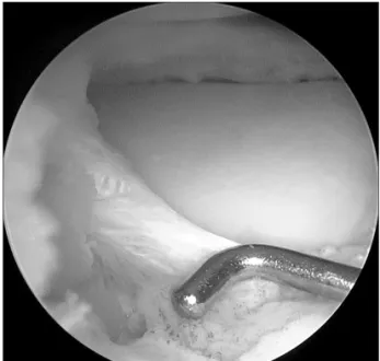
high far anteromedial portal. Fig. 2. During diagnostic arthroscopy, we found degenerated and flap tear in anteromedial portion of complete discoid meniscus.
to allow for unrestricted transverse movement of the blade.
Using a No. 11 long blade holder with a No. 15 blade inserted through the high far anteromedial portal, an initial stab incision approximately 15 mm long was made into the periphery of the anterior segment, leaving about 6 mm rim (Fig. 4). At this time, it is important for the operator to avoid the occurrence of an injury to the anterior cruciate ligament by making the No.
15 blade face anterior. If the blade faces posterior during the approach to the anterior horn, damage to the anterior cruciate ligament can occur. The stab incision in the anterior segment of the meniscus was extended to the corner between the anterior
and middle segments, taking great care to avoid too much extension. Keeping the 30o arthroscope in the high lateral axillary portal, a piecemeal meniscal excision was made in the remaining middle and posterior segments using a basket punch through the high far anteromedial portal and standard anteromedial portal, maintaining a 6 mm rim from anterior to posterior portion (Figs. 5, 6). Occasionally, the arthroscope was placed though the standard anteromedial portal, and we made an additional low anterolateral portal for easy access to the posterior horn of the meniscus. Then, we removed the remaining posterior meniscus and trimmed the peripheral rim to make a smooth contour using Fig. 3. Spinal needle was used for simulation of the pathway of the
instrument through the high far medial portal.
Fig. 4. Initial stab incision approximately 15 mm long was made into the periphery of the anterior segment of the lateral discoid meniscus.
Fig. 5. We performed piecemeal excision of the remaining meniscus on middle and posterior portion.
Fig. 6. The remaining 6 mm rim on overall meniscus could be conform- ed using 5 mm arthroscopic probe.
the basket punch and a motorized shaver.
3. Clinical Evaluation
The clinical results were assessed using the scale of Ikeuchi2). An excellent result was indicated by no limitation of motion, clicking, noise or pain; a good result was indicated by occasional slight pain, but no other symptoms associated with motion; a fair result was indicated by slight pain, clicking or noise with motion as well as limitation of motion; and a poor result was indicated by pain at rest as well as with motion, and limitation of motion. We also evaluated preoperative and postoperative Lysholm scores. The clinical results of Lysholm scores were analyzed using Wilcoxon signed ranks test, and a p-value of less than 0.05 was considered statistically significant.
Results
The mean duration of symptoms before surgery was 5 months (range, 1 to 9 months). The signs included a locking in 22 knees (36.6%), effusion in 14 knees (23.3%), and clunk was noted in 19 (31.6%) knees (Table 1). Meniscal shape according to the classification of Watanabe was complete in 32 knees (53.3%) and incomplete in 28 knees (46.6%), and Wrisberg type could not be found in any of the knees (Table 2). The shape of the tear in complete type lesions was horizontal cleavage in 17 knees (53.1%), flap or complex degenerated tear in 10 knees (31.2%) and radial tear in 5 knees (15.6%). Of the 28 knees that had an incomplete type lesion, 14 knees (50%) had a radial tear, 4 knees (14.3%) had a longitudinal tear and 10 knees (35.7%) had a complex degenerated tear (Table 3). Clinical results assessed using the Ikeuchi’s grading system were excellent in 38 (63.3%), good in 13 (21.6%), fair in 8 knees (13.3%) and poor in 1 knee (1.6%).
Notably, none of our patients required repeated procedures due
to poor results. Complications such as infection, retear of the meniscus, limited joint motion and unrelieved swelling were not demonstrated in any cases. The average Lysholm score of the 60 knees was 82.8 points preoperatively, and the score was improved to 95.4 points at the final follow-up evaluation. A statistically significant difference was found in the Lysholm scoring in the Wilcoxon Signed Ranks test (p=0.00).
Discussion
Kaplan11) speculated that discoid menisci may be caused by abnormal motion of the menisci, but a universally acceptable explanation of discoid lateral meniscus has not yet been developed. A discoid lateral meniscus is more common than a discoid medial meniscus, and has frequently been found in both knees. Surgical treatments for symptomatic discoid lateral meniscus are still controversial. Total meniscectomy had been widely indicated for symptomatic discoid meniscus in the past.
Several studies showed excellent results after total meniscectomy.
In 1995, Washington et al.12) reported on long-term follow-up results of total meniscectomy for 18 cases of discoid meniscus with an average duration of 17 years. They concluded that total meniscectomy might offer the best prognosis, with no evident degenerative change on roentgenograms. Conversely in 1998, Raber et al.13) retrospectively reviewed the long-term results of total meniscectomy for discoid lateral meniscus and reported osteoarthritic changes compared with the untreated contralateral knee in 10 of 11 knees. So, while total meniscectomy was recommended as a treatment for symptomatic discoid lateral meniscus, it caused progressive osteoarthritis, and poor prognosis had been reported in many studies9,10,14). For this reason, a partial meniscectomy (so called “saucerization”) for the treatment of symptomatic discoid meniscus has been recommended to reduce the progressive degeneration of the cartilage in the joint. The goal of a partial meniscectomy is to remove the central portion of the discoid meniscus and leave a stable, balanced rim. By doing so, the remaining portion performs the function of the Table 1. Preoperative Symptoms and Signs of the 58 Patients
Preoperative symptom No. (%)
Locking 22 (36.6)
Effusion 14 (23.3)
Clunk 19 (31.6)
Table 2. Meniscal Shapes According to the Classification of Watanabe
Meniscal shape No. (%)
Complete type 32 (53.3)
Incomplete type 28 (46.6)
Wrisberg type 0 (0)
Table 3. Shapes of Discoid Lateral Meniscus Tear
Tear patients No. (%)
Complete horizontal cleavage tear 17 (53.1)
Complete flap or complex tear 10 (31.2)
Complete radial tear 5 (15.6)
Incomplete radial tear 14 (50.0)
Incomplete complex degenerated tear 10 (35.7)
Incomplete longitudinal tear 4 (14.3)
meniscus, preventing some of the instability resulting from total meniscectomy. The width of the rim of the remaining meniscus is dependent on the degree of the torn meniscus. The posterior and inside of the middle segment are known as the most common tear sites of discoid meniscus. When this site was torn, Hayashi et al.9) left 6−8 mm width for complete and incomplete types of lesions, and Vandermeer and Cunningham10) left 4−5 mm width.
We left the rim 6 mm wide to make a stable rim and to avoid the impingement of the femoral condyle against the rim of the meniscus. There have been many surgical procedures of partial meniscectomy, such as open excision, piecemeal arthroscopic excision, morcellation excision and semiarthroscopic technique.
The one-piece excision as described by Hayashi et al. and Kim et al. seems to be very difficult to perform7,9). The two piece excision of discoid meniscus as described by Ogata8) is not different from the one piece excision methods. Although piecemeal excision is technically less difficult to perform, it is a time-consuming and often frustrating arthroscopic experience8). In my experience of the partial excision of the lateral discoid meniscus, in one-piece, two-piece and piecemeal excision, it is often difficult to cut the anterior segment with scissors or forceps. I also noticed that a stab incision with a knife to the anterior part of the meniscus makes the subsequent cutting using basket forceps much easier.
A high far anteromedial portal, located approximately 1.5 cm medial and 1 cm high to the standard anteromedial portal, is most convenient for making the anterior stab incision by knife.
We penetrated the capsule on the far anteromedial portal using an 18 gauge spinal needle and simulated the passway of a knife.
Then, we conformed the passway and approached the anterior horn of the meniscus again. It is critical that operator face the No.
15 blade anteriorly to avoid the occurrence of injury of anterior cruciate ligament. If the blade is faced posterior during approach to the anterior horn, damage to the anterior cruciate ligament may occur. Although the far anteromedial portal is convenient for the anterior cut, it may be too high to make a satisfactory cut to the posterior part of the meniscus. Once the anterior half of the discoid meniscus is removed, resection of the middle portion becomes very easy to perform through standard medial portal.
Occasionally, additional low anterolateral portal is made to perform the resection of the posterior part of the meniscus after switching the portal. It is important to not hesitate to make an additional portal to enable an easy and safe approach to the target meniscus. Sugawara et al.14) stated that 9 knees required repeat arthroscopic surgery in a group of 139 patients who underwent arthroscopic meniscectomy. Among these, 7 knees exhibited retearing of the remnant meniscus after partial resection of
lateral discoid meniscus. Vandermeer and Cunningham10) reported that retearing of the saucerized rim occurred in 12%
of patients who underwent partial meniscectomy of the discoid lateral meniscus. Radiographs do not play a significant role in the diagnosis of lateral discoid meniscus. Femoral condylar flattening and tibial condylar cupping are the only clues for abnormal meniscal condition. Magnetic resonance imaging can accurately show a discoid meniscus and reveal a tear. Its positive value in diagnosing discoid lateral menisci has been reported as 92%15). A study by Ryu et al.15) involving 77 knees with MRI findings of lateral discoid meniscus tear stated that the most common type of discoid lateral meniscus tear was peripheral tear with horizontal tears. We checked MRIs on the 60 knees preoperatively and found an abnormal shape of the lateral meniscus, with or without tears, in all patients. So, the positive value of our study seems to be higher than that of previous reports. In our series, there is no reoperation for retearing or remnant meniscus. According to the grading system of Ikeuchi, results at follow-up of the 60 knees with symptomatic lateral discoid meniscus were satisfactory.
Conclusions
The clinical outcomes associated with our new arthroscopic technique using knife and high far medial portal in lateral discoid partial meniscectomy suggest successful results. Our surgical technique of arthroscopic excision of the symptomatic discoid meniscus seems to be an easier and convenient method.
Conflict of Interest
No potential conflict of interest relevant to this article was reported.
References
1. Young RB. The external semilunar cartilage as a complete discoid meniscus and memoranda in anatomy. London:
Williams and Nortage; 1889.
2. Ikeuchi H. Arthroscopic treatment of the discoid lateral meniscus: technique and long-term results. Clin Orthop Relat Res. 1982;(167):19-28.
3. Smillie IS. Injuries to the knee joint. 4th ed. Edinburgh:
Churchill Livingstone; 1970.
4. Noble J. Lesions of the menisci: autopsy incidence in adults less than fifty-five years old. J Bone Joint Surg Am.
1977;59:480-3.
5. Casscells SW. Gross pathological changes in the knee joint of the aged individual: a study of 300 cases. Clin Orthop Relat Res. 1978;(132):225-32.
6. Watanabe M, Takeda S, Ikeuchi H. Atlas of arthroscopy.
Tokyo: Igaku-shoin; 1978. p88.
7. Kim SJ, Yoo JH, Kim HK. Arthroscopic one-piece excision technique for the treatment of symptomatic lateral discoid meniscus. Arthroscopy. 1996;12:752-5.
8. Ogata K. Arthroscopic technique: two-piece excision of discoid meniscus. Arthroscopy. 1997;13:666-70.
9. Hayashi LK, Yamaga H, Ida K, Miura T. Arthroscopic meniscectomy for discoid lateral meniscus in children. J Bone Joint Surg Am. 1988;70:1495-500.
10. Vandermeer RD, Cunningham FK. Arthroscopic treatment of the discoid lateral meniscus: results of long-term follow- up. Arthroscopy. 1989;5:101-9.
11. Kaplan EB. Discoid lateral meniscus of the knee joint;
nature, mechanism, and operative treatment. J Bone Joint Surg Am. 1957;39:77-87.
12. Washington ER 3rd, Root L, Liener UC. Discoid lateral meniscus in children: long-term follow-up after excision. J Bone Joint Surg Am. 1995;77:1357-61.
13. Raber DA, Friederich NF, Hefti F. Discoid lateral meniscus in children: long-term follow-up after total meniscectomy. J Bone Joint Surg Am. 1998;80:1579-86.
14. Sugawara O, Miyatsu M, Yamashita I, Takemitsu Y, Onozawa T. Problems with repeated arthroscopic surgery in the discoid meniscus. Arthroscopy. 1991;7:68-71.
15. Ryu KN, Kim IS, Kim EJ, Ahn JW, Bae DK, Sartoris DJ, Resnick D. MR imaging of tears of discoid lateral menisci.
AJR Am J Roentgenol. 1998;171:963-7.