A Novel Fluoroscopic View for Positioning the AO Clavicle Hook Plate Decreases Its Associated in situ Complications
Yoon-Suk Hyun , Gab-Lae Kim, Sang-Min Choi, Woo-Jin Shin, Dong-Yeon Seo
Department of Orthopaedic Surgery, Kangdong Sacred Heart Hospital, Hallym University College of Medicine, Seoul, Korea
Background: The goal of this study was to evaluate whether a modified fluoroscopic technique for positioning a hook plate affected the clinical results of treating Neer type II distal clavicle fractures and Rockwood type V acromioclavicular (AC) joint separations with this de- vice.
Methods: The study was a retrospective consecutive case series with data analysis. Sixty-four patients with a Neer type II distal clavicle fracture or a Rockwood type V AC joint injury treated between March 2009 and June 2013 were divided into 2 groups: traditional fluo- roscopic technique (traditional view, 31 patients) or modified fluoroscopic technique (‘hook’ view, 33 patients). A visual analogue scale (VAS) score, the modified University of California-Los Angeles (UCLA) shoulder scale score, and radiographic osteolysis were the main outcome measures.
Results: The traditional group included a significantly larger number of patients with acromial osteolysis than the hook view group: 23 patients (74.2%) vs. 11 patients (33.3%), respectively (p=0.01). Before plate removal, the hook group reported less pain and higher UCLA shoulder scale scores than the traditional group: average VAS score, 1.55 vs. 2.26, respectively; average UCLA score, 30.88 vs.
27.06, respectively. However, there was no significant difference after plate removal.
Conclusions: The hook view allows more accurate bending of the hook plate around the contour of the acromion, resulting in de- creased osteolysis, decreased pain, and better function with the plate in situ.
(Clin Shoulder Elbow 2016;19(1):25-32)
Key Words: Shoulder; Acromioclavicular joint; Fracture; Dislocation; Fixation
Copyright © 2016 Korean Shoulder and Elbow Society. All Rights Reserved. pISSN 2383-8337
Clinics in Shoulder and Elbow Vol. 19, No. 1, March, 2016 http://dx.doi.org/10.5397/cise.2016.19.1.25
Received October 4, 2015. Revised December 21, 2015. Accepted December 26, 2015.
Correspondence to: Yoon-Suk Hyun
Department of Orthopaedic Surgery, Kangdong Sacred Heart Hospital, Hallym University College of Medicine, 150 Seongan-ro, Gangdong-gu, Seoul 05355, Korea
Tel: +82-2-2224-2230, Fax: +82-2-489-4391, E-mail: [email protected] Financial support: None. Conflict of interests: None.
Introduction
High-grade acromioclavicular (AC) joint separations and distal clavicle fractures accompanied by disruption of the coracocla- vicular ligaments can produce deformity and loss of shoulder function.1,2) Despite controversy regarding whether these injuries should be treated nonoperatively or surgically, the surgical treat- ment remains a challenge because of a variety of factors.
The challenge of treating these injuries is reflected in the wide variety of available surgical techniques.3-9) One of these techniques, the clavicular hook plate, has been advocated by several studies.10-14) The advantage of the hook plate is that it can be used by itself to maintain reduction of the distal clavicle or
in conjunction with ligament reconstructions.6) Its disadvantages are that it can result in pain from acromial osteolysis and that a second operation is required for removal of the plate.15-19)
In our clinical experience with Neer1) type II distal clavicle fractures (Orthopaedic Trauma Association20) type 15-C1.2, 15- C1.3, 15-C2.2, and 15-C2.3) and Rockwood2) type V AC joint separations (Orthopaedic Trauma Association20) type 10-B3.3 and 10-B3.4), we found that the tip of the hook was irritating the acromial undersurface in most cases after conforming the AO hook plate and hook with a conventional shoulder anteropos- terior (AP) radiographic view and altering the shape of the hook portion of the plate to better fit the acromion by assessing the acromial shape with a special fluoroscopic image resulted in less
osteolysis, less pain, and better function with this device in situ than after a traditional placement technique. Our hypothesis is that altering the shape of the hook with our special fluoroscopic image during AO hook plating can cause conforming the plate and the hook to the anatomy for prevention of osteoysis and improvement of its clincal consequences. The goal of this study was to evaluate this observation.
Methods
The Institutional Review Board of The Hallym University granted permission for this study. This study was a consecutive case control series of patients treated at one institution from March 2009 through June 2013. Inclusion requirements for pa- tients included a closed Neer type II distal clavicle fracture or a Rockwood type V AC joint injury, an acute injury to 1 shoulder only, normal function of the shoulder before injury, no other concomitant injuries (via shoulder magnetic resonance imag- ing [MRI] before surgery), and at least 12 months of follow-
up. Exclusion criteria were bilateral injuries, previous shoulder problems on the affected side, a history of previous shoulder surgery on the affected side, or polytrauma of other extremities.
Nineteen patients were excluded: 8 patients had rotator cuff problems, 5 patients had a problem of biceps long head tendon, 3 patients had both, one had other trauma, with a proximal humerus fracture on the same side treated by plating, 1 patient had a history of previous rotator cuff repair on the same side, and 1 patient had bilateral distal clavicle fractures and a 5th cer- vical spine fracture with nerve symptom. Therefore, our study group consisted of 64 patients with a Neer type II distal clavicle fracture (n=40) or a Rockwood type V AC separation (n=24) (Table 1).
All patients were treated with an AO clavicular hook plate (Synthes, Paoli, PA, USA). All patients were placed in the beach chair position, and fluoroscopy was used to verify the plate position and adequate reduction of the deformity. To promote fracture healing, all patients had a superior incision with minimal dissection of the periosteum. The deformity was reduced and
Table 1. Patient Demographics
Parameter Traditional view group Hook view group p-value
Age (yr) 38.3 (18–67) 39.1 (22–62) 0.79
Gender ratio (male:female) 27:4 28:5 0.80
Patients with dominant side involved 20 24 0.13
Follow-up period aft er plate removal (wk) 76.29 (49.43–152.42) 70.47 (49.14–102.71) 0.40
Time between two surgeries (wk) 17.95 (12.14–24.26) 18.23 (12.71–24.86) 0.75
Time from plating to last follow-up (wk) 94.29 (63.14–170.71) 88.57 (67–120) 0.43
Fracture case 20 20
Dislocation case 11 13
Values are presented as median (range) or number only.
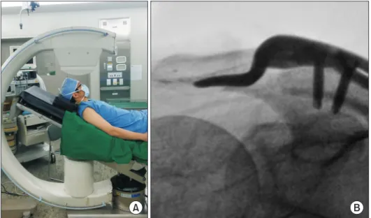
A B
Fig. 1. Hook view. (A) Subject positioning for the fluoroscopic hook view. (B) Hook view can show the optimal contact of the hook plate.
held in position using Kirschner (K)-wires inserted into the distal and proximal fragments for type II clavicle fractures and from the lateral acromion across the AC joint into the distal clavicle for AC joint separations.
For application of the hook plate, the patients underwent 1 of 2 fluoroscopic view techniques: a traditional AP fluoroscopic view of the shoulder (31 patients) or a modified AP fluoroscopic view, which we have termed the ‘hook’ view (Fig. 1; 33 pa- tients). This hook view is obtained by directing the beam from anterior to posterior with the beam placed approximately 30 to 40 degrees caudally; the angle is adjustable depending on anatomic variability. To avoid an impedance of the metal frame of the bed, arm and shoulder should be placed out of the bed as in shoulder arthroplasty (Fig. 1A). The hook view shows the inferior cortex of the posterior portion of the acromion (with its own unique tilt angle) better than the traditional view and thus permits better contouring of the plate to allow closer contact of the hook with the undersurface of the acromion (Fig. 2).
During plating, plate bending was necessary in all cases with traditional shoulder AP view and it was usually done in a bridge part between hook and plate body, not the hook itself. However, the hook view showed us that bending the hook itself as well as the bride part was also absolutely necessary for best fit between hook and acromion in all cases with hook plating (Fig. 3). Under the hook view, plate contour could be adjusted to the acromial undersurface until the tip and rod part of the hook have antomi- cal conformity with the acromion. Regarding the hook bending, we did not realize the necessity of altering of the tip of the hook before the use of the hook view. Develpment of the hook view caused us to appreciate the necessity of bending the hook (tip and rod portion of the hook) as well as the bridge part. Thus,
in some cases with traditional shoulder AP view, we did a blind altering of the hook and rod portion without any concrete idea regarding how to decrease pain after plating. The extent of fit between acromion and hook was not definitely evaluated in the traditional group. In cases with hook view, hook bending was done gradually and accurately in all cases to obtain the best fit.
The tip of the hook was bent downward in both groups. The main point in surgical procedures in both groups is that hook altering and placement under two different types of imaging can make a difference in the extent of altering of hook part and placement.
There are 3 options for hook plating according to hook depth (12, 15, 18 mm). After trying all 3 options, we selected one because it depends on the distance from the upper most part of the most distal part of the clavicle to the undersurface of the posterior acromion. We found that choosing the wrong plate with inappropriate hook depth made plating very difficult.
For all patients, the plates were then applied and their posi- tions were verified. For patients with AC separation, the AC liga- ments were then repaired using a transosseous suture technique.
Once the plate was applied and secured the K-wires were re- moved.
After surgery, all patients were administered an intravenous patient controlled analgesia for 2 days after surgery and took the same nonsteroidal antiinflammatory drugs with the same schedules. All patients were treated with a similar rehabilitation program under the same physical therapeutic team. All patients were allowed to move their fingers, wrists, and elbows imme- diately. Pendulum exercises were allowed as soon as comfort allowed. All patients were discharged with a home rehabilitation program, and active shoulder range of motion was allowed as tolerated. Patients were seen at regular intervals, and shoulder range of motion was measured using hand-held goniometers.
At the preoperative visit for plate removal, all patients under-
Fig. 3. Aft er plate insertion with traditional shoulder anteroposterior view, the hook view shows a sharp contact between hook and acromion.
Fig. 2. Model showing the line of the X-ray beam for the traditional view (dotted arrow) and the hook view (solid arrow). Slanted dotted lines: axis of the posterior acromion.
went a physical examination for evaluation of shoulder range of motion shoulder range of motion. All patients completed a visual analogue scale (VAS) pain assessment and a modified Uni- versity of California-Los Angeles (UCLA) shoulder scale outcome score.21)
For patients with AC separations, plate removal was recom- mended if 3 months had passed after plate application and AC joint tenderness had ended. For all patients with AC separa- tions, plates were removed at an average of 16.82 weeks (range, 13.71–19.14 weeks) after surgery. For patients with fractures, plate removal was recommended as soon as there was radio- graphic evidence of fracture healing; plates were removed at an average of 18.86 weeks (range, 12.14–25.43 weeks) after surgery. All patients with fractures were followed until there was radiographic evidence of fracture healing (average, 17.45 weeks;
range, 12–23.71 weeks). Patients with AC separations were fol- lowed until there was no tenderness at the AC joint.
At the final follow-up after plate removal (average, 74.29 weeks; range, 49.43–152.43 weeks), all patients were again ex- amined for range of motion, completed a VAS pain assessment, completed a modified UCLA shoulder scale outcome score, and underwent radiographic examination (AP and axillary shoulder views for all, plus a hook view for those with that view previ- ously). Pain after plating is usually provoked by motion rather than rest. Patients were asked to assess the average pain level in their daily activity because patients have different activity levels.
The osteolytic lesion is defined as a rod shaped radio-lucent le- sion observed at the same location where the hook was seated.
All radiographs were evaluated for osteolysis of the undersurface of the acromion (Fig. 4) by an independent examiner who was blinded to the type of fluoroscopic view taken at the time of
surgery, but osteolysis could be identified well only on the axil- lary shoulder view. Because the amount of osteolysis could not be quantified, only two categories, whether or not the osteolytic lesion was seen in postoperative axillary view.
Descriptive statistics, including means and standard devia- tions, were calculated for comparison of the traditional and hook view groups. The t-test was used to determine significant difference in radiographic and the identified clinical outcomes and pain between the 2 groups. Because differences of each variable before and after plate removal were not normally dis- tributed, nonparametric univariate analysis was performed with the Wilcoxon signed rank test for comparison of results before and after plate removal. Data analysis was performed using the SPSS software ver. 15.0 (SPSS Inc., Chicago, IL, USA). A p-value of <0.05 was considered statistically significant.
Results
Prior to plate removal, there were significant differences be- tween the groups in terms of pain and function (Table 2): the hook view group had significantly less pain (pain VAS score) and significantly better function (UCLA shoulder scale score, active forward elevation and abduction of the shoulder) than the tra- ditional view group. However, after plate removal at the last fol- low-up, there was no significant difference in shoulder range of motion, pain VAS score, or UCLA shoulder scale score between the 2 groups (Table 2).
There was no occurrence of infection in either group, and all fractures healed. In the traditional view group, 1 patient had dislodgement of the hook that required reoperation (successful), and 1 patient required an arthroscopic release of adhesions dur- ing plate removal because of stiffness. In both groups there was no implant breakage, fracture nonunion, severe displacement of the AC joint, or periprosthetic bone fracture during plate inser- tion, after plate insertion, or after plate removal.
A significantly larger number of patients had acromial osteoly- sis in the traditional view group compared with the hook view group (23/31 patients vs. 11/33 patients, p=0.01) (Table 2).
Both groups showed statistically significant improvements in pain VAS score, UCLA score, and active abduction between the scores prior to plate removal and after plate removal (Table 2).
Discussion
This study has shown that, prior to plate removal, using our hook fluoroscopic technique when inserting the AO clavicular hook plate for a Neer type II distal clavicle fracture or a Rock- wood type V AC separation resulted in less pain, better range of motion, and better function with the plate in situ than a plate inserted using the traditional technique. However, after plate removal we found that the results clinically were equivalent Fig. 4. Acromial osteolysis can be identifi ed on the shoulder axillary view aft er
implant removal.
regardless of the radiographic technique used at the time of sur- gery.
The results of our study are similar to those previously re- ported for hook plates used for these types of injuries. High complications rates, with the use of hook plates, including ac- romial osteolysis, have been reported in the previous literature.
Development of acromial osteolysis after surgery with a hook plate was reported in 86.7% (13/15) by Muramatsu et al.,18) 16.1% (5/31) by Meda et al.,17) 27.8% (5/18) by Tambe et al.,19) and 25.0% (7/28) by Tiren et al.13) Our study confirms those high rates of acromial osteolysis, although the rate was significantly lower in the hook view group than in the traditional view group:
33.3% (11/33 patients) vs. 74.2% (23/31 patients), respectively.
Except the study by Muramatsu et al.,18) the rates of osteolysis were lower compared with our study. We are not sure how os- teolysis was defined in previous studies; we evaluated the status of osteolysis after plate removal. If they checked the status of osteolysis before plate removal, the rate of osteolysis could be less recognized because the hook could conceal the extent of osteolysis.
The literature also documents pain and disability while the hook plate is in place. Impingement pain or pain on shoulder motion after hook plating was reported in 30/44 (68.2%) pa-
tients by Renger et al.,11) 9/10 (90.0%) patients by Bhangal et al.,22) 6/31 (19.4%) patients by Meda et al.,17) all 3 patients of Chandrasenan et al.,15) and 9/28 (32.1%) patients by Tiren et al.13) In our study, pain with shoulder motion with the hook plate in situ was also found in 35/64 (54.7%) patients. However, the hook view group (36.4%, 12/33 patients) showed significantly less pain than patients with a hook plate inserted using the tradi- tional radiographic view (74.2%, 23/31 patients). As reported by the other authors listed above, these symptoms of pain and loss of motion improved significantly once the plate was removed, particularly in the traditional view group. These results support the idea that plate removal should be considered in most cases as the clinical outcome is better with patients having less pain, fewer functional deficits, and better UCLA scores.
The exact cause of the pain when hook plates are in situ is controversial. Hackenberger et al.,23) who evaluated 28 shoul- ders with Tossy type III AC separation treated via hook plates by following-up with ultrasonography and MRI, found no high- grade rotator cuff lesions or signs of impingement. In contrast, Chandrasenan et al.15) reported acromial erosions and damage to the supraspinatus around the hook portion via arthroscopic examination in some patients.
Our study was based on an idea that reshaping of the hook Table 2. Comparison of Clinical and Radiographic Results
Parameter Traditional view group Hook view group p-value
UCLA shoulder scale score
Before plate removal 27.06 ± 2.75 30.88 ± 2.33 <0.001
At last follow-up 31.42 ± 2.01 31.70 ± 1.91 0.547
p-value <0.001 <0.001
VAS score for pain
Before plate removal 2.26 ± 0.96 1.55 ± 1.03 0.006
At last follow-up 0.77 ± 0.62 0.76 ± 0.61 0.914
p-value <0.001 0.006
Range of motion ( o )
Active forward elevation before plate removal 135.00 ± 13.66 147.70 ± 13.75 <0.001
Active forward elevation at last follow-up 153.40 ± 9.34 153.90 ± 10.44 0.825
p-value <0.001 0.001
Range of motion ( o )
Active abduction before plate removal 100.60 ± 13.40 111.82 ± 14.83 0.002
Active abduction at last follow-up 118.59 ± 14.27 117.72 ± 14.25 0.819
p-value 0.001 0.003
Coracoclavicular distance at last follow-up (mm) 2.45 ± 1.04 2.54 ± 1.33 0.545
Percentage acromial osteolysis* 73.7 33.3 0.01
Values are presented as mean ± standard deviation or percent only.
UCLA: University of California-Los Angeles, VAS: visual analog scale.
*Th is mark indicates the number of patients, not the extent of osteolysis.
part of the plate in accordance with contour of the acromial un- dersurface could avoid sharp irritation by the hook tip or edge.
According to the study by ElMaraghy et al.,16) the hook part tends to be located under the posterior part of the acromion (Fig. 5). Impingement, subacromial bursitis, and subacromial osteolysis on X-ray are signs of a mismatch between the plate and the anatomy of the patient.12) These complications can be minimized by performing an anatomic fit of the plate dur- ing the procedure.12) Muramatsu et al.18) found it necessary to bend the hook in 77% of their patients, and found migration of the hook after fixation in most of their patients. It was assumed that forced fixation of the plate to the clavicle caused a pres- sure concentration to the hook itself and then produced hook migration. The appropriate bending of the hook was performed to prevent forced fixation of the plate.18) Lee et al.24) performed arthroscopy during the procedure to verify the position and fit
of the hook and tip besides intra-operative fluoroscopy verifica- tion. However arthroscopic verification can require an unneces- sary sacrifice of healthy bursal tissue and another incisions. We found that by altering the fluoroscopic view as described here, the plate contour could be adjusted to the acromion, potentially lessening the complications. With traditional shoulder AP view we could not determine whether or not placement of the hook on the acromion is important because the relationship between hook and acromial undersurface could not be evaluated, and we could not confirm whether the tip of hook was well placed according to the author’s point, but we found that all hooks performed using traditional shoulder AP view were confirmed as poorly placed with hook view, meaning that all hooks usually had a sharp contact with the undersurface of the acromion with traditional view (Fig. 3).
To the best of our knowledge, there is no previous publica- tion describing a radiologic view that delineates the undersurface of the posterior acromion well. In a review of the 11 previously published studies4,10-12,18,22,25-28) describing the surgical technique of the hook plate, conventional shoulder AP and axillary views and AP AC joint views were usually used for preoperative and postoperative evaluation. Among these studies, fluoroscopy was used during plating in only three studies and plate bending was reported in six studies.12,15,18,24-27) We have found that hook plate application was impossible without plate bending and that bending of the hook part in plate contouring was impossible without fluoroscopic guidance. Traditional radiographic views as reported in the literature may not provide an optimal display of the posterior part of the acromion. The main advantage of our hook view is that it enables the surgeon to contour the plate so that it has a more accurate contact with the posterior acromion (Fig. 6). However, other factors regarding acromial osteolysis should be considered, including that it cannot be completely avoided in cases of AO hook plating because this plate was originally designed as a hook that should have contact with the acromion for maintenance of reduction. And the acromial un- Fig. 5. A hook plate is supposed to be located under the posterior part of the
acromion (circle).
A B
Fig. 6. Postoperative radiographs. (A) A traditional anteroposterior radiograph shows that the hook plate appears to irritate the ro- tator cuff (arrow). (B) Th e hook view shows the plate to be well located (arrow).
dersurface may not be parallel with the motion of the hook dur- ing shoulder motion, so that the best fit between hook and acro- mion cannot avoid acromial osteolysis because the side edge of the hook can erode the acromion despite having the best fit. We confirmed it through arthroscopic exam in some cases with pain after plating (Fig. 7). Therefore, acromial osteolysis could not been eliminated despite the best fit under the author’s standard in the group with H-view despite a significantly low incidence of ostelysis compared with the group with traditional shoulder AP view. Bending of the hook with H-view contributed to reducing the amount of osteolysis, not complete removal.
Our study has several limitations. First, it is a consecutive case series with a historical control group. A prospective, randomized study might produce different results. We did not compare the fluoroscopic techniques in every patient to determine the dif- ference in plate placement using one technique of placement versus another. There was no blinding in this study, but the VAS and UCLA shoulder scale scores were completed by the patients without the assistance or influence of the examiners. Also, the physical examination was not blinded, which might have influ- enced some of the measurements. The limitations of hand-held goniometers are well known, and the use of multiple examiners might have influenced the results.29,30) In the pain evaluation the average pain level in daily activity was used instead of a separate evaluation of resting pain and pain on activity. Unfortunately, we had no method for quantifying the amount of osteolysis. There- fore the relationship between osteolysis and pain could not be analyzed, which was also the weakest point of this study.
Conclusion
Use of a modified fluoroscopic technique (the hook view) for
placement of the AO hook plate enables more accurate place- ment of the hook under the posterior acromion. Our study has shown that this procedure results in less osteolysis, less pain, and less loss of function with the plate in situ. It is important to con- form the plate and the hook to the anatomy in order to prevent osteolysis and its clinical consequences. A prospective and ran- domized study would be recommended to study the use of this fluoroscopic technique for the placement of hook plates in the distal clavicle.
Acknowledgements
The authors would like to thank Edward G. McFarland for his assistance with this manuscript.
References
1. Neer CS 2nd. Fracture of the distal clavicle with detachment of the coracoclavicular ligaments in adults. J Trauma. 1963;3:
99-110.
2. Rockwood CA Jr. Injuries to the acromioclavicular joint. Part II: subluxations and dislocations about the shoulder. In: Rock- wood CA Jr, Green DP, eds. Fractures in adults. Vol. 1. 2th ed.
Philadelphia: JB Lippincott; 1984. 860-5.
3. Boileau P, Old J, Gastaud O, Brassart N, Roussanne Y. All- arthroscopic Weaver-Dunn-Chuinard procedure with double- button fixation for chronic acromioclavicular joint dislocation.
Arthroscopy. 2010;26(2):149-60.
4. De Baets T, Truijen J, Driesen R, Pittevils T. The treatment of acromioclavicular joint dislocation Tossy grade III with a clavi- cle hook plate. Acta Orthop Belg. 2004;70(6):515-9.
5. Macheras G, Kateros KT, Savvidou OD, Sofianos J, Fawzy EA, Papagelopoulos PJ. Coracoclavicular screw fixation for unstable distal clavicle fractures. Orthopedics. 2005;28(7):693-6.
6. Martetschläger F, Kraus TM, Schiele CS, et al. Treatment for unstable distal clavicle fractures (Neer 2) with locking T-plate and additional PDS cerclage. Knee Surg Sports Traumatol Ar- throsc. 2013;21(5):1189-94.
7. Murena L, Vulcano E, Ratti C, Cecconello L, Rolla PR, Surace MF. Arthroscopic treatment of acute acromioclavicular joint dislocation with double flip button. Knee Surg Sports Trauma- tol Arthrosc. 2009;17(12):1511-5.
8. Scheibel M, Dröschel S, Gerhardt C, Kraus N. Arthroscopically assisted stabilization of acute high-grade acromioclavicular joint separations. Am J Sports Med. 2011;39(7):1507-16.
9. Weaver JK, Dunn HK. Treatment of acromioclavicular injuries, especially complete acromioclavicular separation. J Bone Joint Surg Am. 1972;54(6):1187-94.
10. Di Francesco A, Zoccali C, Colafarina O, Pizzoferrato R, Flami- ni S. The use of hook plate in type III and V acromio-clavicular Rockwood dislocations: clinical and radiological midterm Hook
Acromion
Rotator cuff
Fig. 7. Arthroscopic examination showed that the hook moves through pas- sive shoulder motion and the range of hook motion is limited in the eroded portion of the acromion.
results and MRI evaluation in 42 patients. Injury. 2012;43(2):
147-52.
11. Renger RJ, Roukema GR, Reurings JC, Raams PM, Font J, Verleisdonk EJ. The clavicle hook plate for Neer type II lateral clavicle fractures. J Orthop Trauma. 2009;23(8):570-4.
12. Tan HL, Zhao JK, Qian C, Shi Y, Zhou Q. Clinical re- sults of treatment using a clavicular hook plate versus a T- plate in neer type II distal clavicle fractures. Orthopedics.
2012;35(8):e1191-7.
13. Tiren D, van Bemmel AJ, Swank DJ, van der Linden FM. Hook plate fixation of acute displaced lateral clavicle fractures: mid- term results and a brief literature overview. J Orthop Surg Res.
2012;7:2.
14. von Heideken J, Boström Windhamre H, Une-Larsson V, Eke- lund A. Acute surgical treatment of acromioclavicular disloca- tion type V with a hook plate: superiority to late reconstruc- tion. J Shoulder Elbow Surg. 2013;22(1):9-17.
15. Chandrasenan J, Badhe S, Cresswell T, Beer JD. The clavicular hook plate: consequences in three cases. Eur J Trauma Emer- gency Surg. 2007;33(5):557-9.
16. ElMaraghy AW, Devereaux MW, Ravichandiran K, Agur AM.
Subacromial morphometric assessment of the clavicle hook plate. Injury. 2010;41(6):613-9.
17. Meda PV, Machani B, Sinopidis C, Braithwaite I, Brownson P, Frostick SP. Clavicular hook plate for lateral end fractures:- a prospective study. Injury. 2006;37(3):277-83.
18. Muramatsu K, Shigetomi M, Matsunaga T, Murata Y, Taguchi T.
Use of the AO hook-plate for treatment of unstable fractures of the distal clavicle. Arch Orthop Trauma Surg. 2007;127(3):
191-4.
19. Tambe AD, Motkur P, Qamar A, Drew S, Turner SM. Fractures of the distal third of the clavicle treated by hook plating. Int Orthop. 2006;30(1):7-10.
20. Marsh JL, Slongo TF, Agel J, et al. Fracture and dislocation classification compendium - 2007: Orthopaedic Trauma As-
sociation classification, database and outcomes committee. J Orthop Trauma. 2007;21(10 Suppl):S1-133.
21. Nutton RW, McBirnie JM, Phillips C. Treatment of chronic rotator-cuff impingement by arthroscopic subacromial decom- pression. J Bone Joint Surg Br. 1997;79(1):73-6.
22. Bhangal KK, Evans SC, Gibbons CE. Treatment of displaced lateral clavicle fractures with the AO hook plate. Eur J Trauma.
2006;32(5):468-70.
23. Hackenberger J, Schmidt J, Altmann T. The effects of hook plates on the subacromial space: a clinical and MRT study. Z Orthop Ihre Grenzgeb. 2004;142(5):603-10.
24. Lee KW, Lee SK, Kim KJ, Kim YI, Kwon WC, Choy WS. Ar- throscopic-assisted locking compression plate clavicular hook fixation for unstable fractures of the lateral end of the clavicle:
a prospective study. Int Orthop. 2010;34(6):839-45.
25. Flinkkilä T, Ristiniemi J, Lakovaara M, Hyvönen P, Leppilahti J.
Hook-plate fixation of unstable lateral clavicle fractures: a re- port on 63 patients. Acta Orthop. 2006;77(4):644-9.
26. Good DW, Lui DF, Leonard M, Morris S, McElwain JP. Clavicle hook plate fixation for displaced lateral-third clavicle fractures (Neer type II): a functional outcome study. J Shoulder Elbow Surg. 2012;21(8):1045-8.
27. Lee YS, Lau MJ, Tseng YC, Chen WC, Kao HY, Wei JD. Com- parison of the efficacy of hook plate versus tension band wire in the treatment of unstable fractures of the distal clavicle. Int Orthop. 2009;33(5):1401-5.
28. Salem KH, Schmelz A. Treatment of Tossy III acromioclavicular joint injuries using hook plates and ligament suture. J Orthop Trauma. 2009;23(8):565-9.
29. Armstrong AD, MacDermid JC, Chinchalkar S, Stevens RS, King GJ. Reliability of range-of-motion measurement in the el- bow and forearm. J Shoulder Elbow Surg. 1998;7(6):573-80.
30. Riddle DL, Rothstein JM, Lamb RL. Goniometric reliability in a clinical setting. Shoulder measurements. Phys Ther. 1987;
67(5):668-73.