Serial Magnetic Resonance Imaging to Determine the Progression of Neglected Recalcitrant Rotator Cuff Tears: A Retrospective
Multicenter Study
Yon-Sik Yoo, Jin-Young Park1, Chang-Hyuk Choi2, Nam-Su Cho3, Chul-Hyun Cho4, Tae-Gang Lim5, Sang-Don Sim6, Tae-Yon Rhie7, Ho Won Lee8, Jong Ho Jung8, Yong-Beom Lee8
Department of Orthopedic Surgery, Hallym University Dongtan Sacred Heart Hospital, Hwaseong, 1Global Center for Shoulder, Elbow and Sports at Neon Orthopaedic Clinic, Seoul, 2Department of Orthopedic Surgery, Catholic University of Daegu School of Medicine, Daegu, 3Department of Orthopedic Surgery, Kyung Hee University Hospital at Gangdong, Seoul, 4Department of Orthopedic Surgery, Keimyung University Dongsan Medical Center, Keimyung University School of Medicine, Daegu, 5Department of Orthopaedic Surgery, Nowon Eulji Medical Center, Eulji University, Seoul, 6Department of Orthopedic Surgery, Donga Hospital, Gwangju, 7Department of Orthopaedic Surgery, Nalgae Hospital, Seoul, 8Department of Orthopaedic Surgery, Hallym University Sacred Heart Hospital, Anyang, Korea
Background: To determine the natural progression of conservatively treated rotator cuff tears, we evaluated changes in radiologic and clinical parameters in patients whose recalcitrant tears were neglected after conservative treatment.
Methods: A total of 73 patients with recalcitrant rotator cuff tears in spite of conservative treatment were included in this study. We mea- sured changes in tear size and in the extent of fatty infiltration of the rotator cuff by comparing the initial and final follow-up magnetic resonance imagings (MRIs). To determine factors influencing the change in tear size, we collected the medical history of patients taken at the time of initial admission.
Results: The average follow-up period was 20.1 months, and the average increase in tear size across this period was 6.2 mm. In terms of steroid injection, we found that the increases in tear size of the steroid injection group (p=0.049) and of the sub-group that had received more than three steroid injections (p=0.010) were significantly greater than that of the non-steroid injection group.
Conclusions: We found that the increase in cuff tear size was on average 6.2 mm across the follow-up period, indicating that neglecting cuff tears may cause them to progress into more severe tears. We also observed that a history of steroid injection might be a possible risk factor for a worse prognosis of cuff tears. Therefore, we suggest that patients with rotator cuff tears and a history of steroid injection are recommended aggressive modes of treatment such as surgery.
(Clin Shoulder Elbow 2017;20(3):133-137)
Key Words: Rotator cuff injury; Steroid; Injection; Intra-articular
Copyright © 2017 Korean Shoulder and Elbow Society. All Rights Reserved. pISSN 2383-8337
Clinics in Shoulder and Elbow Vol. 20, No. 3, September, 2017 https://doi.org/10.5397/cise.2017.20.3.133
Received February 7, 2017. Revised August 15, 2017. Accepted August 15, 2017.
Correspondence to: Yong-Beom Lee
Department of Orthopaedic Surgery, Hallym University Sacred Heart Hospital, 22 Gwanpyeong-ro 170beon-gil, Dongan-gu, Anyang 14068, Korea
Tel: +82-31-380-3770, Fax: +82-31-380-1814, E-mail: [email protected] IRB approval (No. 2016-I152).
Financial support: None. Conflict of interests: None.
Introduction
Rotator cuff tears are one of the most common tendinous injuries in adults.1,2) Muscle fibers of the rotator cuff become torn because mechanistically they receive persistent tension, which in a rotator cuff tear causes even greater contraction-induced damage. The prognosis of rotator cuff arthropathies resulting
from mechanistic reasons alone has been shown to be difficult to predict.3-5) On one hand, the symptoms of 60% of patients with full-thickness cuff tears were found to be improved after conservative treatment.6) On the other hand, 18 of 50 patients with asymptomatic cuff tears were found to have significantly progressed symptoms and exacerbated cuff tears by the end of a 3-year follow-up.3)
Although it is clear the conservative treatment of rotator cuff tears is associated with resolution of pain and improvement in range of motion, the natural history of patients who receive non- surgical approaches to treatment remains unclear.7) The factors thought to influence the natural history of rotator cuff tears, which may also be important for their treatment, are still contro- versial among orthopedic surgeons.8)
Radiological tests such as magnetic resonance imaging (MRI) have been shown to be most accurate for the diagnosis of rotator cuff tears. It has been reported that the accuracy of MRI in diag- nosing full-thickness rotator cuff tears is greater than 95%.9-14) In this study, we analyzed MRI findings of patients who were initially hospitalized for shoulder pain, which was diagnosed as a rotator cuff tear, were treated conservatively, but were later re-hospital- ized for recalcitrant tears that were neglected without appropri- ate treatment. We investigated the natural history of the rotator cuff tears in terms of the change in cuff tear size, as measured on patients’ MRI scans, and of clinical parameters. In addition, we investigated putative factors at the time of initial admission that could be involved in the progression of cuff tear size.
Methods
We enrolled 73 patients who were hospitalized for shoulder pain and whose condition was diagnosed as a rotator cuff tear through diagnostic MRI. The patients’ condition had been either neglected or conservatively treated initially but were neglected without further treatment because the cuff tears were recalci- trant despite treatment. We conducted a multicenter retrospec- tive study involving ten hospitals across Korea to investigate the change in tear size over time. We analyze putative patient- related factors at the time of initial admission, for which we asked patients for information, such as history of trauma, steroid injections, and shoulder use.
We divided the tears into either partial or full-thickness tears on the basis of tear depth. The extent of fatty infiltration was evaluated on oblique sagittal MRI sections and categorized fol- lowing the Goutallier classification system15) at both the initial and final follow-up examinations. Partial rotator cuff tears were evaluated on oblique coronal MRI sections, and complete rota- tor cuff tears were evaluated on oblique coronal MRI sections.
We measured the average change in tear size and in the extent of fatty infiltration between the initial and final follow-up exami- nations in terms of the extent of tendon retraction. Two ortho- pedic surgeons each analyzed the MRI scans, and the intraclass correlation coefficient was used to determine the interobserver reliability.
We measured change in the clinical parameters–pain and shoulder function–denoted as the difference in visual analogue scale (VAS) score for pain and in the American Shoulder and Elbow Surgeons (ASES) score, respectively, between the initial
and final follow-up examinations. The level of shoulder stiffness was determined on the basis of a 3-item criteria (forward eleva- tion, <120°; internal rotation, <L3; or external rotation at side,
<20°). We used either the chi-square or the Fisher exact test to examine the correlation between two categorical variables and the Pearson correlation coefficient, between two continuous variables. The paired t-test was used to perform a quantitative analysis between two independent variables, and ANOVA was used for more than three independent variables. Statistical sig- nificance was considered as a p-value below 0.05.
Results
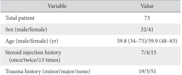
Of the total study participants, 43.8% were male and 56.2%
were female (32 vs. 41 patients). The age of the patients was on average 59.8 years and showed a large range (range, 34–83 years). The proportions of patients with shoulder pain in the absence and in the presence of a history of trauma were 69.9%
(51/73 patients) and 30.1% (22/73 patients), respectively. In the latter group, 19 patients had had light trauma and only 3, severe trauma. All patients received a diagnosis of a rotator cuff tear through diagnostic MRI. The proportion of patients with an oc- cupation involving manual labor was 40.0% (27 patients). Those engaged in sports activities demanding substantial shoulder use was 24.6% of the study population. The proportion of patients who had received at least one steroid injection at the initial ad- mission was 35.6% (26 patients), from which 20.6% (15 patients) had received more than three steroid injections (Table 1).
From initial diagnosis, patients had either not received any sort of treatment or had received various conservative treatments between one month and six year to no avail. The patients were re-hospitalized on average 20.1 months of the initial admis- sion because of recalcitrant and/or exacerbated symptoms. We conducted MRI examinations again at the second admission.
Between the initial and final follow-up examinations, we found that the average VAS score for pain had increased from 5.7 to 6.3 and that the ASES score had decreased from 44.9 to 42.4.
By the final follow-up, the number of patients with shoulder stiffness and with fatty degeneration increased from 11 to 16 pa-
Table 1. Patient Demographics
Variable Value
Total patient 73
Sex (male/female) 32/41
Age (male/female) (yr) 59.8 (34–75)/59.9 (48–83) Steroid injection history
(once/twice/≥3 times) 7/4/15
Trauma history (minor/major/none) 19/3/51 Values are presented as number only or median (range).
tients and from 43 (58.9%) to 61 patients (83.6%), respectively.
Across the same period, we found that the average Goutallier grade denoting the level of fatty degeneration increased from 0 to 2 (Table 2).
The interobserver reliability for the measurements of the change in tear size was 0.88 mm, revealing substantial reliability.
The average increase in tear size depicted on oblique coronal MRI sections between the initial and final follow-up examina- tions was 6.2 mm (from 10.4 to 16.6 mm). The largest increase in tear size was 32.1 mm (Fig. 1). The number of muscles involved in the tear at the final follow-up was greater in ten patients than before (i.e., from an isolated suprapinatus tendon tear to a combined suprapinatus and subscapularis tendon tear).
We also observed that the tear depth was altered in 24 patients (from a partial thickness tear to a full-thickness tear). By gender, the average increase in tear size was 5.66 mm in female patients and 6.85 mm in male patients, but no gender difference was observed. A statistically significant difference was not observed even when we compared by whether or not patient had a labor-
heavy occupation (p=0.986). Although the group that played sports activities demanding substantial shoulder movement had a larger increase in tear size than those that did not, the differ- ence was not significant (p=0.083).
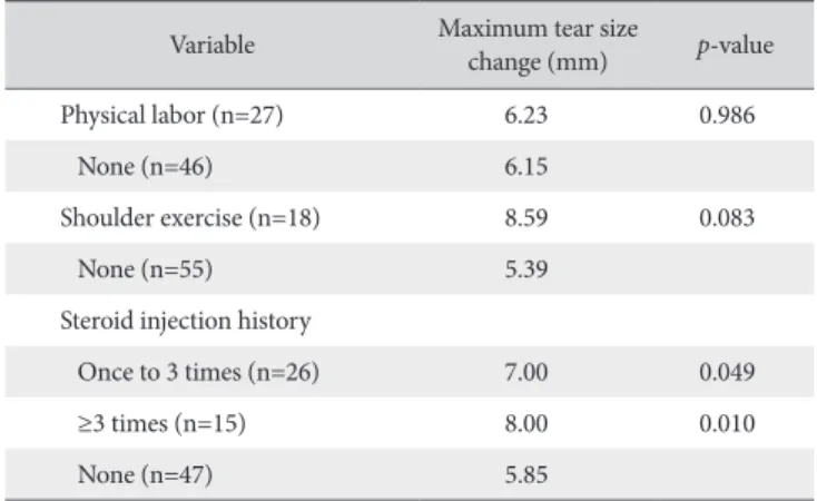
We found that patients who had a history of steroid injection at initial admission had significantly larger change in tear size than those without (p=0.049). This association was even more prominent in patients who had a history of three or more steroid injections at initial admission (p=0.010) (Table 3). The percent- age of patients who went on to receive surgical treatment after the final follow-up was 95.9% (70/73), two of whom underwent shoulder arthroplasty.
Discussion
The natural history of rotator cuff tears remains controversial and is currently being studied by several groups.10) On one hand, Mall et al.5) reported that across a 2-year or longer follow-up full- thickness rotator cuff tears increased by 5 mm in 18% of patients
A B
Fig. 1. (A) Initial oblique coronal magnetic resonance imaging (MRI) section (T2 fat suppression). (B) Final oblique coronal MRI section (T2 fat suppression) at the 5-year follow-up shows an elongated supraspinatus tendon tear.
Table 2. Comparison of Results between Initial and Last Follow-up Data
Variable Initial Last
Mean tear size (mm) 10.4 16.6
Mean VAS (pain) 5.7 6.3
Mean ASES score 44.9 42.4
Stiffness* 11 16
Fatty infiltration* 43 61
Grade 1 28 31
Grade 2 12 20
Grade 3 3 8
Grade 4 0 2
VAS: visual analogue scale, ASES: American Shoulder and Elbow Surgeons.
*The number of patients with shoulder stiffness and with fatty infiltration.
Table 3. Comparison of Maximum Tear Size Change between Initial and Last Follow-up Magnetic Resonance Imaging Oblique Coronal Section
Variable Maximum tear size
change (mm) p-value
Physical labor (n=27) 6.23 0.986
None (n=46) 6.15
Shoulder exercise (n=18) 8.59 0.083
None (n=55) 5.39
Steroid injection history
Once to 3 times (n=26) 7.00 0.049
≥3 times (n=15) 8.00 0.010
None (n=47) 5.85
Values are presented as mean only.
and partial tears progressed into full-thickness tears in 40% of patients (n=195). On the other hand, Fucentese et al.11) reported that the increase in tear size observed on magnetic resonance ar- thrograms after a 3.5-year follow-up was not statistically significant.
The subjects of our study were patients who had received conservative treatment for shoulder pain that had been diag- nosed as a rotator cuff tear through diagnostic MRI. Using pa- tients’ MRI sections, we measured the change in tear size over time and determined factors that may influence these changes.
There has been substantial research on tear size of symptom- atic rotator cuff tears, giving varied results. Previous research has shown that the tear size of full-thickness tears of supraspinatus muscles did not increase with statistical significance across a 3.5- year follow-up in patients younger than 65 years.12) Conversely, full-thickness tears in 39% of patients were found to have signifi- cantly increased by 5 mm on ultrasound findings over a 5-year follow-up.13) Yamanaka and Matsumoto14) also reported that 80%
of patients with partial cuff tears conservatively treated showed an increase in tear size and progression to a full-thickness tear (as determined on joint arthrography) over a 2-year follow-up.
Safran et al.15) reported a 5-mm increase in tear size over a 2- to 3-year period in over half their patients who were aged less than 60 years old and treated conservatively for a rotator cuff tear. With respect to etiology of exacerbated tears, Iannotti and Williams16) have suggested that tears become worse over time because substances that help regeneration at the rotator cuff tear site are washed away by the synovial fluid. In our study, the average follow-up period was 20.1 months across which we ob- served an average 6.2-mm increase in cuff tear size.
Numerous studies have investigated factors influencing the cause of exacerbated or recalcitrant cuff tears. Dean et al.17) reported that steroid injections impair revascularization and pro- duction of hypoxia-inducible factor-1a, which are both involved in the repair response, thereby delaying rotator cuff recovery.
Similarly, Mikolyzk et al.18) demonstrated that steroids weaken the strength of cuff muscles. Our finding that a history of steroid injection at initial admission is associated with a statistically sig- nificant increase in tear size is consistent with these previously reported findings. The same was true for patients who received at least three steroid injections at initial admission. Therefore, we can conclude that conservative treatment in patients with a history of steroid injection may not be as effective as when the same treatment is performed in patients who have no history of steroid injections.
Additionally, we found that the change in tear size did not significantly differ between those who were engaged in sports activities requiring substantial shoulder movement and those who were not, although the former group did tend to have a larger change. Thus, patients with rotator cuff tears should be warned that engaging in too much exercise requiring the shoul- ders could lead to worse prognosis of the cuff tears and that re-
turning to pre-injury level of sports as soon as symptoms alleviate may necessitate surgical treatment later on.
A few studies have shown that conservative treatment of cuff tears improves arm function and alleviates pain at short-to-mid–
term follow-ups. However, other studies have shown that con- servative treatment was associated with low satisfaction levels at long-term follow-ups (more than 6 years).19,20) Many patients with recalcitrant cuff tears in spite of conservative treatment are re-hospitalized within a few years because of recurrent symp- toms. They are found to have exacerbated tears and a signifi- cantly lowered rate of complete and successful cuff repairs.21-23) In our study, we found that over 96% of patients who were re- hospitalized for recalcitrant tears received surgical treatment.
Two patients had severely degenerated tears and therefore underwent arthroplasty. Rotator cuff tears may be neglected de- spite its diagnosis because they are asymptomatic or be treated conservatively and lead to only a temporary relief of symptoms.
But when patients return to unrestrained level of activities with- out completely repairing the rotator cuff tear, we can predict that if symptoms recur patients would most likely require surgi- cal treatment.
There are several limitations that restrict the significance of our study. As a retrospective study with a small study population, the statistical power of this study is limited. Only patients whose symptoms exacerbated or recurred were enrolled in this study, while those whose symptoms had resolved or patients who did not return to our hospital for various reasons (went to a differ- ent hospital or died) were not included; therefore, the study design lends itself to selective bias. These limitations should be addressed in subsequent studies, and a large-scale, randomized controlled prospective study is needed. Yet despite these limita- tions, our study is notable in that it has investigated the natural history of patients with symptomatic rotator cuff tears and un- covered new risk factors of poor prognosis or progression of cuff tear size.
Conclusion
We found that in the absence of appropriate treatment of rotator cuff tears, tear size increased on average 6.2-mm over an average 20.1-month period. We found that a history of steroid injection may be a risk factor for poor prognosis whilst a history of more than three steroid injections was a much stronger in- dicator of progression of rotator cuff tears. Thus, on the basis of our findings, we recommend that when such patients are met at the clinic they should be informed that conservative treatment may be ineffective and be strongly advised to receive more ag- gressive modes of treatment.
References
1. Reilly P, Macleod I, Macfarlane R, Windley J, Emery RJ. Dead men and radiologists don’t lie: a review of cadaveric and ra- diological studies of rotator cuff tear prevalence. Ann R Coll Surg Engl. 2006;88(2):116-21.
2. Rees JD, Wilson AM, Wolman RL. Current concepts in the management of tendon disorders. Rheumatology (Oxford).
2006;45(5):508-21.
3. Moosmayer S, Tariq R, Stiris M, Smith HJ. The natural history of asymptomatic rotator cuff tears: a three-year follow-up of fifty cases. J Bone Joint Surg Am. 2013;95(14):1249-55.
4. Maman E, Harris C, White L, Tomlinson G, Shashank M, Boynton E. Outcome of nonoperative treatment of symptom- atic rotator cuff tears monitored by magnetic resonance imag- ing. J Bone Joint Surg Am. 2009;91(8):1898-906.
5. Mall NA, Kim HM, Keener JD, et al. Symptomatic progression of asymptomatic rotator cuff tears: a prospective study of clini- cal and sonographic variables. J Bone Joint Surg Am. 2010;
92(16):2623-33.
6. Goldberg BA, Nowinski RJ, Matsen FA 3rd. Outcome of non- operative management of full-thickness rotator cuff tears. Clin Orthop Relat Res. 2001;(382):99-107.
7. Beaudreuil J, Bardin T, Orcel P, Goutallier D. Natural history or outcome with conservative treatment of degenerative rotator cuff tears. Joint Bone Spine. 2007;74(6):527-9.
8. Dunn WR, Schackman BR, Walsh C, et al. Variation in ortho- paedic surgeons’ perceptions about the indications for rotator cuff surgery. J Bone Joint Surg Am. 2005;87(9):1978-84.
9. Oh JH, Kim SH, Lee HK, Jo KH, Bin SW, Gong HS. Moder- ate preoperative shoulder stiffness does not alter the clinical outcome of rotator cuff repair with arthroscopic release and manipulation. Arthroscopy. 2008;24(9):983-91.
10. Eljabu W, Klinger HM, von Knoch M. The natural history of ro- tator cuff tears: a systematic review. Arch Orthop Trauma Surg.
2015;135(8):1055-61.
11. Fucentese SF, von Roll AL, Pfirrmann CW, Gerber C, Jost B.
Evolution of nonoperatively treated symptomatic isolated full- thickness supraspinatus tears. J Bone Joint Surg Am. 2012;
94(9):801-8.
12. Brasseur JL, Zeitoun-Eiss D. Ultrasound of acute disorders of
the shoulder. JBR-BTR. 2005;88(4):193-9.
13. Yamaguchi K, Tetro AM, Blam O, Evanoff BA, Teefey SA, Middleton WD. Natural history of asymptomatic rotator cuff tears: a longitudinal analysis of asymptomatic tears detected sonographically. J Shoulder Elbow Surg. 2001;10(3):199-203.
14. Yamanaka K, Matsumoto T. The joint side tear of the rotator cuff. A followup study by arthrography. Clin Orthop Relat Res.
1994;(304):68-73.
15. Safran O, Schroeder J, Bloom R, Weil Y, Milgrom C. Natural history of nonoperatively treated symptomatic rotator cuff tears in patients 60 years old or younger. Am J Sports Med.
2011;39(4):710-4.
16. Iannotti JP, Williams GR Jr. Disorders of the shoulder: diagnosis
& management. 2nd ed. Philadelphia: Lippincott Williams &
Wilkins; 2007.
17. Dean BJ, Franklin SL, Murphy RJ, Javaid MK, Carr AJ. Gluco- corticoids induce specific ion-channel-mediated toxicity in human rotator cuff tendon: a mechanism underpinning the ul- timately deleterious effect of steroid injection in tendinopathy?
Br J Sports Med. 2014;48(22):1620-6.
18. Mikolyzk DK, Wei AS, Tonino P, et al. Effect of corticosteroids on the biomechanical strength of rat rotator cuff tendon. J Bone Joint Surg Am. 2009;91(5):1172-80.
19. Zingg PO, Jost B, Sukthankar A, Buhler M, Pfirrmann CW, Gerber C. Clinical and structural outcomes of nonoperative management of massive rotator cuff tears. J Bone Joint Surg Am. 2007;89(9):1928-34.
20. Itoi E, Hatakeyama Y, Kido T, et al. A new method of immobi- lization after traumatic anterior dislocation of the shoulder: a preliminary study. J Shoulder Elbow Surg. 2003;12(5):413-5.
21. Bishop J, Klepps S, Lo IK, Bird J, Gladstone JN, Flatow EL. Cuff integrity after arthroscopic versus open rotator cuff repair: a prospective study. J Shoulder Elbow Surg. 2006;15(3):290-9.
22. Gladstone JN, Bishop JY, Lo IK, Flatow EL. Fatty infiltration and atrophy of the rotator cuff do not improve after rotator cuff re- pair and correlate with poor functional outcome. Am J Sports Med. 2007;35(5):719-28.
23. Lee E, Bishop JY, Braman JP, Langford J, Gelber J, Flatow EL.
Outcomes after arthroscopic rotator cuff repairs. J Shoulder Elbow Surg. 2007;16(1):1-5.