Correlation between Results of Preoperative Impingement Test and Clinical Outcomes after Arthroscopic Rotator Cuff Repair
Sung Bae Park, Joong Bae Seo , Jee Won Ryu, Yong Eun Shin
Department of Orthopedic Surgery, Dankook University Medical College, Cheonan, Korea
Background: The aim of the present study was to determine the correlation between the amount of pain reduction after local anesthetic injection into the subacromial space preoperatively and clinical outcome after arthroscopic rotator cuff repair.
Methods: A total of 127 patients who underwent arthroscopic rotator cuff repair and followed up at least 1 year were analyzed ret- rospectively. Preoperatively, a visual analogue scale (VAS) for pain was measured in all patients before and after the ultrasound guided impingement test. The participants were divied into four groups according to pain reduntion ater impingement test (Group A: >75%, Group B: 50%–75%, Group C: 25%–50%, Group D: <25%). VAS for pain, shoulder range of motion, shoulder isometric strength, ASES score were evaluated preoperatively and at 3, 6, 9, and 12 months postoperatively.
Results: After surgery, the amount of pain reduction shows significantly at 3, 6 months in Groups A, B as compared to Groups C, D (p<0.05). Among the range of motion of shoulder joint, forward flexion was significantly improved in Group A at 3 months (p<0.05).
The ASES score significantly improved at 3, 6 months in Groups A, B as compared to Group C, D (p<0.05).
Conclusions: Preoperative degree of pain reduction after impingement test correlates with the improvement of pain after arthroscopic rotator cuff repair, especially in the early phase. Therefore, the impingement test could be effectively used.
(Clin Shoulder Elbow 2017;20(3):126-132)
Key Words: Shoulder; Impingement test; Rotator cuff tear; Pain Clinics in Shoulder and Elbow Vol. 20, No. 3, September, 2017 https://doi.org/10.5397/cise.2017.20.3.126
Received March 18, 2017. Revised July 23, 2017. Accepted July 30, 2017.
Correspondence to: Joong Bae Seo
Department of Orthopedic Surgery, Dankook University Medical College, Kinesiologic Medical Science, Dankook University Graduate School, 119 Dandae-ro, Dongnam-gu, Cheonan 31116, Korea
Tel: +82-41-550-3060, Fax: +82-41-556-0551, E-mail: [email protected] IRB approval (No. DKUH 2016-07-035).
Introduction
Rotator cuff tears are a leading cause of prolonged shoulder pain and disability1) that impose a substantial health-economic burden on individual patients and society in general.2) The in- cidence of rotator cuff tears and need for surgical repair have reportedly rapidly increased in selected patient cohorts.3,4)
When a surgeon considers rotator cuff repair, not only ana- tomic repair and healing of the injured lesion, but also improve- ment of pain is important.5,6) Substantial postoperative pain interrupts intensive postoperative rehabilitation, leading to long- term dissatisfaction. Because reduction of pain is known to be the most important reason patients have surgery, pain improve- ment after rotator cuff repair has become the most important
parameter of outcome measurement.7) However, it is difficult to predict how much pain will be reduced after surgical repair in each patient.8)
Although numerous diagnostic tools, such as magnetic reso- nance imaging (MRI) and magnetic resonance arthrogram, have been developed with advances in medical science, a number of physical examinations are performed to increase the accuracy of diagnosis. Moreover, several studies have shown that the im- pingement test, originally introduced by Neer,9) can also be used to predict the outcome of arthroscopic subacromial decompres- sion.10,11)
The present study was conducted to identify the correla- tion between the amount of pain reduction after injection of lidocaine into the subacromial space preoperatively and the
symptomatic and functional outcomes after arthroscopic rotator cuff repair and subacromial decompression. We hypothesized that the amount of pain reduction after preoperative injection of lidocaine into the subacromial space relates to the pain reduc- tion and functional improvement after arthroscopic rotator cuff repair and subacromial decompression, and can be a predictor of the outcome following surgery.
Methods
Subjects
After approval from the Institutional Review Board of the Dankook University Medical College, a total of 261 patients who underwent arthroscopic rotator cuff repair under general anesthesia for full thickness rotator cuff tears were assessed retro- spectively from January 2012 to June 2015.
This study included individuals with full thickness small- to medium-sized supraspinatus tears, who had failed at least 6 months of conservative treatment. The anteroposterior dimen- sion (size) and retraction of the cuff tear was measured with a probe during the arthroscopic procedures. A rotator cuff tear was suspected clinically when the patient complaint of pain localized to the anterolateral aspect of the shoulder and when physical examination elicited a positive Neer12) and Hawkins and Kennedy13) impingement sign. Routine radiographic evalu- ation including an anteroposterior view of the glenohumeral joint, a supraspinatus outlet view, an axillary view, a Rockwood view, and MRI were conducted to provide adequate diagnosis and preoperative plan. An impingement test was performed the day before surgery in all cases, and the visual analogue scale (VAS) for pain was measured before and after the preoperative impingement test. Following arthroscopic rotator cuff repair and subacromial decompression, patients who were available for
functional outcome assessment at least 1 year postoperatively were included.
However, 38 patients refused to undergo the impingement test or had missing data. Furthermore, 96 patients with radio- graphic evidence of glenohumeral or acromioclavicular arthritis, previous fractures around the shoulder joint, clinical evidence of instability or infection, partial thickness, or large to massive tears were also excluded. As a result, a total of 127 patients were evaluated in this study. The study group comprised 73 men and 54 women with a mean age of 55.1 ± 7.8 years (range, 38–74 years). The mean follow-up period for the evaluation was 16.9
± 7.9 months (range, 12–48 months). The dominant shoulder was involved in 108 patients (85.0%). The mean duration of symptoms before surgery was 12.7 ± 8.3 months (range, 6–25 months). There were 61 small (<1 cm) and 66 medium (1–3 cm) rotator cuff tears (Table 1).
Pre- and Postoperative Evaluation 1) Impingement test
A pre-injection VAS for pain was measured by the Neer and Hawkins impingement test. The VAS for pain was scaled from 0 to 10, with 10 indicating the highest level of pain and 0 indicating free of pain. After physical examination, the im- pingement test9) was performed in a standardized manner in all cases. The patients were positioned upright and sitting with the arm dropped at the side and the affected shoulder was pre- pared using a sterile technique. The ultrasound assessment was performed with the arm in the internal rotation position. The arm was positioned in pronation on the patients’ backs. The long axis of the transducer was located between the acromion and the greater tuberosity of humerus, and was also positioned perpendicular to the supraspinatus tendon. The impingement test was performed from the lateral side of the transducer and
Table 1. Demographic Data
Variable Total Group*
p-value
Group A Group B Group C Group D
Subject 127 41 36 34 16
Gender (male/female) 73/54 20/21 21/15 23/11 9/7 0.436
Age (yr) 55.1 ± 7.8 (38–74) 56.5 ± 8.3 (38–74) 54.1 ± 7.1 (43–73) 54.3 ± 8.1 (38–72) 55.2 ± 7.8 (41–73) 0.542
Tear size 0.188
Small (<1 cm) 61 22 17 16 6
Medium (1–3 cm) 66 19 19 18 10
Diabetes mellitus 17 4 6 4 3 0.742
Hypertension 47 17 14 11 5 0.819
Values are presented as number only or mean ± standard deviation (range).
*Four groups were divided according to the amount of pain reduction (%) after the impingement test; Group A: 75%–100%, Group B: 50%–74%, Group C: 25%–
49%, Group D: 0%–24%.
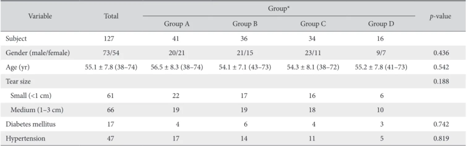
ultrasonography was used to ensure appropriate positioning of the needle in the subacromial space. Ultrasonography was per- formed using a Siemens Acuson S2000 (Siemens Medical Solu- tions USA, Inc., Malvern, PA, USA) with a linear array transducer at a frequency of 9 MHz. In the next stage, 8 ml of 2% lidocaine (Huons, Seongnam, Korea) were injected using a 23-gauge needle (Fig. 1). The time interval between the pre- and post- injection VAS for pain evaluation was about 30 to 60 minutes to ensure diffusion of the agent in the subacromial space and allow for onset time.14) Post-injection VAS for pain was measured using the same maneuver as was used before the impingement test.
2) Other functional outcome evaluations
The range of motion of the shoulder joint, including forward flexion, external rotation, and internal rotation by the level of the vertebral body, were measured. Shoulder isometric strength of the forward flexion, external rotation, and internal rotation were checked using the Oxford scale15) and a digital dynamometer (NIDEC-SHIMPO Corp., Kyoto, Japan). The American Shoulder and Elbow Surgeons (ASES) standardized form16) was used as an outcome evaluation tool. All evaluations were assessed preop- eratively and at 3, 6, 9, and 12 months postoperatively.
3) Group classification
According to the results of impingement test, the participants were divided into four groups: Group A, pain reduction of 75%
or more after the impingement test; Group B, pain reduction of 50% or more, but less than 75%; Group C, pain reduction of 25% or more, but less than 50%; Group D, pain reduction of less than 25%. No significant differences among groups were observed with respect to gender (p=0.436), age (p=0.542), or number of days from onset (p=0.286). In addition, according to the classification of DeOrio and Cofield,17) the distribution of tear size did not differ significantly among groups (p=0.188). Finally, no significant differences among groups were noted for diabetes mellitus (p=0.742) and hypertension (p=0.819) (Table 1).
Surgical Procedure
Under general anesthesia, each patient was placed in the beach chair position. The anteroposterior dimension and re- traction of the cuff tear were measured with a probe during arthroscopic procedures and categorized according to the clas- sification of DeOrio and Cofield.17)
All repairs were performed by the senior author with the ar- throscopic technique using suture anchors (double row suture bridge technique) for full coverage according to tear configura- tion. The number of medial anchors was decided upon tear size.
Arthroscopic acromioplasty was conducted using techniques similar to those described by Altchek et al.18) and Gartsman19) in all cases. At the end of the procedure, 20 ml of 0.75% ropiva- caine (AstraZeneca, Södertälje, Sweden) were injected into the subacromial space.
Postoperative Rehabilitation
All shoulders were immobilized with an abduction brace for 6 weeks. Pendulum exercise was started on the day after sur- gery. Immediate controlled passive motion of forward elevation, abduction, external rotation, and internal rotation was allowed from 5 days postoperatively with the brace off. Muscle strength- ening exercise was encouraged after weaning off the brace. All sports activities were permitted at 6 months after the operation.
Statistical Analyses
The repeated measures ANOVA was used to compare the symptomatic and functional outcomes after arthroscopic rotator cuff repair with acromioplasty between the four groups under study. Univariate regression analysis was used to evaluate the correlation between the pre- and post-injection VAS for pain difference and improvement of VAS for pain after surgery. The analyses were performed using IBM SPSS ver. 19.0 (IBM Co., Armonk, NY, USA), with a p<0.05 considered statistically signifi- cant.
Fig. 1. Ultrasound Guided Impingement Test.
DT: deltoid, SSP: suraspinatus tendon, HH:
Results
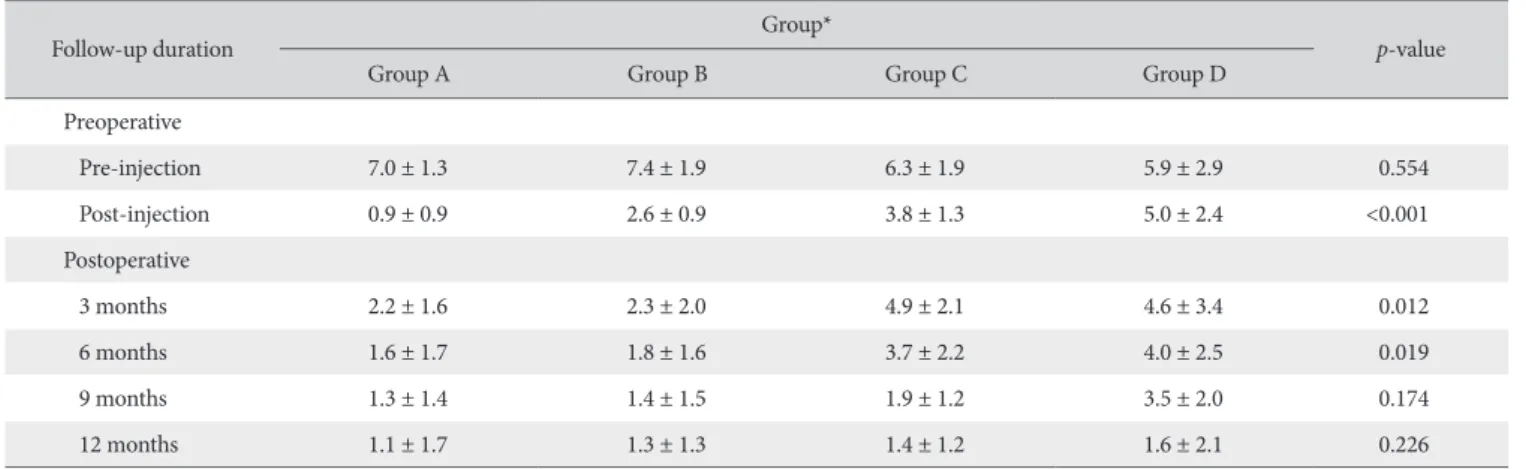
The mean pre-injection VAS for pain of Group A was 7.0 ± 1.3 and the mean post-injection VAS for pain of Group A was 0.9
± 0.9, while those of Group B were 7.4 ± 1.9 and 2.6 ± 0.9, those of Group C were 6.3 ± 1.9 and 3.8 ± 1.3, and those of Group D were 5.9 ± 2.9 and 5.0 ± 2.4, respectively (Fig. 2). Al- though different degrees of pain reduction were observed, most participants reported pain reduction after the impingement test.
The VAS for pain was improved significantly at postoperation 3 and 6 months than preoperation in group A and B, at 9 months in Groups A, B, and C (p<0.05), and at 12 months in all groups (p<0.05). The mean VAS for pain also improved significantly at 3 and 6 months postoperatively in Groups A and B when
compared to Groups C and D (p<0.05) (Table 2, Fig. 2). These results demonstrate that the amount of pain reduction after the impingement test is significantly related to improvement of pain postoperatively.
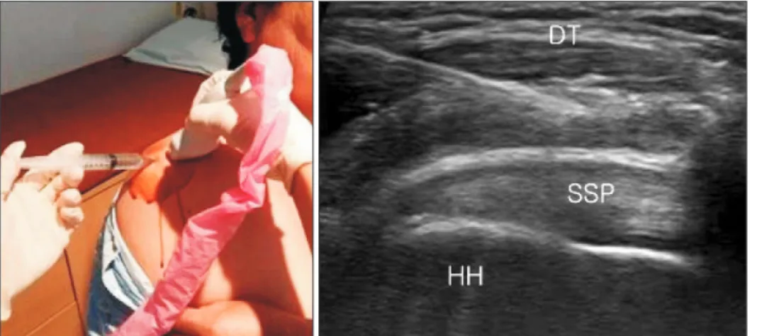
Among groups, the range of forward flexion significantly im- proved in Group A at 3 months postoperatively (p<0.05) (Fig.
3). The correlation of the difference between preoperative and postoperative range of external rotation and internal rotation between groups was not significantly different (p=0.789, 0.725).
The correlation of the difference between preoperative and postoperative isometric strength of the forward flexion, external rotation, and internal rotation between the four groups under study also did not differ significantly (p=0.825, 0.764, 0.723, respectively).
The ASES score improved at 3 and 6 months postoperatively
Preoperative 160
140
120
100
80
0
Forwardflexion()
PO 3 mo PO 6 mo PO 9 mo PO 12 mo Group A Group B Group C Group D
Fig. 3. Range of forward flexion. The range of forward flexion significantly improved in Group A at 3 months postoperatively. Four groups were divided according to the amount of pain reduction (%) after the impingement test;
Group A: 75%–100%, Group B: 50%–74%, Group C: 25%–49%, Group D:
0%–24%.
PO: postoperative.
Pre-Inj Post-Inj 8
7 6 5 4 3 2 1 0
VAS
PO 3 mo PO 6 mo PO 9 mo PO 12 mo Group A Group B Group C Group D 7.4
7 6.3
5.9 5
3.8
2.6
0.9
4.9
4.6
2.3 2.2
4
3.7
1.8 1.6
3.5
1.9 1.4 1.3
1.6 1.4 1.3 1.1
Fig. 2. VAS pain scores from preoperative day to 12 months postoperative day. The mean VAS for pain improved significantly at 3 and 6 months post- operatively in Groups A and B relative to Groups C and D. Four groups were divided according to the amount of pain reduction (%) after the impingement test; Group A: 75%–100%, Group B: 50%–74%, Group C: 25%–49%, Group D:
0%–24%.
VAS: visual analogue scale, Pre-Inj: pre-injection, Post-Inj: post-injection, PO:
postoperative.
Table 2. Visual Analogue Scale Pain Scores from Preoperative Day to 12 MonthsPostoperative Day
Follow-up duration Group*
p-value
Group A Group B Group C Group D
Preoperative
Pre-injection 7.0 ± 1.3 7.4 ± 1.9 6.3 ± 1.9 5.9 ± 2.9 0.554
Post-injection 0.9 ± 0.9 2.6 ± 0.9 3.8 ± 1.3 5.0 ± 2.4 <0.001
Postoperative
3 months 2.2 ± 1.6 2.3 ± 2.0 4.9 ± 2.1 4.6 ± 3.4 0.012
6 months 1.6 ± 1.7 1.8 ± 1.6 3.7 ± 2.2 4.0 ± 2.5 0.019
9 months 1.3 ± 1.4 1.4 ± 1.5 1.9 ± 1.2 3.5 ± 2.0 0.174
12 months 1.1 ± 1.7 1.3 ± 1.3 1.4 ± 1.2 1.6 ± 2.1 0.226
Values are presented as mean ± standard deviation.
*Four groups were divided according to the amount of pain reduction (%) after the impingement test; Group A: 75%–100%, Group B: 50%–74%, Group C: 25%–
49%, Group D: 0%–24%.
in Groups A and B, at 9 months in Groups A, B, and C, and at 12 months in all groups (p<0.05). The score also significantly improved at 3 and 6 months postoperatively in Groups A and B when compared to Groups C and D (p<0.05) (Table 3).
According to the results of univariate regression analysis, the following equation was established: (reduction of VAS for pain in impingement test)×0.216+5.64=postoperative 12 months VAS for pain.
Discussion
In rotator cuff disorders, pain is usually the most serious symptom that concerns patients.20,21) Many patients have paid attention to pain itself, and pain is thought to be the main cause of patients’ decision for undergoing surgery.21)
If the amount of pain reduction can be predicted preopera- tively, it could be helpful when discussing the outcome of sur- gery with patients. However, it is difficult to predict how much pain will be reduced after surgical repair in each patient.8) No definite clinical factors have been found to reliably account for and determine the amount of pain reduction after rotator cuff surgery.22)
We found that patients whose VAS for pain was improved in Groups A and B were more likely to experience a reduction of pain than patients whose VAS for pain was improved in Groups C and D, especially at 3 months and 6 months postoperatively (p=0.012, 0.019, respectively). The VAS for pain at 12 months follow-up among the four groups was not significantly different (p=0.226) (Table 2). The ASES score also improved at 3 and 6 months postoperatively in patients whose pain was improved in Groups A and B with pain reduction (p=0.026, 0.037, respec- tively). We also demonstrated that the range of forward elevation improved in patients whose pain was improved in Group A at 3 months postoperatively (p=0.038). Our data revealed a signifi-
preoperative impingement test and the decrease in VAS for pain after surgery, especially 3 and 6 months postoperatively.
The impingement test9) is one of the best known examina- tions for predicting symptomatic and functional outcome after rotator cuff surgery and subacromial decompression.8,10,11) This test was initially designed to confirm the diagnosis of subacro- mial impingement syndrome. Oh et al.8) modified the original impingement test for their study and concluded that the amount of pain reduction after a modified impingement test was related to pain reduction following rotator cuff repair. Furthermore, Skedros and Pitts14) suggested that there is temporal variation in the onset of effect in the impingement test. The authors claimed that assessing pain at 10 minutes for a Neer-type impingement test can fail to accurately determine a positive test in a substan- tial percentage of patients. Therefore, we waited for about 30 to 60 minutes to ensure diffusion of the agent in the subacromial space and to allow for onset time, after which we subsequently re-checked the visual analog scale for pain. We also regulated the time of the impingement test to the day before the surgery.
In this way, the patients’ precise preoperative pain degree could be appropriately reflected.
Conversely, Kirkley et al.10) analyzed 30 patients with rotator cuff tendinosis and argued that the impingement test was a poor tool for predicting the success of subacromial decompression.
The authors found no correlation between the reduction of pain after the impingement test and the change in Western Ontario Rotator Cuff Index and the ASES score following subacromial decompression.
In contrast, Oh et al.8) demonstrated that there is a significant correlation between the amount of pain reduction after the modified impingement test and the change in VAS for pain at the final follow-up visit, in agreement with our results. Further- more, Mair et al.11) categorized 55 patients with impingement syndrome based on the degree of pain reduction following the impingement test. The authors defined a positive impingement test as a patient with more than 75% pain relief and demon- strated that patients with a positive impingement test had a suc- cessful outcome after surgery. They further purported that the impingement test could be an effective tool to predict outcome after arthroscopic subacromial decompression. In the present study, we divided the patients into four groups according to this reference and obtained similar results. Similarly, Altchek et al.18) concluded that a positive impingement test was the best predic- tor of the postoperative functional Hospital for Special Surgery score, and that this score was correlated with the total postop- erative score.
The authors performed the impingement test under the guid- ance of ultrasonography to increase the accuracy. The accuracy of the blind impingement test was 70% to 80%,23-25) and the accurate placement of the injection was confirmed through the Table 3. American Shoulder and Elbow Surgeons Scores from Preoperative
Day to 12 MonthsPostoperative Day Follow-up
duration
Group*
Group A Group B Group C Group D
Preoperative 43.8 ± 8.6 45.1 ± 9.2 44.2 ± 7.8 41.6 ± 8.8 Postoperative
3 months 72.7 ± 7.4 69.3 ± 8.6 42.8 ± 9.1 43.6 ± 7.5 6 months 78.2 ± 8.9 75.6 ± 8.8 52.7 ± 8.5 50.2 ± 6.7 9 months 80.7 ± 6.8 78.7 ± 8.1 70.2 ± 8.6 56.9 ± 6.9 12 months 82.1 ± 6.9 83.0 ± 9.3 78.9 ± 7.3 79.1 ± 9.2 Values are presented as mean ± standard deviation.
*Four groups were divided according to the amount of pain reduction (%) after the impingement test; Group A: 75%–100%, Group B: 50%–74%, Group C: 25%–49%, Group D: 0%–24%.
21.6% of patients experienced worsening or no change in the level of pain after the modified impingement test. However, in the present study, only 2 of 96 patients (2.1%) felt no change in pain level after the preoperative impingement test. Moreover, no patients reported worsening of pain after injection.
It should be noted that our study has the following limitations.
First, the participants were limited to those with small- to medi- um-sized rotator cuff tears, making it difficult to apply the results to entire rotator cuff tears or other shoulder disorders. Second, we investigated VAS for pain only at passive motion and did not evaluate it during patients’ active motion of shoulder joints.
Third, the participants were divided into four groups according to the pain reduction amount after the impingement test. There- fore, other factors for grouping that could have affected func- tional outcomes were not considered. Fourth, although pain is closely related to healing of the rotator cuff, healing after rotator cuff repair was not evaluated in the present study. However, the significance of our results lies in the fact that the present study sought to analyze the correlation between the preoperative im- pingement test and postoperative clinical outcomes in rotator cuff tears.
Conclusion
Preoperative degree of pain reduction after the impingement test was found to correlate with the improvement of pain after arthroscopic rotator cuff repair, especially in the early phase.
Therefore, the impingement test could be used as a reliable predictor of outcome following arthroscopic rotator cuff repair within 6 months postoperatively.
References
1. Mitchell C, Adebajo A, Hay E, Carr A. Shoulder pain: diagno- sis and management in primary care. BMJ. 2005;331(7525):
1124-8.
2. Oh LS, Wolf BR, Hall MP, Levy BA, Marx RG. Indications for rotator cuff repair: a systematic review. Clin Orthop Relat Res.
2007;455:52-63.
3. Colvin AC, Egorova N, Harrison AK, Moskowitz A, Flatow EL.
National trends in rotator cuff repair. J Bone Joint Surg Am.
2012;94(3):227-33.
4. Judge A, Murphy RJ, Maxwell R, Arden NK, Carr AJ. Temporal trends and geographical variation in the use of subacromial decompression and rotator cuff repair of the shoulder in Eng- land. Bone Joint J. 2014;96(1):70-4.
5. Lee E, Bishop JY, Braman JP, Langford J, Gelber J, Flatow EL.
Outcomes after arthroscopic rotator cuff repairs. J Shoulder Elbow Surg. 2007;16(1):1-5.
6. Galatz LM, Ball CM, Teefey SA, Middleton WD, Yamaguchi K.
The outcome and repair integrity of completely arthroscopi-
cally repaired large and massive rotator cuff tears. J Bone Joint Surg Am. 2004;86(2):219-24.
7. O’Holleran JD, Kocher MS, Horan MP, Briggs KK, Hawkins RJ.
Determinants of patient satisfaction with outcome after rotator cuff surgery. J Bone Joint Surg Am. 2005;87(1):121-6.
8. Oh JH, Kim SH, Kim KH, Oh CH, Gong HS. Modified im- pingement test can predict the level of pain reduction after rotator cuff repair. Am J Sports Med. 2010;38(7):1383-8.
9. Neer CS 2nd. Impingement lesions. Clin Orthop Relat Res.
1983;(173):70-7.
10. Kirkley A, Litchfield RB, Jackowski DM, Lo IK. The use of the impingement test as a predictor of outcome following subacro- mial decompression for rotator cuff tendinosis. Arthroscopy.
2002;18(1):8-15.
11. Mair SD, Viola RW, Gill TJ, Briggs KK, Hawkins RJ. Can the impingement test predict outcome after arthroscopic subacro- mial decompression? J Shoulder Elbow Surg. 2004;13(2):150- 3.
12. Neer CS 2nd. Anterior acromioplasty for the chronic impinge- ment syndrome in the shoulder: a preliminary report. J Bone Joint Surg Am. 1972;54(1):41-50.
13. Hawkins RJ, Kennedy JC. Impingement syndrome in athletes.
Am J Sports Med. 1980;8(3):151-8.
14. Skedros JG, Pitts TC. Temporal variations in a modified Neer impingement test can confound clinical interpretation. Clin Orthop Relat Res. 2007;460:130-6.
15. Clarkson HM. Musculoskeletal assessment: joint range of mo- tion and manual muscle strength. 2nd ed. Philadelphia: Lip- pincott Williams & Wilkins; 2000.
16. Richards RR, An KN, Bigliani LU, et al. A standardized method for the assessment of shoulder function. J Shoulder Elbow Surg. 1994;3(6):347-52.
17. DeOrio JK, Cofield RH. Results of a second attempt at surgi- cal repair of a failed initial rotator-cuff repair. J Bone Joint Surg Am. 1984;66(4):563-7.
18. Altchek DW, Warren RF, Wickiewicz TL, Skyhar MJ, Ortiz G, Schwartz E. Arthroscopic acromioplasty. Technique and results.
J Bone Joint Surg Am. 1990;72(8):1198-207.
19. Gartsman GM. Arthroscopic acromioplasty for lesions of the rotator cuff. J Bone Joint Surg Am. 1990;72(2):169-80.
20. Tetzlaff JE, Brems J, Dilger J. Intraarticular morphine and bu- pivacaine reduces postoperative pain after rotator cuff repair.
Reg Anesth Pain Med. 2000;25(6):611-4.
21. Modi CS, Veillette CJ, Gandhi R, Perruccio AV, Rampersaud YR. Factors that influence the choice to undergo surgery for shoulder and elbow conditions. Clin Orthop Relat Res. 2014;
472(3):883-91.
22. Oh JH, Kim SH, Ji HM, Jo KH, Bin SW, Gong HS. Prognos- tic factors affecting anatomic outcome of rotator cuff repair and correlation with functional outcome. Arthroscopy. 2009;
25(1):30-9.
23. Kang MN, Rizio L, Prybicien M, Middlemas DA, Blacksin MF.
The accuracy of subacromial corticosteroid injections: a com- parison of multiple methods. J Shoulder Elbow Surg. 2008;
17(1 Suppl):61S-6S.
24. Mathews PV, Glousman RE. Accuracy of subacromial injec-
tion: anterolateral versus posterior approach. J Shoulder Elbow Surg. 2005;14(2):145-8.
25. Yamakado K. The targeting accuracy of subacromial injection to the shoulder: an arthrographic evaluation. Arthroscopy.
2002;18(8):887-91.