The Effect of Left Subclavian Artery Coverage During Endovascular Repair of the Thoracic Aortic Aneurysm on Cerebral Hemodynamics: Two Cases of Flow
Measurement by using 2D Phase Contrast Magnetic Resonance Imaging
Seung-Hoon Baek1, Sung Won Youn1, 2, Ho Kyun Kim2, Oh-Choon Kwon3, Sub Lee3, Jongmin Lee4
1Vascular and Aging Research Group, Catholic University of Daegu School of Medicine, Daegu, Korea
2Department of Radiology, Catholic University of Daegu School of Medicine, Daegu, Korea
3Department of Cardiothoracic Surgery, Catholic University of Daegu School of Medicine, Daegu, Korea
4Department of Radiology, Kyungpook National University School of Medicine, Daegu, Korea
The proximity of thoracic aortic aneurysm to the left subclavian artery (LSA) has made the coverage of LSA during tho- racic endovascular aortic repair (TEVAR) be essential. Despite controversy concerning the safety of LSA coverage and the indications for LSA revascularizations, the cerebral hemodynamic change after LSA coverage has not been demonstrated.
We prospectively examined two patients who would undergo TEVAR with LSA coverage by using 2D cine phase contrast MR imaging. After LSA coverage, the left subclavian steal was properly compensated by the increased flow volumes of both carotid arteries and right vertebral artery, which is the major collateral supply. The total brain supply after TEVAR did not lessen, which showed good correlation with uneventful clinical outcome. Therefore, 2D phase contrast MR imaging can be recommended as a useful technique to evaluate the hemodynamic change of the LSA coverage during TEVAR and to triage the candidate for LSA revascularization.
Index words : Aortic aneurysm∙Thoracic endovascular aortic repair (TEVAR)∙Left subclavian artery (LSA) 2D cine phase contrast MR imaging∙Subclavian steal
Thoracic endovascular aortic repair (TEVAR) has
proven to be an effective option replacing open surgery for treating thoracic aortic lesions including aneurysm, dissection, and blunt traumatic injury (1-6).
The aortic arch lesions especially of zone 2 necessitate proximal extension of the stent-graft to achieve an adequate landing zone, and the coverage of left subclavian artery (LSA) becomes essential (2-4). The antegrade flow through LSA may be critical for brain, spinal cord, and arm perfusion, and the concerns on the risk of posterior circulation stroke or vertebrobasi- lar insufficiency, spinal cord ischemia, and left distal arm ischemia has emerged when LSA is covered.
However, the previous reports have offered conflicting data of whether LSA coverage increase risk or INTRODUCTION
�Received; June 7, 2012�Revised; August 27, 2012
�Accepted; August 28, 2012
This work was supported by Basic Science Research Program through the National Research Foundation of Korea (NRF) funded by Ministry of Eduation, Science and Technology (grant number 20119123). Otherwise, the authors have no conflicts of interests and/or disclosure.
Corresponding author : Sung Won Youn, M.D., Department of Radiology, Catholic University of Daegu School of Medicine, 3056-6 Daemyung-4 dong, Nam-gu, Daegu 705-718, Korea.
Tel. 82-53-650-4309, Fax. 82-53-650-4339 E-mail : [email protected]
Case Report
preoperative LSA revascularization reduced the risk of stroke or spinal cord injury (2-6). Furthermore, the hemodynamic feature of LSA coverage has not been demonstrated systematically so far.
2D cine phase contrast MR imaging has been useful to evaluate the variety of cerebrovascular disease and their treatment response including carotid artery stenosis, moyamoya disease, and subclavian steal (7, 8). The quantitative flow data can be obtained within a few minutes of scanning in a noninvasive manner and without contrast agent injection. However, there have been no observations of 2D phase contrast MR imaging describing the hemodynamic change after LSA coverage during TEVAR. In addition, the hemodynamic change of venous drainage and the cerebrospinal fluid (CSF) flux have not been addressed until recently.
In this case report, we present cerebral hemody- namic change of intentional LSA coverage during TEVAR in patients with traumatic aortic dissection or recurred aortic aneurysm by using 2D cine phase contrast MR imaging.
2D cine phase contrast MR imaging
The 2D cine phase contrast MR imaging was prospectively performed in two patients who were waiting for elective TEVAR by using a 1.5-T MR imaging unit (Signa, GE Medical, Milwaukee,WI,
USA) before and 2 days after TEVAR, and the detailed scanning protocols are described at Table 1.
The acquisition planes for flow quantification were selected perpendicular to the presumed direction of the flow (Fig. 1). The region of interest (ROI) was manually extracted at each level by using an image processing software (ReportCARD, GE Medical, Milwaukee,WI, USA), and its flow curves of arterial, venous and CSF were generated versus time over the 30 segments of one cardiac cycle (Fig. 2). The flow volumes (FVs) of artery, vein, and CSF were recorded in mL/min, and the FVs of total cerebral blood flow (FVTCBFs) were calculated as sum of both internal carotid arteries (ICA) and both vertebral arteries (VAs) at the level between first and second cervical spine (C1-2) or basilar artery (BA) at midbasilar artery (MBA) level. The arteriovenous transit time (AVTT) was defined as the difference between the time points (msec) that arterial and venous curve meet maximal slope.
Patient 1
A 76-year-old man had multiple fractures of left ribs, left scapular, left femur after bike accident. He complained of chest pain, and 3D chest CT angiogra- phy showed Stanford type B aortic dissection at arch distal to LSA origin (Fig. 3). There were left side hemothorax and hemoretroperitoneum. He had no neurologic deficit at admission. He had no previous history of hypertension or diabetes mellitus. Blood pressure was 80/50, and pulse rate was 110. Cerebral CASE REPORT
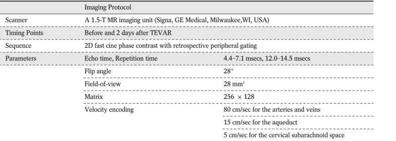
Table 1. Scanning Parameters of 2D Cine Phase Contrast MR Imaging
Imaging Protocol
Scanner A 1.5-T MR imaging unit (Signa, GE Medical, Milwaukee,WI, USA) Timing Points Before and 2 days after TEVAR
Sequence 2D fast cine phase contrast with retrospective peripheral gating
Parameters Echo time, Repetition time 4.4-7.1 msecs, 12.0-14.5 msecs
Flip angle 28
Field-of-view 28 mm2
Matrix 256 128
Velocity encoding 80 cm/sec for the arteries and veins 15 cm/sec for the aqueduct
5 cm/sec for the cervical subarachnoid space Note.─ TEVAR, thoracic endovascular aortic repair.
CT angiography show no cerebral vascular abnormal- ity. The posterior communicating artery was not seen, and the diameter of left side VA appears to be larger than that of right side. TEVAR with LSA coverage was designed in consideration of the location of aortic dissection near to LSA. A spinal catheter was inserted at third lumbar space to monitor CSF pressure to be under 10 mmHg. Heparin of 100 units per body weight (kg) was administered intravenously.
Procedures were performed under general anesthesia in an operation suite equipped with a C-arm machine
(Series 9800, OEC Medical Systems, Salt Lake City, Utah, USA) having fluoroscopic and angiographic capabilities. The pig tail catheter through 5F right femoral sheath was used to monitor lesion segment and landing zone. A 24 × 110 mm-sized endovascular stent-graft (S & G biotech, Sungnam-si, Korea) through 18F right femoral access was advanced, and the distal end of stent-graft was employed to cover LSA. Preoperative LSA revascularization or coil placement to prevent retrograde flow or endoleak was not performed. On angiogram immediately after
a b c
d e f
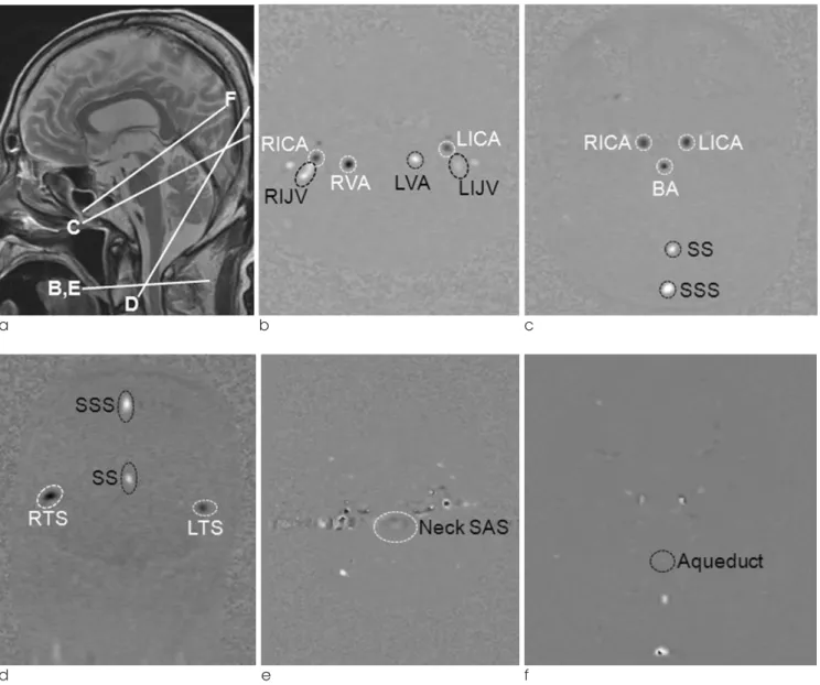
Fig. 1. Scanning protocol of 2D cine phase contrast MR imaging and data extraction.
(a) T2-weighted midline sagittal scan was used as localizers to select the anatomic levels for flow quantification. Ellipsoid region of interest was drawn on each arteries and veins of C1-2 (b) and midbasilar artery level (c). The same process was done for venous drainage at coronal scan (d), for cervical subarachnoid space of C1-2 level (e), and for cerebrospinal fluid at aqueduct (f). RICA, right internal carotid artery; LICA, right internal carotid artery; RVA, right vertebral artery; LVA, left vertebral artery; RIJV, right internal jugular vein; LIJV, left internal jugular vein; BA, basilar artery; SS, straight sinus; SSS, superior sagittal sinus; RTS, right transverse sinus;
LTS, left transverse sinus; SAS, subarachnoid space.
TEVAR, aortic dissection was successfully excluded from vascular lumen, and the origin of LSA was covered.
The FVTCBF after TEVAR were 109.2% of those before TEVAR at C1-2 level and 105.9% at MBA level, respectively (Table 2). The flow direction of left VA was reversed, and the FV of right VA (FVRVA) was 698.31% of those before TEVAR. The FVs of BA (FVBAMBA), right ICA (FVRICAMBA) and left ICA (FVLICAMBA) at MBA level after TEVAR was 83.03%, 111.04%, and 120.85% of those before TEVAR, respectively. The FVs of axial SS+SSS (FVSS+SSSax), coronal SS+SSS (FVSS+SSScor), both side TS (FVTS), and both side IJV(FVIJV) were 107.7%, 90.06%, 85.37%, and 134.18% of those before TEVAR, respectively. The baseline FV of CSF at C1-2 level (FVCSFC1-2) and at aqueduct (FVCSFaqueduct) showed
caudocephalad direction in averaging. The FVCSFC1-2 decrease in volume, and the FVCSFaqueductchanged into craniocaudal direction in averaging after TEVAR. The AVTTs after TEVAR were slightly shortened as comared to the AVTTs before (the change of AVTT 100 msec; Table 3).
The patient was assessed for neurologic and left arm symptoms, and did not show sign of paraplegia or cerebral ischemia. He was uneventful for 6 months of follow-up period, and 6 months follow-up CT angiog- raphy showed stable occlusion of aortic aneurysm.
Patient 2
Type A intramural hematoma was detected in a 61- year-man with acute onset back pain and dyspnea, and graft replacement of ascending aorta was performed for it six months ago. On six months
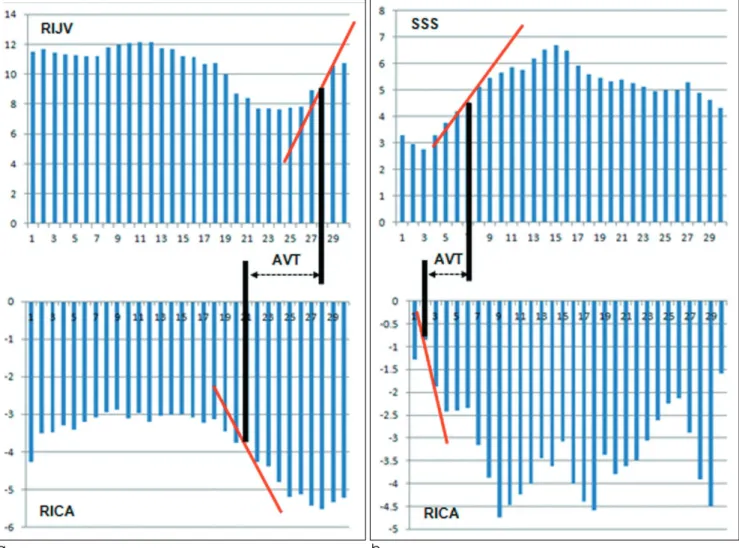
a b
Fig. 2. Generation of time-volume curves and the definition of arteriovenous transit time (AVTT). AVTT is defined as the difference of the time points between arterial and venous curve meet its maximal slope (msec) at C1-2 level (a) and midbasilar artery level (b). RICA, right internal carotid artery; RIJV, right internal jugular vein; SSS, superior sagittal sinus.
follow-up 3D CT angiography, a 35 × 20 mm-sized aneurysmal sac was newly found at distal aortic arch (Fig. 4). Cerebral MR angiography before TEVAR revealed no significant cerebral vascular abnormality and paralleling both side VAs. He had controlled diabetes mellitus. Blood pressure was 100/70, and heart rate was 76. Under general anesthesia, a spinal catheter was placed to monitor CSF pressure.
Procedures were performed in C-arm machine along with the systemic heparinization of 100 units per body weight (kg). The pig tail catheter via left femoral
access was used to measure lesion length and landing zone. A 35 × 130 mm-sized endovascular stent-graft was delivered via 18F right femoral access. The proximal and distal landing zone was confirmed on fluoroscopy with contrast injection, and the distal end of stent-graft was deployed to cover LSA. Left CCA origin was confirmed to be intact on immediate post- angiogram.
As compared to FVTCBFs before TEVAR, the FVTCBFs after TEVAR were 118.50% at C1-2 level and 169.19%
at MBA level, respectively (Table 2). The flow direction
a
b c
d e
Fig. 3. A 76-year-old man (patient 1) with type B aortic dissection after bike accident.
a.Iodine-enhanced CT angiography shows an 18 x 7 mm-sized focal dissecting aneurysm at arch 3 mm apart from left subclavian artery origin. There is no significant cerebral vascular abnormality. The diameter of left side vertebral artery appears to be larger than that of right side.
b, c.On the 2 days after thoracic endovascular repair, axial magnitude and phase image of C1-2 level shows reversed flow at left vertebral artery (arrowhead). Arrow indicates right vertebral artery.
d, e.Six months follow-up CT angiography shows covered left subclavian artery origin but visible left vertebral and subclavian arteries, suggesting development of left subclavian steal. There is no endoleak or recurrence of aneurysm.
f g
Fig. 3. f, g. The origin of left common carotid artery (arrow) is intact just in front of proximal end of stent-graft.
Table 2. Flow Volumes (FVs) Before and 2 Days after Left Subclavian Artery Coverage During TEVAR. The Each FVs of Arteries, Veins, and Cerebrospinal Fluid at C1-2 or Midbasilar Artery Level were Measured in mL/min, and the % of Difference in the FVs after TEVAR in Comparison with Those Before (%�) were Calculated
Location Patient 1, mL/min Patient 2, mL/min
Before After % � Before After % �
C1-2 level
FVRICAC1-2 190.25 226.13 +18.86 218.18 298.93 +37.01
FVLICAC1-2 171.31 221.54 +29.32 295.90 379.41 +28.22
FVRVAC1-2 44.35 309.70 +598.31 149.58 299.09 +99.95
FVLVAC1-2 133.24 -168.60 -226.54 131.73 -34.90 -126.50
FVTCBFC1-2 539.15 588.77 +9.20 795.39 942.53 +18.50
FVIJVC1-2 322.07 432.15 +34.18 653.5 713.14 +9.13
FVCSFC1-2 -31.01* -16.80* -45.82 6.26 -30.00* -579.23
Midbasilar Artery level
FVRICAMBA 292.72 325.04 +11.04 190.13 354.56 +86.48
FVLICAMBA 155.95 188.47 +20.85 278.22 509.88 +83.27
FVBAMBA 167.79 139.31 -16.97 183.26 238.00 +29.87
FVTCBFMBA 616.46 652.82 +5.90 651.61 1102.44 +69.19
FVSS+SSSax 289.35 311.62 +7.70 425.65 451.3 +6.03
FVSS+SSScor 437.86 394.34 -9.94 561.49 510.02 -9.17
FVTS 206.08 175.93 -14.63 461.53 504.66 +9.35
FVCSFaqueduct -2.34* 3.19 -236.32 0.32 -1.38* -531.25
Note.─ TEVAR, thoracic endovascular aortic repair; %� = (FV after TEVAR - FV before TEVAR) / FV before TEVAR × 100 (%);
C1-2, between first and second cervical vertebral level; MBA, midbasilar artery level; RICA, right internal carotid artery; LICA, left internal carotid artery; RVA, right vertebral artery; LVA, left vertebral artery; TCBF, total cerebral blood flow; IJV, internal jugular vein; CSF, cerebrospinal fluid; BA, basilar artery; FVSS+SSSaxor FVSS+SSScor, sum of FVs at straight sinus and superior sagittal sinus measured at axial or coronal plane; FVTS, sum of FVs measured at right and left transverse sinus; FVIJV, sum of FVs measured at right and left internal jugular vein; FVCSFC1-2or FVCSFaqueduct, CSF flow measured at C1-2 or aqueduct; *, minus means caudocephalad direction of averaged FVCSFC1-2and FVCSFaqueduct.
of left VA was reversed, and the FVRVA was 199.95% of that before TEVAR. The FVRICAMBA, FVLICAMBA, and
FVBAMBAwere 186.48%, 183.27%, 129.87% of those before TEVAR, respectively. The FVSS+SSSax,
FVSS+SSScor, FVTS, and FVIJV were 106.03%, 90.83%, 109.35%, and 109.13% of those before TEVAR, respec- tively. The baseline FVCSFC1-2 and FVCSFaqueductwere craniocaudal direction in averaging, but they changed into caudalcephalad direction in averaging after TEVAR.
The AVTTs after TEVAR were shortened as comared to the AVTTs before (the change of AVTT 200 msec;
Table 3).
The patient was uneventful for 6 months of follow- up period, and 6 months follow-up CT angiography showed stable occlusion of aortic aneurysm. Left CCA origin was spared, and the origin of LSA was covered without endoleak.
The proximity of thoracic aortic aneurysm or dissec- tion to the LSA, especially located at landing zone 2, has occasionally required intentional coverage (2-4).
Controversy continues concerning the safety of LSA
coverage and the indications for LSA revasculariza- tions, with the importance of antegrade flow through the LSA for brain and spinal cord perfusion. The stroke and spinal cord ischemia associated with the intentional LSA coverage during TEVAR have been reported to be developed in 3.1-8.6% and in 2.5- 2.8% of the patients, respectively (4-6). However, the primary revascularization of LSA did not offer protec- tive effect against stroke, considering that the stroke rate between the revascularization and non-revascular- ization groups was not different (4, 5). Interestingly, the most strokes were embolic and occurred in the anterior circulation (5). The distal end of devices, wires, and catheters are likely to increase atheroma manipulation at arch, which can subsequently dislodge embolic material at the orifices of the great vessels.
Our study results are consistent with these in that coverage of the LSA origin is usually well tolerated as a result of the collateral flow via the right VA (3-5). In our two patients, the total brain supply after TEVAR did not lessen despite subclavian steal, being actually 105.9-169.19% of that before TEVAR, which were well correlated with uneventful clinical outcome.
Among them, the FV of right VA was most signifi- cantly increased among other arteries.
DISCUSSION
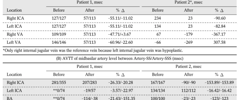
Table 3. The Arteriovenous Transit Time (AVTT) Before and After TEVAR. The AVTT is Defined as the Difference between the Time Points having Maximal Slope of Arterial and Venous Time-Volume Curve
(A) AVTT of C1-2 level between Artery-right IJV/Artery-left IJV (msec)
Patient 1, msec Patient 2*, msec
Location Before After % � Before After % �
Right ICA 127/127 57/113 -55.11/-11.02 234 23 -90.60
Left ICA 127/127 57/113 -55.11/-11.02 134 23 -82.84
Right VA 109/109 57/113 -47.71/+3.67 67 -179 -367.17
Left VA 146/146 57/113 -60.96/-22.60 -66 -269 307.58
*Only right internal jugular vein was the reference vein because left internal jugular vein was hypoplastic.
(B) AVTT of midbasilar artery level between Artery-SS/Artery-SSS (msec)
Patient 1, msec Patient 2, msec
Location Before After % � Before After % �
Right ICA 281/355 207/283 -26.33/-20.28 167/167 -90/-90 -153.89/-153.89
Left ICA **0/74 -19/57 -3.57/-22.97 134/134 112/112 -16.42/-16.42
BA **0/74 -114/-38 -21.43/-151.35 100/100 -23/-23 -123/-123
Note.─ TEVAR, thoracic endovascular aortic repair; % � = (AVTT after TEVAR - AVTT before TEVAR) / AVTT before TEVAR 100 (%); ICA, internal carotid artery; VA, vertebral artery; IJV, internal jugular vein; BA, basilar artery; SS, straight sinus; SSS, superior sagittal sinus.**0, Total trigger delay at this plane was 532 msec in patient 1.
From this viewpoint, the primary LSA revasculariza- tion is not mandatory before TEVAR, but should be considered in selected patients. Indications for primary LSA revascularization included a patent left internal mammary artery-left anterior descending artery bypass, a functioning dialysis fistula in the left arm, need for long segment aortic coverage, and prior or concomitant infrarenal aortic replacement. The secondary revascularization can be performed in patients that became symptomatic or present vertebrobasilar insufficiency after LSA coverage (3).
An atretic or stenotic right VA may increase risk for spinal cord ischemia, provided that the anterior spinal artery is partly formed from branches of the thyrocer- vical trunk. Therefore, cerebral CT or MR angiography before TEVAR should be recommended to visualize both vertebral and the basilar arteries and to exclude concomitant VA trauma or anomaly (9). Duplex ultrasound may be helpful to evaluate the patency of the extracranial circulation especially of the right VA and presence of subclavian steal (10). 2D phase contrast MRI may be useful in identifying candidate of
a
b d
c
e
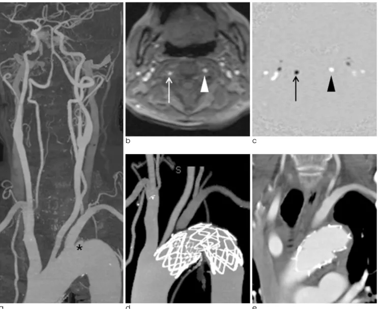
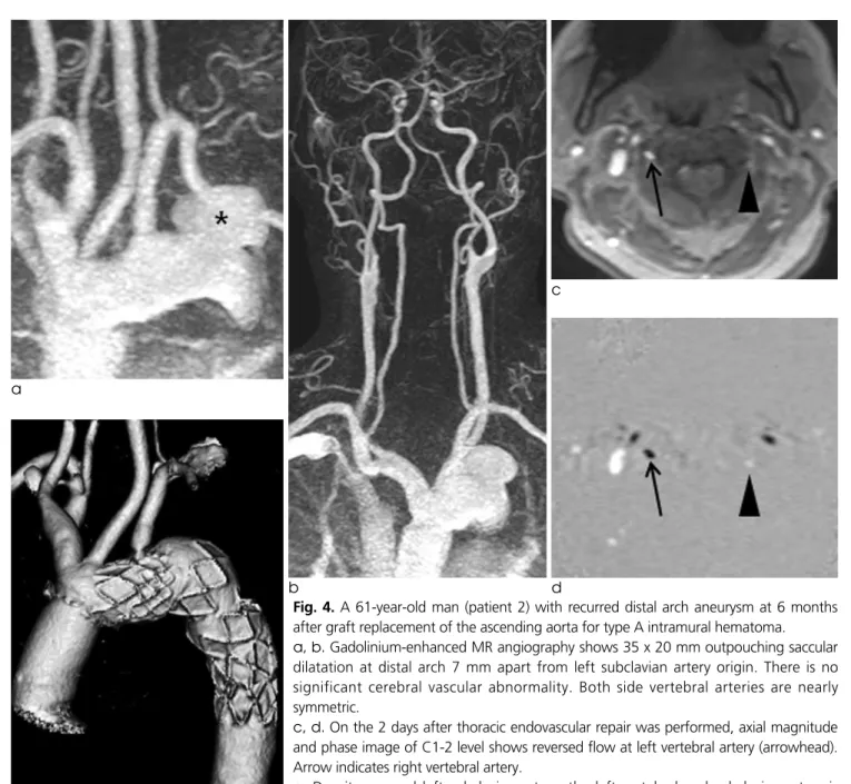
Fig. 4. A 61-year-old man (patient 2) with recurred distal arch aneurysm at 6 months after graft replacement of the ascending aorta for type A intramural hematoma.
a, b.Gadolinium-enhanced MR angiography shows 35 x 20 mm outpouching saccular dilatation at distal arch 7 mm apart from left subclavian artery origin. There is no significant cerebral vascular abnormality. Both side vertebral arteries are nearly symmetric.
c, d.On the 2 days after thoracic endovascular repair was performed, axial magnitude and phase image of C1-2 level shows reversed flow at left vertebral artery (arrowhead).
Arrow indicates right vertebral artery.
e. Despite covered left subclavian artery, the left vertebral and subclavian artery is demonstrated possibly due to left subclavian steal on 6 months follow-up CT angiography. There is no endoleak or recurrence of aneurysm.
primary or secondary LSA revascularization with an inadequate collateral vertebral circuit (7-9). In our patients, the direction of left VA flow changed to retrograde fashion after LSA coverage. The amount of FV of retrograde left VA seems to be dependent upon pre-TEVAR VA dominancy, as seen from 168.60 mL/min at dominant left VA of patient 1 and 34.90 mL/min at equal both side VAs of patient 2. The subclavian steal behave like an arteriovenous shunt requiring supplies for left VA flow as well as basilar flow, which was compensated by the increased FV of other cerebral arteries including both ICAs and contralateral VA. The right VA is a major collateral supply, and the degree of increasing % of the patient 1 with non-dominant right VA was as high as 6 times than that of the patient 2 with equally dominant both VAs. It seems that non-dominant right VA may be rather insufficient to compensate the reversed dominant contralateral VA, although it became enlarged remarkably to supply collateral flow, as seen from that the FVBA after TEVAR of patient 1(non- dominant right VA) and patient 2 (equally dominant VA) was 83.03% and 129.87% of FVBA before TEVAR, respectively.
To the best of our knowledge, this is the first report to measure prospectively the FV changes of patients who undergone LSA coverage by using 2D cine phase contrast MR imaging. Even in a few literatures on the flow measurement of VA in the patients with subcla- vian steal by using Doppler ultrasound or 2D phase contrast MR imaging (7, 8, 10), the direct and quanti- tative measurement of venous and CSF has not been made. In comparison to arterial FVs, the venous FVs after TEVAR showed rather inconsistent results. The
FVSS+SSSaxafter TEVAR were 106.03-107.70% of those before TEVAR, but the FVSS+SSScor after TEVAR were 90.06-90.83% of those before TEVAR.
The FVIJV and FVTS after TEVAR were 109.13- 134.18% and 85.37-109.35% of those before TEVAR, respectively. Different angle and location of the scanning plane, and anatomical complexity of venous drainage might be considered. We could not find any consistent trend in the change of FVCSF.
AVTT seemed to be shortened with varying degree, and the order of arterial and venous peak was sometimes reversed, which remains to be further investigated.
In terms of limitation, as being case report of two
patients, the method of analysis depends only upon the comparison between before and after TEVAR.
Whether preoperative evaluation of the vertebrobasi- lar system would allow selective LSA revasculariza- tion remains to be further investigated in a future prospective study of larger population. The patients were clinically observed during 6 months period, but there was no available 6 months follow-up 2D phase contrast MR imaging, although the chronic hemody- namic feature at 6 months may be different from that of immediate post-TEVAR.
In summary, the total brain supply after TEVAR is properly compensated despite subclavian steal, most significantly by increased FV of right VA, which show good correlation with favorable clinical outcomes. 2D phase contrast MR imaging can be recommended as a useful technique to evaluate the hemodynamic change of the LSA coverage during TEVAR and to triage the candidate for LSA revascularization.
References
1. Dake MD, Miller DC, Semba CP, Mitchell RS, Walker PJ, Liddell RP. Transluminal placement of endovascular stent-grafts for the treatment of descending thoracic aortic aneurysms. N Engl J Med 1994;331:1729-1734
2. Peterson BG, Eskandari MK, Gleason TG, Morasch MD. Utility of left subclavian artery revascularization in association with endoluminal repair of acute and chronic thoracic aortic pathol- ogy. J Vasc Surg 2006;43:433-439
3. Riesenman PJ, Farber MA, Mendes RR, Marston WA, Fulton JJ, Keagy BA. Coverage of the left subclavian artery during thoracic endovascular aortic repair. J Vasc Surg 2007;45:90-94 4. Woo EY, Carpenter JP, Jackson BM, et al. Left subclavian
artery coverage during thoracic endovascular aortic repair: a single-center experience. J Vasc Surg 2008;48:555-560
5. Cooper DG, Walsh SR, Sadat U, Noorani A, Hayes PD, Boyle JR. Neurological complications after left subclavian artery coverage during thoracic endovascular aortic repair: a system- atic review and meta-analysis. J Vasc Surg 2009;49:1594-1601 6. Buth J, Harris PL, Hobo R, et al. Neurologic complications
associated with endovascular repair of thoracic aortic pathology:
incidence and risk factors. a study from the European Collaborators on Stent/Graft Techniques for Aortic Aneurysm Repair (EUROSTAR) registry. J Vasc Surg 2007;46:1103-1110 7. Akin K, Kosehan D, Kirbas I, Yildirim M, Koktener. Diagnosis
and percutaneous treatment of partial subclavian steal: Doppler ultrasonography and phase contrast magnetic resonance angiog- raphy findings and a brief review of the literature. Jpn J Radiol 2011;29:207-211
8. Langer DJ, Lefton DR, Ostergren L, et al. Hemispheric revascu- larization in the setting of carotid occlusion and subclavian steal:
a diagnostic and management role for quantitative magnetic resonance angiography? Neurosurgery 2006;58:528-533 9. Tay KY, U-King-Im JM, Trivedi RA, et al. Imaging the vertebral
artery. Eur Radiol 2005;15:1329-1343
10. Youn SW, Yu SW, Lee NJ, Kim JH. Neurological picture. Acute middle cerebral artery stroke and an innominate steal from a
ruptured brachiocephalic trunk atheroma. J Neurol Neurosurg Psychiatry 2009;80:492-493
통신저자 : 윤성원, (705-718) 대구 남구 대명 4동 3056-6번지, 대구가톨릭대학교 의과대학 영상의학과 Tel. (053) 650-4309 Fax. (053) 650-4339 E-mail: [email protected]
흉부대동맥류의 혈관내치료 도중 좌측 쇄골하동맥 폐색이 뇌혈류역동에 미치는 효과: 2차원 위상차 대조 자기공명영상을 이용한 혈류 측정 2례 보고
1대구가톨릭대학교 의과대학 혈관과 노화 연구회
2대구가톨릭대학교 의과대학 영상의학과
3대구가톨릭대학교 의과대학 흉부외과
4경북대학교 의과대학 영상의학과
백승훈1∙윤성원1,2∙김호균2∙권오춘3∙이 섭3∙이종민4
흉부대동맥류는 좌측 쇄골하동맥에 가까이 위치해 있어서 흉부대동맥내치료를 시행할 때 좌측 쇄골하동맥의 폐색 이 불가피한 경우가 많다. 좌측 쇄골하동맥 폐색의 안전성과 좌측 쇄골하동맥 재생술의 필요성에 대해 논란이 계속되 고 있음에도 불구하고, 좌측 쇄골하동맥의 폐색후 뇌혈류역학적인 변화에 대한 기존의 체계적인 연구는 없었다. 저자 들은 좌측 쇄골하동맥폐색을 동반한 흉부대동맥내치료의 대상이 되는 2명의 환자에서 치료 전후의 2차원 위상차 대 조 자기공명영상을 시행하였다. 좌측 쇄골하동맥의 폐색 후 좌측 쇄골하도주가 발생하였으며, 이로 인한 뇌혈류의 소 실을 양측 경동맥과 주된 부행혈류인 오른쪽 추골동맥이 적절하게 보상하였다. 전체 뇌혈류는 감소하지 않았으며 이 는 임상적으로 뇌졸증이나 척수마비의 증상이 나타나지 않은 것과 잘 일치하였다. 2차원 위상차 대조 자기공명영상은 좌측 쇄골하동맥 폐색을 동반한 흉부대동맥내치료의 뇌혈류역학적인 평가와 좌측 쇄골하동맥 재생술이 필요한 환자 의 선별에 유용할 것으로 사료된다.
대한자기공명의과학회지 16:159-168(2012)