Anesthetic Management for Lung Adenocarcinoma Experienced Acute Neurocardiogenic Syncope and Cardiac Arrest
Jin Hye Han, Youn Jin Kim, Jong Hak Kim, Dong Yeon Kim, Guie Yong Lee, Chi Hyo Kim
Department of Anesthesiology and Pain Medicine, Ewha Womans University School of Medicine, Seoul, Korea
Introduction
Vasovagal syncope (neurocardiogenic syncope) is considered the most common cause of repetitive syncope [1]. Severe pain, emotional stress, anxiety, erect postural change in a patient who is in a hypovolemic state, invasive medical procedures such as intravenous catheter insertion, venipuncture and neuraxial anes- thesia are regarded as triggering factors for this syndrome. Ad- ditionally, the resulting sympathetic tone increase and the positive inotropic state that result from the aforementioned triggers are considered potent vagal reflex responses leading to sympathetic inhibition, bradycardia, acute drop in blood pressure and ensuing syncope and circulatory collapse [2]. Generally, these conditions resolve spontaneously or treated with conservative methods, such as volume replacement, without any significant sequelae [2,3].
In this case report, the suspected vasovagal syncope that devel- oped immediately after the transcutaneous fine-needle aspiration biopsy of the right lung nodule did not respond to the conserva- tive therapy and abruptly progressed to fatal cardiovascular col- lapse with cardiac arrest, and resulted in cerebrovascular accident (CVA). Five weeks after the event, when the patient’s neurological state was stabilized, the elective lung lobectomy was performed after prophylactic pacemaker insertion and was completed safely.
Herein, we report this case experience and review the literature, followed by a discussion.
Case
A 59-year-old female admitted our clinic for evaluation of persistent cough over 2 months, foul smelling sputum, mild
Report
pISSN 2234-3180 • eISSN 2234-2591Vasovagal syncope is one of the most common causes of transient syncope during an- esthesia for elective surgery in patients with a history of syncope and requires special attention and management of anesthetics. The causes and pathophysiological mecha- nism of this condition are poorly understood, but it has a benign clinical course and recovers spontaneously. However, in some cases, this condition may cause cardiovas- cular collapse resulting in major ischemic organ injury and be life threatening. Herein we report a case and review literature, regarding completing anesthesia safely during an elective surgery of a 59-year-old female patient with history of loss of consciousness due to suspected vasovagal syncope followed by cardiovascular collapse and cardiac arrest, which required cardiopulmonary resuscitation and insertion of a temporary pacemak- er and intra-aortic balloon pump immediately after a fine-needle aspiration biopsy of a lung nodule located in the right middle lobe. (Ewha Med J 2014;37(Suppl):S28-S32)
Received May 1, 2014 Accepted June 27, 2014 Corresponding author Youn Jin Kim
Department of Anesthesiology and Pain Medicine, Ewha Womans University School of Medicine, 1071 Anyangcheon-ro, Yangcheon-gu, Seoul 158-710, Korea
Tel: 82-2-2650-2885, Fax: 82-2-2655-2924 E-mail: ankyj@ewha.ac.kr
Key Words
Anesthesia; Lung neoplasms; Syncope, vasovagal
Copyright ⓒ 2014, The Ewha Medical Journal
cc This is an Open Access article distributed under the terms of the Creative Commons Attribution Non Commercial License (http://creativecommons.org/licenses/by-nc/3.0/) which permits unrestricted non commercial use, distribution, and reproduction in any medium, provided the original work is properly cited.
tachypnea, and dyspnea. She was under teratment for type II dia- bets mellitus, hypothyroidism, hypertension and major depressive disorder.
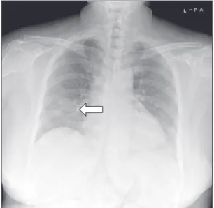
A nodule in the right middle lobe of the lung was found and considered malignant due to increased size (about 2.8×1.5 cm) and change in shape compared to the past images (Fig. 1). On the second day of admission, a fine-needle aspiration biopsy of the lung lesion was conducted. Approximately 5 minutes after the procedure, while in a sitting position waiting to be transferred to the ward, the patient was found loss of consciousness. The patient was transferred to the emergency room, emergency endotracheal intubation, and CPR were performed and spontaneous circula- tion was recovered. However, the ST segment elevation suggested acute myocardial infarction was found (Fig. 2). Additionally, consistent decrease of blood pressure was observed and an emer- gency coronary angiography was performed to rule out variant angina showing normal results. Also emergency transthoracic echocardiography found no specific abnormal finding. Therefore,
Fig. 1. Simple chest X-ray. It shows a mass (about 2.8×1.5 cm) in the right middle lobe (arrow) suggesting malignancy.
Fig. 2. Electrocardiography. (A) Electrocardiography before attack shows normal sinus rhythm (86 beats per minute [bpm]). (B) Electrocardiogra- phy immediately after cardiopulmonary resuscitation for cardiac arrest resulting from fatal vasovagal syncope shows wide QRS tachycardia (124 bpm), right bundle branch block (RBBB), and ST elevation. (C) Acute myocardial infarction was suspected with junctional rhythm (49 bpm) and ST elevation. (D) Preoperative electrocardiography showed normal sinus rhythm (76 bpm) 5 weeks after fatal vasovagal syncope.
A B
C D
variant angina and stress induced cardiomyopathy can be ruled out in the way that the absence of definite stenosis on coronary angiography and wall motion abnormality or apical balloon find- ing on echocardiography which is specific for the stress induced cardiomyopathy and it makes the vasovagal reflex as the cause of syncope and circulatory collapse more probable.
Due to consistent bradycardia of 40~50 bpm and hypotension of systolic blood pressure 55~80 mmHg despite infusion of dopa- mine, dobutamine and norepinephrine, a temporary pacemaker and intra-aortic balloon pump (IABP) was inserted into the patient. The patient regained consciousness after approximately 30 minutes but was drowsy and the vital signs continued to be degraded.
Subsequent neurological examination revealed upper limb weakness accompanied by facial palsy of the left side.(mild, grade I) The brain computed tomography (CT) showed no hemorrhagic lesions and magnetic resonance angiogram (MRA) showed no definite stenosis of medium to large vessel, but diffusion magnetic resonance imaging (MRI) revealed acute infarction in the bilat- eral frontal lobes and parietal lobe
On day 4 after admission, the patient’s vital signs stabilized.
The patient’s endotracheal tube was extubated, Intra-Aortic Bal- loon Pump(IABP)removed and oral administration of aspirin 100 mg was started. Neurological exams showed the patient’s facial palsy had completely disappeared and the upper limb weakness grade I improved to grade IV without additional abnormal neu- rological findings.
On day 5 after admission, the patient was transferred to the general ward. The pathological biopsy result revealed adeno- carcinoma; thus, preoperative evaluation for elective lobectomy was started. Considering the patient’s general condition, the lung lobectomy was scheduled for 5 weeks later when the patient’s neurological state would be stabilized.
Preoperative tests and evaluations were performed on an out- patient basis, and the results were within normal limit. Regarding the neurological consultation for operability, the paralytic symp- toms of the patient after the recent CVA were improved and the patient’s general neurological state was stabilized for surgery with close monitoring of vital signs and avoidance of perioperative ce- rebral hypoperfusion. On cardiovascular consultation, preopera- tive prophylactic transcutaneous temporary cardiac pacemaker insertion was recommended, thus the patient was admitted on the day before surgery and a prophylactic transcutaneous temporary
pacemaker was inserted. No premedication was administered and the blood pressure on arrival to the operating room was 168/72 mmHg and the heart rate was 112 bpm.
The patient was preoxygenated with 100% oxygen through a simple mask. Anesthesia was induced by intravenous administra- tion of midazolam (3 mg), glycopyrrolate (0.2 mg) followed by pentothal sodium (250 mg), fentanyl (100 μg), and rocuronium (50 mg) and inhalation of 2 vol% sevoflurane. Ninety seconds after rocuronium administration, a 37-F double-lumen endotra- cheal tube intubation failed and a single-lumen endotracheal tube with a bronchial blocker was intubated. Soon after the intubation, the patient’s hemodynamic state was stabilized and a left radial artery catheter for continuous arterial pressure monitoring, an ultrasound imaging guided right internal jugular venous catheter for fluid infusion, and a central venous pressure (CVP) monitor- ing were inserted. The patient’s position was then changed to left lateral and one-lung ventilation was applied.
Anesthesia during surgery was maintained with 4~6 vol% of desflurane and additional administration of fentanyl (50 μg) to maintain the bispectral index score (BIS) between 40 and 60. Lo- bectomy through video-assisted thoracotomy was attempted first, but the procedure for lung mass excision was converted to right open thoracotomy because the patient was obese, had a small intrathoracic volume, ambiguous interlobar fissure and a positive finding of a mediastinal node from the frozen biopsy result.
The mass diameter was about 3 cm. The mass was located in the right middle lobe but a bilobectomy with mediastinal lymph node dissection, which excised both the right middle lobe and lower lobe, was performed because the mass was a central lesion adjacent to the right hilum and very close to the bronchus and bronchial artery of the right lower lobe. For postoperative pain control, an intercostal nerve block was injected and a localized pain-relieving device (ON-Q SilverSoaker, B Braun, Melsungen, Germany) was inserted in the incision site.
During anesthesia, hemodynamic status and arterial blood gas analysis findings were relatively stable and CVP was maintained at 10~13 mmHg. The fluid therapy consisted of 2,200 mL of fluid (crystalloid 1,900 mL, fresh frozen plasma 300 mL). Estimated blood loss was about 400 mL, and 300 mL of urine output was measured.
Total operation time was 3 hours 30 minutes and anesthesia time was 5 hours 5 minutes. Muscle relaxation was reversed with 10 mg of pyridostigmine and 0.5 mg of glycopyrrolate. There-
after, the patient responded properly to the verbal commands and recovered consciousness with BIS over 90. However, a tidal volume of 300~350 mL was less than satisfactory; therefore, the patient was transferred to the recovery room under intubation inhaling oxygen. Fifteen minutes after the patient was moved to the recovery room, the arterial blood gas analysis showed a fraction of inspired oxygen (FiO2) of 0.6, oxygen partial pressure (PaO2) of 69 mmHg and carbon dioxide tension (PaCO2) of 46 mmHg. The chest X-ray was normal except for the chest tube insertion after bi-lobectomy. Due to the lack of tidal volume and oxygenation status, the patient was transferred to the intensive care unit (ICU) under intubation and mechanical ventilation with FiO2 of 0.4 pressure control mode was applied. During and after the surgery, no symptoms or signs of vasovagal syndrome (such as hypotension or bradycardia) were found.
After being transferred to ICU, the patient showed stable vi- tal signs with stable oxygenation including an arterial PaO2 of 142 mmHg (FiO2 of 0.5 spontaneous respiration with 5 cmH2O continuous positive airway pressure) and tidal volume increase to 500~600 mL. On postoperative day (POD) 2, the endotracheal tube was extubated and the temporary pacemaker was removed.
The patient was transferred to the general ward on POD 3 and discharged after conservative treatment on POD 17.
Discussion
Vasovagal syncope is the most common cause for syncope but generally does not require any particular treatment [1]. However, when symptoms recur or circulatory collapse appears, it can cause permanent damage to the nervous system due to central nervous system ischemia. Especially in patients who are sched- uled for surgery and anesthesia, the physical and psychological stimulation and the stress from anesthesia and surgery can be a trigger for vasovagal syncope and shock; therefore, it is essential that there is thorough prevention or prompt treatment when it occurs [4]. Regarding this, there was a domestic reports of a cases in which vasovagal syncope and subsequent cardiac arrest oc- curred during a cesarean section under epidural anesthesia in a puerpera with a history of vasovagal syncope [5,6].
Vasovagal syncope is the occurrence of bradycardia and pe- ripheral vasodilation because of an autonomic nerve reflex caused by various stimulations [3]. The mechanism has not been clearly revealed yet; however, the excessive response of the autonomic
nervous system to stimulation that activates the C-fiber in the heart [2,3].
The most common stimulation causing vasovagal syncope is the standing position for a prolonged period or psychological stress; however, it has also been reported that it can be caused by medical procedures [4,7]. In addition, it has been reported that it can occur from a direct stimulation to the vagus nerve branches from a tumor in the lungs or in the bronchial tubes [8-10].
Three factors are suspected as stimulations causing vasovagal syncope. First, the invasive biopsy procedure may have caused emotional anxiety and stress [11]. Second, patient may failed to compensate the decreased venous return due to venous pooling in the lower extremities after prolonged sitting position as a result of automonic neuropathy in long standing diabetes [12]. And, a direct mechanical stimulation to the vagus nerve by cancer mass near the right lung entrance could lead to vasovagal syncope [8- 10].
Shimizu et al. [8] reported 4 cases of cardiac arrest due to va- sovagal syncope which occurred in patients with lung cancer. In the above case report, the patient had no history of syncope and the syncope had newly developed after a lung cancer diagnosis;
thus, neurogenic paraneoplastic syndrome was suspected as the cause for the vasovagal syncope. Especially in all 4 cases, the location of the tumor was near the hilum of the left bronchial tube. After tumor reduction after chemotherapy, it was found that the vasovagal response had disappeared with the head up tilt test.
Hence, it was assumed that the stimulation from the mechanical pressure of the cancer mass on the vagus nerve branch near the left bronchial tube hilum caused the cardiac C-fiber response. In this case, the patient also had no history of syncope and the cir- culatory response was more severe than usual vasovagal syncope which led to circulatory collapse. Thus, it is believed that para- neoplastic syndrome might play a role in vasovagal syncope in addition to the major vasovagal response caused by the mechani- cal stimulation on the vagus nerve branch near the cancer mass.
The patient in this case showed syncope accompanied by hy- potension and a bradycardia after the invasive procedure for the neoplastic lung lesion; there were no abnormalities shown in the emergency coronary angiography and the subsequent echocar- diography in order to rule out possible causes like variant angina, or stress-induced cardiomyopathy; thus, it could be supposed that the syncope was from the vasovagal reflex shock caused by the invasive transcutaneous procedure.
A brain diffusion MRI and MRA was performed for the left facial paralysis and the weakened upper extremities, and an acute cerebral infarction was observed. Considering that there were no neurological deficit symptoms or prior signs, it is believed to be caused by hypotension syncope rather than being the reason for it. Because surgery was scheduled for excision of the lung lesion after the patient became stabilized, there was need for thorough prevention and preparation for the possibility of vasovagal shock and subsequent ischemic tissue damage from the anesthesia and surgical stimulation [13].
In conclusion, the authors experienced a case where a lung cancer patient developed fatal suspected vasovagal syncope which did not respond to inotropic medication and vasoconstric- tors and even reached cardiac arrest. It was severe to the point of needing the insertion of an artificial cardiac pacemaker. Consid- ering the above, when fatal vasovagal syncope occurs in a patient with a mass of a certain size located at the hilum of the bronchial tubes, the feasible cause should be perceived early, and aggressive treatment should be performed.
References
1. Soteriades ES, Evans JC, Larson MG, Chen MH, Chen L, Benja- min EJ, et al. Incidence and prognosis of syncope. N Engl J Med 2002;347:878-885.
2. White CM, Tsikouris JP. A review of pathophysiology and therapy of patients with vasovagal syncope. Pharmacotherapy
2000;20:158-165.
3. Chen-Scarabelli C, Scarabelli TM. Neurocardiogenic syncope.
BMJ 2004;329:336-341.
4. Kinsella SM, Tuckey JP. Perioperative bradycardia and asystole:
relationship to vasovagal syncope and the Bezold-Jarisch reflex.
Br J Anaesth 2001;86:859-868.
5. Jang YE, Do SH, Song IA. Vasovagal cardiac arrest during spinal anesthesia for Cesarean section: a case report. Korean J Anesthe- siol 2013;64:77-81.
6. Park SY, Kim SS. An anesthetic experience with cesarean section in a patient with vasovagal syncope: a case report. Korean J An- esthesiol 2010;59:130-134.
7. Tsai PS, Chen CP, Tsai MS. Perioperative vasovagal syncope with focus on obstetric anesthesia. Taiwan J Obstet Gynecol 2006;45:208-214.
8. Shimizu K, Yoshii Y, Watanabe S, Hosoda C, Takagi M, Tominaga T, et al. Neurally mediated syncope associated with small cell lung cancer: a case report and review. Intern Med 2011;50:2367- 2369.
9. Angelini P, Holoye PY. Neurocardiogenic syncope and Prinzmet- al’s angina associated with bronchogenic carcinoma. Chest 1997;111:819-822.
10. Koga T, Kaseda S, Miyazaki N, Kawazoe N, Abe I, Sadoshima S, et al. Neurally mediated syncope induced by lung cancer--a case report. Angiology 2000;51:263-267.
11. Gracie J, Baker C, Freeston MH, Newton JL. The role of psycho- logical factors in the aetiology and treatment of vasovagal syn- cope. Indian Pacing Electrophysiol J 2004;4:79-84.
12. Grubb BP. Neurocardiogenic syncope and related disorders of orthostatic intolerance. Circulation 2005;111:2997-3006.
13. Parry SW, Matthews IG. Update on the role of pacemaker therapy in vasovagal syncope and carotid sinus syndrome. Prog Cardiovasc Dis 2013;55:434-442.
