https://doi.org/10.4174/astr.2018.94.1.52 Annals of Surgical Treatment and Research
Surgery of multiple lymphangioma in small bowel:
a rare case report of chronic gastrointestinal bleeding
Dae Ro Lim, Jung Cheol Kuk, Taehyung Kim, Eung Jin Shin
Section of Colon and Rectal Surgery, Department of Surgery, Soonchunhyang University Bucheon Hospital, Soonchunhyang University College of Medicine, Bucheon, Korea
INTRODUCTION
Small bowel lymphangioma is a rare benign tumor of the lymphatic system, characterized by the presence of dilated lymphatic spaces and significant gastrointestinal bleeding [1].
Lymphangioma lesions are generally located in the head and neck region and the axilla, rarely developing intra-abdominally.
The most common locations for lymphangiomas of the intra- abdominal cavity are the mesentery, omentum, mesocolon, and retroperitoneum [2]. The diagnosis of a lymphangioma in the small bowel is very difficult preoperatively, even if biopsy is performed by gastroenteroscopy. Small bowel lymphangiomas are rare in adults and published case reports are few in number.
The patient reported here had an unusual case of multiple lymphangiomas in the small bowel, presenting as chronic anemic symptoms in an adult.
CASE REPORT
A 70-year-old South Korean man was admitted presenting with anemia, dizziness, and intermittent melena. He had a history of iron deficiency anemia beginning 3 years prior. He had been admitted to hospital 4 times over the past 3 years because of anemia (hemoglobin count < 5 g/dL) and melena, and he had undergone transfusion with packed RBCs. He had undergone a clipping of a hemangioma on the duodenum by gastroenteroscopy 1 year previously. Laboratory testing revealed a white blood cell count of 5,500 cells/mm3 and a hemoglobin count of 5.2 g/dL. Other routine lab tests revealed no specific findings. His clinical symptoms were dizziness, general weakness, and melena. A video capsule endoscopy was performed and revealed mucosal erosion with a blood clot at the jejunum (at about 60–70 cm distal to the ligament of Treitz) (Fig. 1). Gastroenteroscopy revealed multiple polypoid Small bowel lymphangioma is a rare benign tumor of the lymphatic system, characterized by the presence of dilated lymphatic spaces and significant gastrointestinal bleeding. Small bowel lymphangiomas are rare in adults and case reports are few. Lymphangiomas in the jejunum or ileum are extremely rare and account for less than 1% of all lymphangiomas.
The case reported herein is of an older patient (70-year-old male) with melena and chronic anemia (hemoglobin count
< 5 g/dL) who had small-sized multiple lymphangiomas in his small bowel (jejunum). Surgical resection was performed after failure of treatment by gastroenteroscopy. Final pathological analysis revealed lymphangioma with thrombus and hemorrhage. After surgery, he no longer had decreased hemoglobin count, nor symptoms of anemia and melena. Also, at the last follow-up visit, the patient’s hemoglobin count patient was normal and he returned to normal daily functions.
[Ann Surg Treat Res 2018;94(1):52-56]
Key Words: Lymphangioma, Endoscopy, Surgery
Reviewed January February March April May June July August September October November December
Received January 12, 2017, Revised March 15, 2017, Accepted April 1, 2017 Corresponding Author: Eung Jin Shin
Section of Colon and Rectal Surgery, Department of Surgery, Soonchunghyang University Bucheon Hospital, Soonchunhyang University College of Medicine, 170 Jomaru-ro, Wonmi-gu, Bucheon 14584, Korea
Tel: +82-32-621-6267, Fax: +82-32-621-6950 E-mail: [email protected]
ORCID code: https://orcid.org/0000-0002-2029-4136
Copyright ⓒ 2018, the Korean Surgical Society
cc Annals of Surgical Treatment and Research is an Open Access Journal. All articles are distributed under the terms of the Creative Commons Attribution Non- Commercial License (http://creativecommons.org/licenses/by-nc/4.0/) which permits unrestricted non-commercial use, distribution, and reproduction in any medium, provided the original work is properly cited.
masses, with bleeding, in the long segment of the jejunum.
Histology from endoscopic biopsies did not provide a conclusive diagnosis but revealed a chronic inflammation pattern with lymphangiectasia.
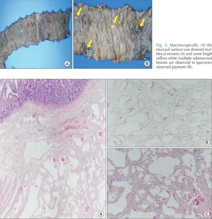
The patient underwent repeated gastroendoscopy (4 times) for bleeding control by clipping due to low his hemoglobin count and chronic bleeding (Fig. 2). However, his symptoms did not improve. He continued to show chronic anemia and melena even after bleeding control by gastroendoscopy. He underwent segmental resection of the long segment of the jejunum (about 70 cm) that had been marked (tattooing) previously during gastroendoscopy. The intraoperative finding was not remarkable except for the tattoo and no palpable masses. Macroscopically, the mucosal surface showed multifocal erosions, and multiple
bright yellow-white submucosal lesions were observed in the resected jejunum (Fig. 3). Histologically, numerous dilated lymphatics were identified in the submucosa by hematoxylin and eosin (H&E) staining. The lymphatics were positive for D2-40, based on immunohistochemical staining (Fig. 4). The final pathological analysis was consistent with lymphangioma with thrombus and hemorrhage. After surgery, the patient no longer experienced drops in hemoglobin count or symptoms of anemia and melena. At the last follow-up visit (after 3 months), his hemoglobin count was normal (10.4 g/dL) and he was living a normal life. He then visited the outpatient clinic for hypertension on February, 2017 (6 months after surgery). His hemoglobin count was 12.9 g/dL and he had no symptoms.
Ethical approval was not necessary as this case report was
A B
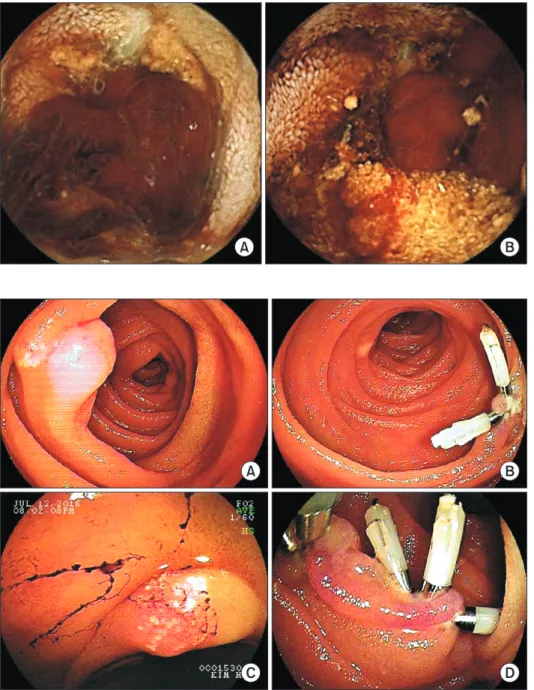
Fig. 1. Video capsule endoscopy revealed mucosal erosion (A) with blood clot at jejunum (B) (at about 60 cm distal to the ligament of Treitz).
A B
C D
Fig. 2. Gastroendoscopy revealed mucosal erosions (A) on jejunum and performed bleeding control (C) by clipping due to chronic bleeding (B-D).
based on retrospective review.
DISCUSSION
Lymphangiomas are rare lymphatic system tumors and represent 6% of small bowel tumors found in children and 1.4%–2.4% of those tumors in adults [3]. Approximately 60%
of patients with lymphangiomas are younger than 5 years;
however, a significant number of abdominal lymphangiomas do not manifest until adulthood [3]. The common sites of lymphangiomas are the head, neck, and axillary regions.
Tumors at other locations, such as the abdominal or media- stinal cavity, are rare and account for approximately 5% of all lymphangiomas [2,4]. Among these, lymphangiomas of the small bowel are very rare, and lymphangiomas in the jejunum or ileum are extremely rare, accounting for less than A
B
C Fig. 4. (A) Histologically, numerous dilated lymphatics are identified in submucosa in hematoxylin and eosin staining (×40). (B) The lymphatics are positive for D2-40 in immunohistochemical staining (×100). (C) Hematoxylin and eosin staining (×100).
A B
Fig. 3. Macroscopically, (A) the mucosal surface was showed mul- tifocal erosion (A) and some bright yellow-white multiple sub mucosal lesions are observed in specimen (resected jejunum) (B).
1% of all lymphangiomas [3]. A literature review of studies on lymphangiomas revealed that only 19 cases of small bowel lymphangioma were reported from 1960 to 2009 [5].
Similarly, small bowl lymphangiomas with gastrointestinal bleeding were reported in only 15 cases from 1967 to 2012 [6].
Two recent case reports of small bowel lymphangioma were published: one, reported in 2014, was a 24-year-old woman with clinical presentation of iron deficiency anemia who had a lymphangioma in the second portion of the duodenum [7];
the other case report, published in 2015, was a 28-year-old man with melena and anemia who had a lymphangioma in the third duodenal segment [8]. The present case is an elderly patient with melena and clinical presentation of iron deficiency anemia who had a lymphangioma in the long segment of the jejunum.
The clinical presentation of lymphangioma is unspecific and often presents with nausea, vomiting, and abdominal discomfort or pain. Other symptoms of lymphangioma are acute or chronic hemorrhage, infection, perforation, intussusceptions, torsion, rupture, and protein-losing gastroenteropathy [8]. The most common complications of lymphangioma are spontaneous or traumatic bleeding, hemorrhage, rupture and infection [9]. Histologically, lymphangioma consist of blood vessels and lymphatic channels of various sizes. The pathogenesis of spontaneous hemorrhage of lymphangioma is unknown. The present case had symptoms of chronic hemorrhage, dizziness, and melena. Diagnosis of small bowel lymphangioma is
difficult and requires pathological confirmation of the lesion.
Histopathological examination of lymphangioma demonstrates dilated lymphatic vessels and space by H&E staining and positive D2-40 immunohistochemical staining [7-9]. The size and number of lymphangiomas varies. One case report published in 2012 described a single lesion 5.0 cm × 4.0 cm in size in the duodenum [10]. The present case involved multiple small-sized lesions (<10 mm) in the long segment of the jejunum.
The first-line treatment of small bowel lymphangiomas is bleeding control by gastroenteroscopy or double-balloon enteroscopy [6]. Surgery is necessary when bleeding control by gastroenteroscopy fails. Surgical segmental resection of the bowel, including the lesion, is the optimal treatment for avoiding recurrence (Table 1). Our patient underwent surgical resection of a small bowel segment including the lesion.
CONFLICTS OF INTEREST
No potential conflict of interest relevant to this article was reported.
ACKNOWLEDGEMENTS
This work was supported by the Soonchunhyang University Research Fund.
REFERENCES
1. Gerosa Y, Bernard B, Lagneau M, You K, Hoang C, Brasseur JL, et al. Cystic lymph- angioma of the duodenum revealed by digestive hemorrhage and associated with exudative enteropathy. Gastroenterol Clin Biol 1993;17:591-3.
2. Enzinger FM, Weiss SW. Tumors of lymph
vessels. In: Enzinger FM, Weiss SW, edi- tors. Soft tissue tumors. 3rd ed. St. Louis (MO): Mosby; 1995. p. 679-99.
3. Suthiwartnarueput W, Kiatipunsodsai S, Kwankua A, Chaumrattanakul U. Lymph- angioma of the small bowel mesentery: a case report and review of the literature.
World J Gastroenterol 2012;18:6328-32.
4. Jang JH, Lee SL, Ku YM, An CH, Chang ED. Small bowel volvulus induced by mesenteric lymphangioma in an adult: a case report. Korean J Radiol 2009;10:319- 22.
5. Morris-Stiff G, Falk GA, El-Hayek K, Vargo Table 1. Previous cases and present case of small bowel lymphangioma
Study Age/sex Symptom Location Size Treatment
Fang et al. [10] 57/F Anemia,
melena Small intestine 30 cm distal
to the flexor tendon 5.0 cm × 4.0 cm Surgical resection
Bucciero et al. [8] 28/M Anemia,
melena Third duodenal segment Multiple sized masses Surgical resection
Morris-Stiff et al. [5] 34/M Anemia Jejunum Multinodula tumor
(5.3 cm × 4 cm × 1.5 cm) Surgical resection
Antonino et al. [7] 24/F Anemia 2/3 of duodenum 40 mm × 15 mm Surgical resection
Present case 70/M Anemia,
melena Jejunum Multiple sized masses Surgical resection
J, Bronner M, Vogt DP. Jejunal cavernous lymphangioma. BMJ Case Rep 2011 May 12;2011. pii: bcr0320114022. https://doi.
org/10.1136/bcr.03.2011.4022.
6. Kida A, Matsuda K, Hirai S, Shimatani A, Horita Y, Hiramatsu K, et al. A pedun cu- lated polyp-shaped small-bowel lym ph- angioma causing gastrointestinal bleed ing and treated by double-balloon entero- scopy. World J Gastroenterol 2012;18:4798-
800.
7. Antonino A, Gragnano E, Sangiuliano N, Rosato A, Maglio M, De Palma M. A very rare case of duodenal hemolymph- an gioma presenting with iron deficiency ane mia. Int J Surg Case Rep 2014;5:118-21.
8. Bucciero F, Marsico M, Galli A, Tarocchi M.
Small bowel lymphangioma: a rare case of intestinal bleeding. Dig Liver Dis 2015;47:
815.
9. Woo YS, Joo KR, Kim KY, Oh WT, Kim YH.
Unusual presentation of cystic lymph an- gioma of the gallbladder. Korean J Intern Med 2007;22:197-200.
10. Fang YF, Qiu LF, Du Y, Jiang ZN, Gao M.
Small intestinal hemolymphangioma with bleeding: a case report. World J Gastroenterol 2012;18:2145-6.