ISSN 2234-3806 • eISSN 2234-3814
https://doi.org/10.3343/alm.2019.39.5.493 www.annlabmed.org 493
Ann Lab Med 2019;39:493-495
https://doi.org/10.3343/alm.2019.39.5.493
Letter to the Editor
Diagnostic Hematology
First Report of a Patient With Natural Killer- Lymphoblastic Leukemia/Lymphoma in Korea
Jiwon Lee , M.D.1, Ho Young Kim , MD, Ph.D.2, Boram Han , M.D.2, Miyoung Kim , M.D., Ph.D.1, Ji-Young Park , M.D., Ph.D.3, Yousun Chung , M.D.3, and Young Kyung Lee , M.D., Ph.D.1
Departments of 1Laboratory Medicine and 2Internal Medicine, Hallym University Sacred Heart Hospital, Anyang; 3Department of Laboratory Medicine, Kangdong Sacred Heart Hospital, Seoul, Korea
Dear Editor,
Natural killer (NK)-lymphoblastic leukemia/lymphoma is a rare hematopoietic neoplasm designated by the WHO as an acute leukemia of ambiguous lineage in 2008 and recategorized as a precursor lymphoid neoplasm in 2016 [1-3]. Patients with this disease are positive for CD56 and immature T-cell markers but negative for B-cell and myeloid markers; moreover, the TCR and IG genes are in the germline configuration [3, 4]. However, the features of this disease overlap with those of other hematologi- cal malignancies, and markers specific to NK cell progenitors such as CD94 and CD161 are not commonly tested [4-6].
Only three patients have met the 2008 WHO criteria [5, 6].
The morphologic and cytogenetic/molecular genetic features of their diseases remain unknown. We report the first Korean pa- tient with NK-lymphoblastic leukemia/lymphoma diagnosed ac- cording to the 2016 WHO classification and describe her leuke- mia-related translocation pattern, genetic profile, and clinical course. Ours is a single case report which does not meet the definition of human subject research, thus was exempted from approval by an Institutional Review Board.
A 51-year-old woman with leukopenia that developed six months prior was referred to Hallym University Sacred Heart Hospital, Anyang, Korea in May 2018. She had experienced transient pan-
cytopenia, presumably caused by a toxic insult to her bone mar- row (BM), in February 2017. After spontaneous recovery, her white blood cell count was 1.6×10⁹/L; absolute neutrophil count, 0.25×10⁹/L; hemoglobin, 119 g/L; and platelet count, 176×109/L.
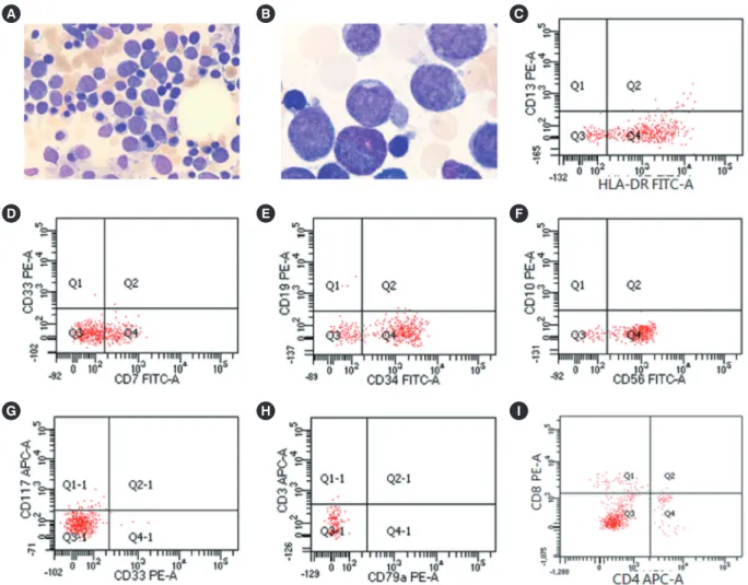
Her peripheral blood smear showed a few large lymphocytes with abnormal morphologies. BM aspirate showed a reduced number of normal trilineage hematopoietic cells and an incre- ased number of blasts (63.0% of total nucleated cells) that were large with high nuclear/cytoplasmic ratios, agranular cytoplasm, very fine nuclei with indentation, and conspicuous nucleoli (Fig.
1A and 1B). All cells were negative for peroxidase and Sudan Black B but positive for periodic acid-Schiff; some were also posi- tive for acid alpha-naphthyl acetate esterase. Biopsy revealed BM hypocellularity (10%), while immunohistochemistry showed an increased number of immature cells positive for CD34 and terminal deoxynucleotidyl transferase (TdT). Most blasts were positive for HLA-DR, CD7, CD34, and CD56 but negative for myeloid antigens (CD13, CD33, CD14, CD117, and myeloper- oxidase), B-cell antigens (CD10, CD19, CD20, and CD79a), and other T-cell antigens (CD2, cytoplasmic CD3, surface CD3, CD4, CD5, and CD8) (Fig. 1C-1I). The karyotype in BM was 46,XX, -7,del(7)(q35),add(11)(q24),-12,+1mar,+2mar[10]/46,XX[10].
The results of multiplex reverse-transcriptase PCR tests for 28
Received: January 27, 2019 Revision received: March 4, 2019 Accepted: April 16, 2019
Corresponding author: Miyoung Kim, M.D., Ph.D.
Department of Laboratory Medicine, Hallym University Sacred Heart Hospital, 22 Gwanpyeong-ro 170beon-gil, Dongan-gu, Anyang 14068, Korea
Tel: +82-31-380-1795, Fax: +82-31-380-1798 E-mail: [email protected]
© Korean Society for Laboratory Medicine
This is an Open Access article distributed under the terms of the Creative Commons Attribution Non-Commercial License (http://creativecommons.org/licenses/by-nc/4.0) which permits unrestricted non-commercial use, distribution, and reproduction in any medium, provided the original work is properly cited.
1 / 1 CROSSMARK_logo_3_Test
2017-03-16 https://crossmark-cdn.crossref.org/widget/v2.0/logos/CROSSMARK_Color_square.svg
Lee J, et al.
NK-lymphoblastic leukemia/lymphoma profile
494 www.annlabmed.org https://doi.org/10.3343/alm.2019.39.5.493 leukemia-causing chromosomal translocations were all nega-
tive. Targeted next-generation sequencing of 54 genes, includ- ing BRAF, IKZF1, KRAS, MYD88, NRAS, and TP53, showed no oncogenic mutations; tests for TCRB, IGH, and IGK gene rear- rangement showed no dominant clonotypes. Hence, she was diagnosed as having NK-lymphoblastic leukemia/lymphoma.
Subsequent physical examinations and computed tomography revealed no organomegaly, lymph node enlargement, or skin le- sions. The patient underwent three cycles of fludarabine and cy- clophosphamide and remains stable six months post diagnosis.
Jain et al. [7] reported a 23-year-old man with lymphadenop- athy with large BM blasts with moderate-to-abundant agranular cytoplasm and inconspicuous or single nuclei. He was positive for CD2, CD5, CD7, CD56, TdT, and HLA-DR and negative for
CD34; however, he underwent neither BM karyotyping nor ge- netic profiling. The authors posited that previously reported leu- kemias possibly arising from immature NK cells were likely acute myeloid leukemias expressing NK cell markers. Kontogeorgi et al. [8] reported a 24-year-old woman with a brain lesion and in- filtrating intermediate-sized agranular lymphocytes expressing TdT and CD56 but lacking myeloid, B-cell, and T-cell markers;
she was negative for TCR and IGH gene rearrangement and re- sponded poorly to chemotherapy. Sedick et al. [9] reported a 19-year-old man with lymphadenopathy; intermediate-to-large blasts positive for CD45, HLA-DR, CD16/56, CD2, CD7, CD34, and CD38; variable cytoplasm; nuclear indentation or clefting;
and vesicular chromatin. BM karyotyping and FISH showed MYC translocation and TP53 deletion.
Fig. 1. Immunohistochemical and flow cytometric analyses of the patient’s blasts. (A) Large blasts with a high nuclear/cytoplasmic ratio and agranular cytoplasm on the bone marrow aspirate (Wright stain, ×400). (B) Blasts showed very fine nuclei with indentation and con- spicuous nucleoli (Wright stain, ×1,000). (C) Flow cytometric analysis showed that the blasts were HLA-DR positive and CD13 negative, (D) CD7 positive and CD33 negative, (E) CD34 positive and CD19 negative, (F) CD56 positive and CD10 negative, (G) CD33 negative and CD117 negative, (H) cytoplasmic CD3 negative and CD79a negative, and (I) CD4 negative and CD8 negative.
A
D
G
B
E
H
C
F
I
Lee J, et al.
NK-lymphoblastic leukemia/lymphoma profile
https://doi.org/10.3343/alm.2019.39.5.493 www.annlabmed.org 495
Our patient’s blast profile and the lack of specific TCR and IG clonotypes ruled out T-acute lymphoblastic leukemia/lymphoma and mixed-phenotype acute leukemia. She was negative for CD2, although NK-lymphoblastic leukemia/lymphoma cases are usu- ally positive for the immature T-cell marker CD2 and pan-T-cell marker CD7 [9]. NK-lymphoblastic leukemia/lymphoma should also be differentially diagnosed from blastic plasmacytoid den- dritic cell neoplasm, a rare and deadly myeloid neoplasm that morphologically resembles lymphoblasts with blasts characteris- tically positive for CD4, CD56, and CD123 [2]. Our patient’s blast morphology was not indicative of blastic plasmacytoid dendritic cell neoplasm, and CD4 negativity ruled it out altogether. Never- theless, she is the first patient to undergo both cytogenetic and molecular genetic profiling.
Acute myeloid/lymphoid leukemias expressing NK cell mark- ers are generally aggressive and have poor prognoses [3]. Three of the four existing cases, including ours, responded well to treat- ment and had stable clinical courses at the time of reporting.
However, long-term follow-up beyond six months remains nec- essary.
Authors’ Disclosures of Potential Conflicts of Interest
There are no conflicts of interest to declare.
ORCID
Jiwon Lee https://orcid.org/0000-0002-9232-9342
Ho Young Kim https://orcid.org/0000-0003-0024-7452 Boram Han https://orcid.org/0000-0001-7913-413X Miyoung Kim https://orcid.org/0000-0002-8903-5044 Ji-Young Park https://orcid.org/0000-0002-5706-8810 Yousun Chung https://orcid.org/0000-0002-5197-6340 Young Kyung Lee https://orcid.org/0000-0003-0433-8028
REFERENCES
1. Swerdlow SH, Campo E, et al. eds. WHO classification of tumours of hematopoietic and lymphoid tissues. Lyon: IARC, 2008:155.
2. Swerdlow SH, Campo E, et al. eds. WHO classification of tumours of hematopoietic and lymphoid tissues, 4th ed. Lyon: IARC, 2017:213.
3. Oshimi K. Progress in understanding and managing natural killer-cell malignancies. Br J Haematol 2007;139:532-44.
4. Koita H, Suzumiya J, Ohshima K, Takeshita M, Kimura N, Kikuchi M, et al. Lymphoblastic lymphoma expressing natural killer cell phenotype with involvement of the mediastinum and nasal cavity. Am J Surg Pathol 1997;21:242-8.
5. Petrella T, Bagot M, Willemze R, Beylot-Barry M, Vergier B, Delaunay M, et al. Blastic NK-cell lymphomas (agranular CD4+CD56+ hematoder- mic neoplasms): a review. Am J Clin Pathol 2005;123:662-75.
6. Petrella T, Comeau MR, Maynadié M, Couillault G, De Muret A, Mal- iszewski CR, et al. ‘Agranular CD4+ CD56+ hematodermic neoplasm’
(blastic NK-cell lymphoma) originates from a population of CD56+ pre- cursor cells related to plasmacytoid monocytes. Am J Surg Pathol 2002;
26:852-62.
7. Freud AG and Caligiuri MA. Human natural killer cell development. Im- munol Rev 2006;214:56-72.
8. Jain S, Kumar R, Purohit A, Pati HP. Precursor NK cell lymphoblastic leukemia/lymphoma-report of a case with literature review. Indian J He- matol Blood Transfus 2014;30(S1):283-5.
9. Sedick Q, Alotaibi S, Alshieban S, Naheet KB, Elyamany G. Natural kill- er cell lymphoblastic leukaemia/lymphoma: Case report and review of the recent literature. Case Rep Oncol 2017;10:588-95.