- 48 -
고신대학교 의과대학 학술지 제23권 제2호Kosin Medical Journal
Vol. 23. No. 2, pp. 48∼51, 2008
자궁경부 소세포암 4예의 임상적 특징과 예후에 관한 연구
오영림, 김흥열, 어완규1, 김홍배2
고신대학교 의과대학 산부인과학 교실, 경희대학교 의과대학 내과학 교실
1, 한림대학교 의과대학 산부인과학 교실
2Small Cell Carcinoma of the Uterine Cervix : a Clinical Characteristics and Prognostic Factors of the 4 Cases
Young-Lim Oh, Heung-Yeol Kim, Wan-Kyu Eo1, Hong-Bae Kim2
Department of Obstetrocs & Gynecology, Kosin University College of Medicine, Busan, Korea,
1
Kyung Hee University College of Medicine, Seoul, Korea
2
Department of Obstetrocs & Gynecology, College of Medicine, Hallym University
――― Abstract ――――――――――――――――――――――――――――――――――――――――
Background : To investigate clinicopathologic finding of patients with small cell carcinoma of uterine cervix, and to evaluate the recurrence pattern and survival time of small cell carcinoma of uterine cervix
Methods : The medical records of four patients who were diagnosed with small cell carcinoma of the uterine cervix and whose initial treatment was between January 1990 and December 2006 were studied retrospectively
Results : Patient ages ranged between 43 and 50 years. The clinical stages at diagnosis were Ib, IIa, IIb, IIIa. All patients presented with abnormal vaginal bleeding. Tumor size at diagnosis was under 2cm in 1 patient and over 2cm in 3 patients. Disease recurred in 3 patients at 5~26 months and all of them died. Through analyzing overall survival time, FIGO stage and tumor size were significant prognostic factors in small cell carcinoma of the uterus
Conclusion : Small cell carcinoma of uterine cervix revealed poor prognosis. Our study found FIGO stage and tumor size were significant prognostic factors in small cell carcinoma of the uterine cervix. Because of limitation of number of patients, further large scaled multicenter studies are needed.
――――――――――――――――――――――――――――――――――――――――――――――――― Key words : Small cell carcinoma of uterine cervix
교신저자:김 흥 열
주소 : 602-702, 부산광역시 서구 암남동 34번지 고신대학교 의과대학 산부인과학교실 TEL : 051-990-6463, FAX : 051-244-6939 E-mail : hykyale@yahoo.com
서 론
소세포암은 주로 폐에서 많이 발견되지만 그 외, 위, 직장, 유방, 난소 등에서도 발견되며 매우 드물게 자궁 경부에서 발견되기도 한다. 자궁경부 소세포암(small cell carcinoma of the uterine cervix)은 전체 자궁경부암 중에 서 0.31~5%를 차지하는 매우 드문 질환이다.
1-6자궁경 부 소세포암은 다른 세포 종류에 비해 비교적 젊은 연령 에서 호발하고 공격적이고 예후가 불량한 것으로 알려져 있다.
7-10진단 초기 림프절 전이나 원격 전이가 많고 이 로 인하여 대부분의 경우 치료 실패에 이르게 되며 사망 률이 높고, 재발이 조기에 일어나 80%에서 진단 후 1년
이내에 주로 간, 폐, 뼈, 뇌, 림프선 등에 전이되므로, 초 기라 하더라도 수술적 치료에 추가하여 보조적으로 항암 화학 요법이나 방사선 치료를 병행하는 것이 예후를 향 상시키고 재발을 줄일 수 있다.
7,11-14자궁경부 소세포암 의 예후는 극히 불량하다고 알려져 있으며, 예후인자로 는 병소크기, 세포형태, 기질 침윤 깊이, 림프혈관강 침윤 유무, 림프절 전이 유무 등이 중요하다고 알려져 있다.
1,6이에 본 연구에서는 조직학적으로 진단받은 4예의 자 궁경부 소세포암을 대상으로 임상 병리학적 특성과 재발 양상, 생존율 등을 살펴보고자 하였다.
연구대상 및 방법
1990년 1월부터 2006년 12월 까지 본원 산부인과에 내
원하여 조직학적으로 자궁경부 소세포암으로 진단되고
치료 받았던 4예를 대상으로 임상병리학적 특성, 생존율
자궁경부 소세포암 4예의 임상적 특징과 예후에 관한 연구
- 49 -
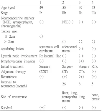
Table 1. Clinicopathologic characteristics of the patients with small cell carcinoma of uterine cervix
1 2 3 4
Age (yrs) 49 50 49 43
Stage Ib IIb IIa IIIa
Neuroendocrine marker (NSE, synaptophysin,
chromogranin) (-) NSE(+) (-) (-)
Tumor size
≦ 2cm ○
> 2cm ○ ○ ○
coexisting lesion squamous cell
carcinoma adenocarci
noma (-) (-)
Lymph node involvement Rt internal iliac (-) (-) (-) lymphovascular invasion (-) (-) (+) (-) Initial treatment Surgery Surgery Surgery RTx
Adjuvant therapy CCRT CTx CTx (-)
Recurrence (-) (+) (+) (+)
Interval to
recurrence(month) 26 8 5
Site of recurrence liver, lung, retroperito
neum lung bone, breast
Survival (+)
*(-) (-) (-)
NSE : nueron-specific enolase RTx : radiation therapy
CCRT : concurrent chemoradiotherapy CTx : chemotherapy
*생존 기간 : 13년
을 살펴 보고자 환자의 의무기록 열람을 통하여 연령, 병 기, 치료 방법 등을 분석하였으며 전화를 통하여 생존여 부를 확인하였다. 병기의 결정은 International Federation of Obstetrics and Gygnecology (FIGO)의 분류에 따랐다.
환자의 치료는 병기가 IIb 이하인 경우는 광범위 자궁 적출술을 시행하였고, IIIa 이상인 경우에는 방사선 요법 및 항암화학요법을 시행하였다. 조직학적 검사는 광학 현미경 관찰 및 neuron-specific enolase(NSE), synaptophysin, chromaogranin의 신경내분비 표지자를 이 용한 면역화학염색(immunohistochemistry)을 시행하였다.
광범위 자궁적출술을 시행한 모든 환자에서 골반 림프절 절제술을 시행하였고 수술 후 조직학적 검사에서 암세포 의 종류와 크기, 림프절 전이 여부를 확인하였으며 림프 혈관강의 침윤 또는 골반림프절에 전이가 있거나 침윤의 깊이가 자궁경부 벽의 2/3 이상인 경우에는 수술 후 항암 화학요법 또는 방사선 요법을 시행하였다. 항암화학요 법은 cisplatin과 etoposide의 병합요법이나 cisplatin과 5-FU의 병합 요법을 사용하였고 재발 환자에서는 ifosfamide, cisplatin, etoposide의 병합요법을 사용하기도 하고 cisplatin, paclitaxel 항암화학요법을 사용하기도 하 였다.
대상 환자는 1차 치료 후 골반진찰, 자궁경부 세포진 검사, 종양표식자 등을 매 3개월마다 시행하였으며 단순 흉부촬영술 및 골반 컴퓨터 단층 촬영은 매년 시행하여 추적관찰 하였다. 재발이 의심되는 경우에는 의심 부위 의 전산화 단층촬영 및 골 조사를 시행하였으며 필요시 조직 생검을 통하여 재발의 여부를 확인하였다.
결 과
1990년 1월부터 2006년 12월까지 자궁 경부 소세포암 으로 진단 받은 사람은 4예로 확인되었다. 환자의 연령 분포는 43~50세였으며 FIGO병기는 각각 Ib, IIa, IIb, IIIa 였다. 조직학적 검사결과에서 1예만 NSE에 양성인 신경 내분비 소세포암으로 확인되었고 두 예는 미분화 소세포 암, 1예는 편평상피 소세포암이었다.
자궁경부 소세포암의 진단 당시 1예에서 국소 골반 림 프절 전이를 보였으며 원격 전이는 없었다. 수술적 치료 를 시행한 3예 중 3예 모두 수술 후 추가 치료를 시행하 였고 그 중 1예는 항암화학치료와 방사선 치료를 병행하 였고 2예는 항암 화학치료만 하였다. 위 4예의 환자를 추적 관찰한 결과 3예에서 재발이 확인되었고 재발 기간
은 5~26개월이었고 현재까지의 생존 환자는 1명 이었다.
재발 부위는 간 1예, 폐 2예, 뼈 1예, 유방 1예, 후복막 부
위 1예 등이었다. 현재까지 생존한 환자는 병기가 Ib였 고 IIb 이상은 모두 사망하였다. (Table 1)
고 찰
자궁경부 소세포암은 1972년 Albores-Saavedra 등에 의 해 카르시노이드(carcinoid)로 불리워져 처음 보고된 이 후
15수많은 이름으로 불리워져 왔는데 소세포 신경내분 비암(small cell neuroendocrine tumor), 소세포 미분화암 (small cell undifferentiated carcinoma), 오트 세포암(oat cell carcinoma), 호은성 세포암(argynophilic carcinoma), 내분비암(endocrine carcinoma, intermediated cell type) 등
이다.
16,17그러다가 1996년 자궁경부 내분비 종양을 조직
학적으로 소세포(small cell), 대세포(large cell), 전형적인 카르시노이드(classic carcinoid), 비전형적 카르시노이드 (atypical carcinoid)로 분류하면서 자궁경부 소세포암은 자궁경부 내분비 종양의 일부로 분류되었다.
18자궁경부 소세포암의 진단은 Veda 등과 Michels 등이
고신대학교 의과대학 학술지 제23권 2호, 2008
- 50 - 보고한 바와 같이 광학현미경으로 신경내분비성 과립 (neurosecretory granule)을 확인하는 방법과 신경내분비 표지자(neurendocrine marker)를 이용하여 진단하는 방법 을 혼합하여 사용하고 있다.
19,20광학현미경적 관찰시 보 이는 세포의 특징은 세포가 작고 둥글거나 방추형을 이 루며, 세포질이 적고, 과염색성 핵을 띄고, 미세 과립 염 색질이 있고, 핵소체가 보이지 않는 것, 세포 분열이 활 발한 것 등이다.
18면역화학적 방법에 이용하는 신경내 분비 표지자에는 CD56, cytokeratin, synaptophysin, chromogranin, nueron-specific enolase(NSE), epithelial membrane antigen(EMA) 등이 있으며 각각의 표지자에 대한 양성율은 다양하다.
10편평상피암이나 선암과 동 시에 존재하는 경우도 21~77%에서 보고되고 있다.
21-25자궁경부 소세포암은 전체 자궁 경부 침윤성 암의 0.31~5%
1-6,16를 차지하는 매우 드문 질환이다. 연령별로 살펴보면 10대에서 90대까지 넓은 연령층에서 발생하지
만
1-4,6평균 나이는 40대이며 본 연구에서도 주로 40대에
발생한 것으로 나타났다.
비정상 질출혈이 가장 흔한 증상이며 무증상인 경우는 매우 드물다.
6,22-24,29본 연구에서도 모든 환자에서 첫 증 상이 비정상 질출혈로 나타났다. 자궁경부 소세포암의 임상적 특징으로 몇몇 환자에 있어 부신피질 자극 호르 몬에 의한 쿠싱 증후군과 같은 임상적 증상을 나타내기 도 한다고 알려져 있으며 신경내분비 표지자에 양성을 보인 경우에 내분비계 이상에 의한 임상적 증상을 나타 내는 것은 아니다. 본 연구에서는 내분비계 증상을 나타 내는 환자는 없었으며 신경내분비 소세포암과 관련 호르 몬인 ACTH, Calcitonin, Gastrin, Serotonin, Pancreatic polypeptide 등에 의한 증상이 있을 수 있으나 이같은 호 르몬이 분비되어도 비활성형 이거나 그 양이 증상을 나 타낼 정도로 많지 않고 혈액 내에서 빠르게 비활성화 하 기 때문에 대부분 증상이 없다고 알려져 있다.
16,27자궁경부 소세포암은 발생 빈도가 낮아 병기에 따른 치료 방법이 정립되어 있지는 않지만 대부분의 경우 편 평상피암의 치료와 같이 치료하고 있으며 이는 진단 당 시 병기와 범위에 따라 결정되는데 초기 단계에서는 광 범위 자궁적출술과 골반 림프절 절제술을 시행하며 이 질병의 경우 진행이 공격적이므로 질병 초기라 하더라도 수술 후 항암화학요법과 방사선 치료를 추가하여야 한 다.
28예후는 매우 불량한 것으로 알려져 있으며
5,17,23-24그 이유는 림프절 전이가 많고
29, 폐 등에 원격 전이가 일찍
나타나기 때문이다.
17,24본 연구에서도 Ib기인 환자 한 명만이 생존해 있고 나머지 3예에서 모두 재발하여 사망 한 것으로 나타났다. Sheeet 등
30은 종양 크기가 2cm 이하 인 경우가 2cm 보다 큰 경우에 비해 생존율이 좋고 무병 생존율이 길다고 하여 진단 당시 종양 크기가 중요한 예 후인자임을 주장하였고, Silva 등
31은 조직학적으로 편평 세포암이나 선암이 같이 있을 경우 소세포암 성분만 있 는 경우에 비해 예후가 좋다고 하여 주된 예후인자가 FIGO 병기와 조직소견이라고 발표하였는데, 본 연구에 서는 병기가 Ib기였던 환자 1명 외에는 2기 이상의 3예 모두가 사망하였고 역시 병소의 크기도 2cm이상인 환자 3명 모두가 사망하여 병기와 종양의 크기가 예후에 영향 을 미치는 것을 알 수 있었으나 증례 수가 적어 병소 크 기나 조직소견에 따른 무병생존기간에 의미 있는 차이를 알 수는 없었다.
본 연구에서는 대상 환자의 수가 제한되어 자궁경부 소세포암의 생존율 및 예후에 관한 비교 분석을 하기에 는 한계가 있었으며 향후 더 많은 환자를 대상으로 한 다 기관 연구를 통하여 여러 가지 예후인자에 따른 최선의 치료 방법에 대하여 연구한다면 향후 자궁경부 소세포암 의 치료 결과 및 생존율이 더 좋아질 것으로 사료된다.
결 론
1990년 1월부터 2006년 12월까지 고신대학교 산부인 과에 내원하여 조직학적으로 자궁경부 소세포암으로 진 단되고 치료받았던 4예를 대상으로 임상병리학적 특성 과 예후를 분석한 결과 자궁경부 소세포암 환자에서 질 병의 FIGO 병기와 병소의 크기가 생존에 영향을 미치는 주요 예후인자로 밝혀졌다. 그러나 본 연구는 연구 대상 환자 수가 적고 자궁경부 소세포암의 예후에 관여하는 인자 각각에 대해 다양한 비교분석이 이루어지지 않아 한계가 있었다. 향후 대규모 연구를 통하여 계속적인 연 구를 통해 자궁경부 소세포암의 치료 결과 및 예후와 생 존율에 큰 도움이 되리라 생각한다.
참고문헌
