ISSN: 2233-601X (Print) ISSN: 2093-6516 (Online)
Received: October 10, 2017, Revised: February 2, 2018, Accepted: February 7, 2018, Published online: June 5, 2018
Corresponding author: Puwadon Thitivaraporn, Cardiovascular and Thoracic Surgery Unit, King Chulalongkorn Memorial Hospital, 1873 Rama 4 Pathumwan, Bangkok, Thailand 10330
(Tel) 66-2-256-4944 (Fax) 66-2-256-4905 (E-mail) [email protected]
© The Korean Society for Thoracic and Cardiovascular Surgery. 2018. All right reserved.
This is an open access article distributed under the terms of the Creative Commons Attribution Non-Commercial License (http://creativecommons.org/
licenses/by-nc/4.0) which permits unrestricted non-commercial use, distribution, and reproduction in any medium, provided the original work is properly cited.
Thrombocytopenia in Moderate- to High-Risk Sutureless Aortic Valve Replacement
Puwadon Thitivaraporn, M.D. 1 , Sarun Chiramongkol, M.D. 1 , Dittapol Muntham, M.S. 2 , Nopporn Pornpatrtanarak, M.D. 1 , Chanapong Kittayarak, M.D. 1 , Jule Namchaisiri, M.D. 1 ,
Seri Singhatanadgige, M.D. 1 , Pat Ongcharit, M.D. 1 , Vichai Benjacholamas, M.D. 1
1
Cardiovascular and Thoracic Surgery Unit, King Chulalongkorn Memorial Hospital,
2
Section of Mathematic, Faculty of Science and Technology, Rajamangala University of Technology Suvarnabhumi
Background: This study aimed to compare preliminary data on the outcomes of sutureless aortic valve re- placement (SU-AVR) with those of aortic valve replacement (AVR). Methods: We conducted a retrospective study of SU-AVR in moderate- to high-risk patients from 2013 to 2016. Matching was performed at a 1:1 ra- tio using the Society of Thoracic Surgeons predicted risk of mortality score with sex and age. The primary outcome was 30-day mortality. The secondary outcomes were operative outcomes and complications. Results:
A total of 277 patients were studied. Ten patients (50% males; median age, 81.5 years) underwent SU-AVR.
Postoperative echocardiography showed impressive outcomes in the SU-AVR group. The 30-day mortality was 10% in both groups. In our study, the patients in the SU-AVR group developed postoperative thrombocytopenia. Platelet counts decreased from 225×10
3/μL preoperatively to 94.5, 54.5, and 50.1×10
3/μL on postoperative days 1, 2, and 3, respectively, showing significant differences compared with the AVR group (p=0.04, p=0.16, and p=0.20, respectively). The median amount of platelet transfusion was higher in the AVR group (12.5 vs. 0 units, p=0.052). Conclusion: There was no difference in the 30-day mortality of moderate- to high-risk patients depending on whether they underwent SU-AVR or AVR. Although SU-AVR is associated with favorable cardiopulmonary bypass and cross-clamp times, it may be associated with postoperative thrombocytopenia.
Key words: 1. Sutureless valve replacement 2. Moderate to high risk patient 3. Postoperative thrombocytopenia 4. Aortic valve replacement
Introduction
Aortic stenosis is the most common form of valvu- lar heart disease [1]. The standard treatment for aortic stenosis is surgical aortic valve replacement (AVR) [2]. This operation involves sternotomy with cardiopulmonary bypass (CPB) during the operation.
The diseased aortic valve (especially calcific pathol- ogy) needs to be resected and replaced with a new valve. However, with the increased age of the at-risk population, older patients with aortic stenosis are at a higher risk during conventional operations because of comorbid disease and a heavily calcified annulus [3]. Up to 30% of aortic stenosis patients are classi-
https://doi.org/10.5090/kjtcs.2018.51.3.172
One reason for the high incidence of complications in TAVR is that the replacement of a tissue valve over a diseased calcified native valve can cause un- predictable damage to the calcified structure.
Moreover, in patients who decide to undergo com- bined cardiac surgery (e.g., myocardial revasculariza- tion, other valve repair/replacement, or the maze procedure), TAVR is not a suitable solution.
Momentum within the field returned to the pre- vious concept of AVR. Therefore, AVR needs to be performed more rapidly and safely in order to avoid compromised hemodynamic and clinical outcomes in intermediate- to high-risk patients, who are consid- ered to be in a gray zone regarding this procedure.
There have been many studies of the rapid deploy- ment of sutureless aortic valve replacement (SU-AVR), especially after the Food and Drug Administration approved this method in 2009. This technique has the benefit of rapid deployment without leaving any knots in the aortic valve annulus. SU-AVR also in- volves resecting and decalcifying the diseased valve, which is known to be best for patients.
Despite the benefits of SU-AVR, which include bet- ter hemodynamics in patients with a small aortic an- nulus [7], shorter CPB and cross-clamp times [7-12], and facilitating minimally invasive valve surgery [7,13], strong indications for SU-AVR remain unclear.
Prospective clinical studies of high-risk patients showed that the 30-day and 1-year mortality rates were not inferior to conventional AVR [8,14], and no significant differences were found in terms of compli- cations [15].
Methods
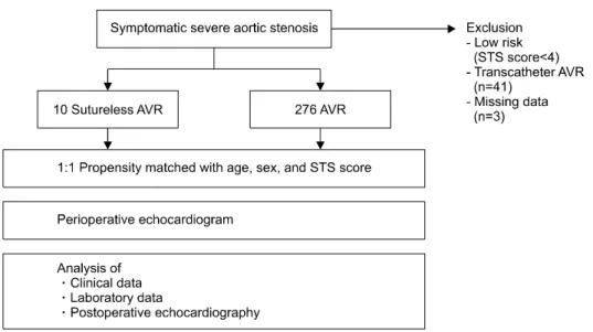
Symptomatic patients with severe aortic stenosis who underwent SU-AVR were matched with patients
perioperative and intraoperative outcomes and com- plications, including echocardiographic data. Echocar- diographic findings, complications, and clinical out- comes were compared between the groups.
1) Definitions
AVR was defined as conventional AVR with a stented aortic tissue valve replacement. SU-AVR was defined as sutureless AVR. TAVR was defined as transfemoral or transapical TAVR. Low-risk patients had a Society of Thoracic Surgeons (STS) score of
<4. Intermediate-risk patients had an STS score of
≥4–8. High-risk patients had an STS score of ≥8.
2) Data collection
This was a retrospective cohort study that was performed from January 2013 to May 2016. We car- ried out 1:1 matching of the SU-AVR and AVR groups, using the STS predicted risk of mortality (PROM) score with sex and age. All patients with symptomatic aortic stenosis were included. The ex- clusion criteria were as follows: (1) inoperable pa- tients who were assessed by the heart team; (2) pa- tients who underwent TAVR; (3) low-risk patients (STS score <4); (4) patients with a bicuspid aortic valve; (5) patients with a mechanical aortic valve; (6) patients with dissection or dilatation of the ascending aorta; (7) patients with a sinotubular junction/annu- lus ratio >1.3 (as a contraindication for the Perceval valve); (8) patients with known hypersensitivity to nickel alloys; (9) patients with an aortic annulus
<19 millimeter or >27 millimeter, which was not
compatible with the Perceval valve; (10) patients
with previous or concomitant root aneurysm; and
(11) patients with a recent history of stroke.
Fig. 1. Schema of 1:1 propensity matching. STS, Society of Thoracic Surgeons; AVR, aortic valve re- placement.
3) Operative procedure
All AVR operations included in this study were performed via the full median sternotomy approach.
CPB was established with the cardioplegic arrest technique. An aortic incision was made in either an oblique or transverse fashion above the sinotubular junction. The diseased aortic valve was meticulously excised. The aortic annulus was decalcified by ron- geur forceps or a no. 11 knife for proper valve positioning. The new valve was prepared after meas- urement of the aortic annular diameter. Either a su- tured or sutureless valve was replaced. Double-lay- ered aortotomy was performed for closing the incision.
(1) Conventional aortic valve replacement: All AVR procedures were performed with a bovine pericardial tissue valve replacement. The suture technique de- pended on the surgeon’s preference for either multi- ple simple stitches or a mattress suture with pledgets.
Supra-annular valve positioning was achieved in all AVR procedures.
(2) Sutureless valves: All SU-AVRs were performed with a Perceval S prosthesis (Sorin/LivaNova Group, Saluggia, Italy) and this sutureless valve was made of bovine pericardium. The valve was prepared to shrink before insertion and expansion. Three threads were sutured at the nadir of each sinus of Valsalva to the button hole at the Perceval valve. These threads were used as a reference line for alignment and to avoid malrotation. The valve was parachuted through the aortotomy incision to the aortic annulus.
After the valve was released from mounting, aug- mented expansion with pneumatic balloon dilatation pressure at 4 atm for 40 seconds was performed at the level of the annulus. The 3 hanging sutures were removed later.
4) Statistical analysis
After matching was performed, demographics, co- morbidities, and outcomes of interest were compared using the chi-square test or the Fisher exact test and the t-test for categorical and continuous variables, respectively. Results are expressed as mean±standard deviation or median with interquartile range (IQR).
Repeated continuous variables were compared using 1-way analysis of variance. All analyses were per- formed using IBM SPSS for Windows ver. 22.0 (IBM Corp., Armonk, NY, USA). All p-values <0.05 were considered to indicate statistical significance.
Results
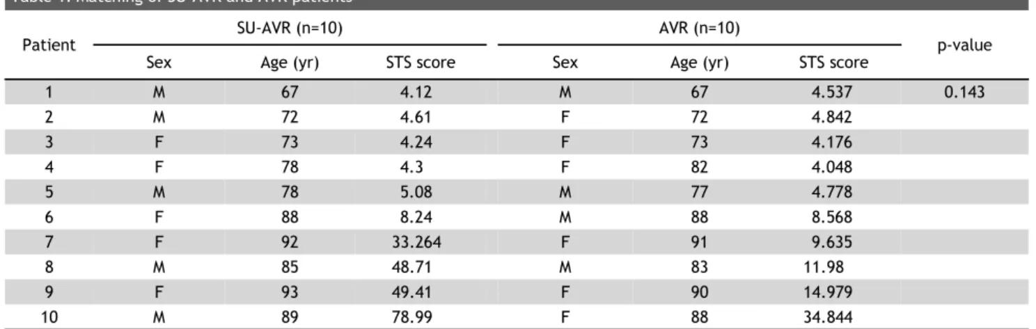
The results of 318 patients who were diagnosed with symptomatic severe aortic stenosis were initially analyzed. After the exclusion criteria were applied, there were 267 patients in the AVR group and 10 in the SU-AVR group (Fig. 1). STS PROM matching anal- ysis was performed at a 1:1 ratio (Table 1).
Demographic data, including sex, age, STS score, underlying disease, urgency of surgery, and type of surgery, are shown in Table 2. A total of 10 (5 men;
median age, 81.5 years) patients underwent SU-AVR,
9 F 93 49.41 F 90 14.979
10 M 89 78.99 F 88 34.844
SU-AVR, sutureless aortic valve replacement; AVR, aortic valve replacement; STS, Society of Thoracic Surgeons; M, male; F, female.
Table 2. Demographic data
Variable Sutureless AVR (n=10) AVR (n=10) p-value
Sex (male) 5 (50) 4 (40) 0.655
Age (yr) 81.5±9.1 81.1±8.4 0.758
Society of Thoracic Surgeons score 6.66 (4.28 –48.88) 6.70 (4.44 –12.72) 0.143
Diabetes mellitus 5 (50) 6 (60) 0.705
Hypertension 10 (100) 8 (80) 0.168
Renal failure 0 (0) 1 (10) NA
History of smoking 3 (30) 1 (10) 0.317
Coronary artery disease 7 (70) 6 (60) 0.608
New York Heart Association class ≥3 5 (50) 5 (50) 1.0
Echo: ejection fraction 55.10±17.57 57.06±17.64 0.84
Aortic valve area (cm
2) 0.67±0.34 0.51±0.33 0.313
Timing, emergency 5 (50) 2 (20) 0.25
Redo surgery 2 (20) 0 (0) NA
Combined valve surgery 2 (20) 2 (20) 1
Combined coronary artery bypass graft 5 (50) 6 (60) 0.705
Values are presented as number (%), mean±standard deviation, or median (interquartile range).
AVR, aortic valve replacement; NA, not applicable.
and were matched at a 1:1 ratio using the STS PROM score to 10 patients who underwent AVR in the study period. Sutureless valves were successfully im- planted in 9 of the 10 patients. The valve size was small in 1, medium in 5, and large in 4 patients in the SU-AVR group. The valve size was 21 mm in 4 patients, 23 mm in 5 patients, and 25 mm in 1 pa- tient in the AVR group. The median STS score was 6.66 (IQR, 4.28–48.88) in the SU-AVR group and 6.70 (IQR, 4.44–12.72) in the AVR group. The most com- mon presenting symptom was progressive dyspnea with a mean functional class of 3. The median CPB time was 120 minutes (IQR, 102–176.7 minutes) in
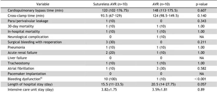
the SU-AVR group and 148 minutes (IQR , 113–175.5 minutes) in the AVR group (p=0.607) (Fig. 2). The median cross-clamp duration was 93.5 minutes (IQR , 67–129 minutes) in the SU-AVR group and 124 mi- nutes (IQR, 98.5–149.5 minutes) in the AVR group (p=0.140) (Fig. 2). The 30-day mortality was the same in the SU-AVR and AVR groups (10%).
One-year mortality was also the same in the SU-AVR
and AVR groups (10%). Two patients in the SU-AVR
group required a redo operation because of para-
valvular leakage. At 24 months of follow-up, the
rates of freedom from valve-related mortality, stroke,
acute myocardial infarction, endocarditis, and pros-
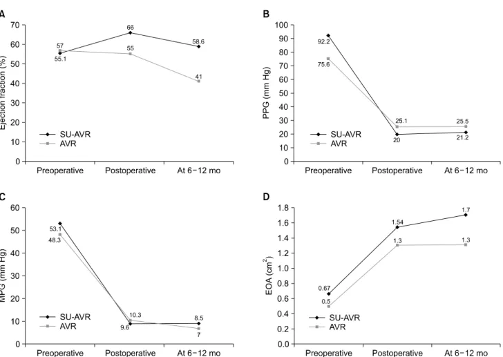
Fig. 2. (A) Ejection fraction, (B) PPG (mm Hg), (C) MPG (mm Hg), and (D) EOA (cm2). SU-AVR, sutureless aortic valve replacement; AVR, aortic valve replacement. PPG, peak pressure gradient; MPG, mean pressure gradient; EOR, effective orifice area.
Fig. 3. Comparison of platelet counts between the SU-AVR and AVR groups. SU-AVR, sutureless aortic valve replacement; AVR, aortic valve replacement; D, postoperative day. *p <0.05.
thesis regurgitation were 100%, 100%, 100%, 100%, and 90%, respectively, in the SU-AVR group. One pa- tient in the SU-AVR group developed moderate re- gurgitation during follow-up.
At postoperative follow-up, patients in the SU-AVR and AVR groups had a median functional class of I.
Postoperative echocardiography showed impressive outcomes in the SU-AVR group, with a reduced mean pressure gradient from 53.1 to 9.6 mm Hg post- operatively and 8.5 mm Hg at follow-up without left ventricular impairment (ejection fraction: 55.1% pre- operatively, 66% postoperatively, and 58.6% at 6–12 months of follow-up; p=0.41) (Fig. 3). The effective orifice area of the aortic valve increased from 0.67 to 1.54 cm
2in the SU-AVR group and from 0.51 to 1.3 cm
2in the AVR group (Fig. 3).
The median blood transfusion (packed red cells)
was 2 units (IQR, 1–3.25 units) in the SU-AVR group
Liver failure 0 0 NA
Tracheostomy 1 (10) 1 (10) 1.00
Atrial fibrillation 1 (10) 3 (30) 0.582
Pacemaker implantation 0 0 NA
Bleeding dysfunction
a)10 (100) 1 (10) <0.001
Length of hospital stay (day) 15.5 (11 –23.5) 20.5 (14 –27.75) 0.057
Intensive care unit stay (day) 3.82±1.75 3.59±1.81 0.89
Values are presented as median (interquartile range), number (%), or mean±standard deviation.
AVR, aortic valve replacement; NA, not applicable.
a)