ISSN: 2233-601X (Print) ISSN: 2093-6516 (Online)
Received: May 25, 2017, Revised: July 10, 2017, Accepted: July 11, 2017, Published online: February 5, 2018
Corresponding author: Byung-Chul Chang, Department of Cardiovascular Surgery, Severance Cardiovascular Hospital, Yonsei University College of Medicine, 50-1 Yonsei-ro, Seodaemun-gu, Seoul 03722, Korea
(Tel) 82-2-2228-8481 (Fax) 82-2-313-2992 (E-mail) [email protected]
*Current affiliation: Department of Thoracic and Cardiovascular Surgery, CHA Bundang Medical Center, CHA University, 59 Yatap-ro, Bundang-gu, Seongnam 13496, Korea
© The Korean Society for Thoracic and Cardiovascular Surgery. 2018. All right reserved.
This is an open access article distributed under the terms of the Creative Commons Attribution Non-Commercial License (http://creativecommons.org/
licenses/by-nc/4.0) which permits unrestricted non-commercial use, distribution, and reproduction in any medium, provided the original work is properly cited.
Early Clinical Experience with Sutureless Aortic Valve Replacement for Severe Aortic Stenosis
Do Jung Kim, M.D., Hyo-Hyun Kim, M.D., Shin-Young Lee, M.D., Sak Lee, M.D., Ph.D., Byung-Chul Chang, M.D., Ph.D.*
Department of Cardiovascular Surgery, Severance Cardiovascular Hospital, Yonsei University College of Medicine
Background: Sutureless aortic valve replacement (SU-AVR) has been developed as an alternative surgical treatment for patients with symptomatic severe aortic stenosis (AS). The aim of this study was to evaluate the clinical outcomes of SU-AVR through an assessment of hemodynamic performance and safety. Methods:
From December 2014 to June 2016, a total of 12 consecutive patients with severe AS underwent SU-AVR.
The endpoints were overall survival and valve-related complications (paravalvular leakage, valve thrombosis, migration, endocarditis, and permanent pacemaker implantation). The mean follow-up duration was 18.1±8.6 months. Results: The mean age of the patients was 77.1±5.8 years and their mean Society of Thoracic Surgeons score was 9.2±17.7. The mean cardiopulmonary bypass and aortic cross-clamp times were 94.5±37.3 minutes and 54.9±12.5 minutes, respectively. Follow-up echocardiography showed good prosthesis function with low transvalvular pressure gradients (mean, 13.9±8.6 mm Hg and peak, 27.2±15.0 mm Hg) at a mean of 9.9±4.2 months. No cases of primary paravalvular leakage, valve thrombosis, migration, or endo- carditis were reported. A new permanent pacemaker was implanted in 1 patient (8.3%). The 1-year overall survival rate was 83.3%±10.8%. Conclusion: Our initial experience with SU-AVR demonstrated excellent early clinical outcomes with good hemodynamic results. However, there was a high incidence of permanent pace- maker implantation compared to the rate for conventional AVR, which is a problem that should be solved.
Key words: 1. Aortic valve stenosis 2. Bioprosthesis
3. Heart valve prosthesis implantation
Introduction
Conventional aortic valve replacement (AVR) for patients with symptomatic severe aortic stenosis (AS) is recommended as the gold-standard treatment to alleviate symptoms and to improve survival [1-5].
The outcomes of AVR have improved over the past decades, but the incidence of mortality and morbidity
after surgical management remain high among pa-
tients with older age and multiple comorbidities
[6-8]. In recent years, as the number of high-risk eld-
erly patients has increased, technological advances
have led to less invasive alternative treatment modal-
ities, including sutureless AVR (SU-AVR) and trans-
catheter aortic valve implantation (TAVI), which have
expanded the indications for surgery to include high-
https://doi.org/10.5090/kjtcs.2018.51.1.1
or prohibitive-risk patients [4]. SU-AVR, which in- volves a rapidly deploying aortic valve device, has the advantages of reducing the cardiopulmonary by- pass (CPB) duration and aortic cross-clamp (ACC) time, thereby minimizing the operative risk, and en- abling the straightforward implantation of an aortic biologic valve prosthesis without sutures through a minimally invasive approach [4,9,10]. Several studies have reported that these sutureless valves resulted in a lower incidence of postoperative complications, es- pecially misplacement and paravalvular leakage, be- cause they involve the direct removal of the calcified valve and allow accurate debridement of the diseased aortic annulus [9,11,12]. The aim of this study was to evaluate retrospectively the 1-year clinical and echocardiographic outcomes of SU-AVR through an assessment of hemodynamic performance and safety.
Methods
Between December 2014 and June 2016, a total of 12 consecutive patients with severe AS who under- went SU-AVR at Severance Cardiovascular Hospital, Yonsei University College of Medicine were reviewed.
All patients were implanted with the Perceval valve system (Sorin Group Srl, Saluggia, Italy), which is a self-expanding, self-anchoring, sutureless bioprosthetic valve [13]. Patients with a congenital bicuspid aortic valve, an asymmetric aortic annulus, an annulus-to-si- notubular-junction ratio greater than 1.3, and an aortic annulus diameter less than 19 mm or greater than 27 mm were excluded from this study [12-14].
Transthoracic echocardiography (TTE) and coro- nary-valve computed tomography were routinely per- formed preoperatively to assess valvular morphology.
Preoperative and perioperative data were obtained from a review of the patients’ hospital charts, and follow-up was performed when patients returned for follow-up visits or by conducting telephone interviews.
The collection of follow-up data for at least 1 year was complete (100%). All patients underwent a clin- ical evaluation, blood tests, and TTE at each fol- low-up visit. The preoperative variables included in the analysis were age, sex, body surface area, hyper- tension, diabetes mellitus, coronary artery disease, chronic obstructive pulmonary disease, chronic kid- ney disease, arrhythmia, a history of a previous per- cutaneous coronary intervention or cardiac operation,
New York Heart Association (NYHA) functional class, Society of Thoracic Surgeons (STS) score, the logistic European System for Cardiac Operative Risk Evalua- tion (Euroscore), and echocardiographic indicators (left ventricular ejection fraction, aortic valve area, peak and mean systolic pressure gradient, left atrial diameter, left ventricular end systolic and diastolic dimension, left atrial volume index, left ventricular mass index, and right ventricular systolic pressure).
High-risk patients were defined as those with an STS operative risk score of 8% or higher.
The following perioperative variables were re- corded: CPB and ACC time, valve size, and any con- comitant cardiac procedures. Clinical outcomes were assessed in terms of all-cause mortality and post- operative valve-related complications. Valve-related events were defined as valve thrombosis, embolism, and bleeding events (formerly anticoagulant hemor- rhage) according to the American Association for Thoracic Surgery guidelines for reporting morbidity and mortality after cardiac valve interventions [15].
1) Operative techniques
All operations were performed under CPB using systemic hypothermia through a median or minimal invasive upper sternotomy. A transverse aortotomy in a relatively high position, approximately 3.5 cm above the aortic annulus, was performed to accom- modate the height of the prosthetic stent. After de- calcification of the aortic annulus, the expandable stent was implanted in the appropriate annular posi- tion without any permanent suture. We routinely used specific sizers for the optimal valve size and 3 guiding sutures between 2 commissures to correctly insert the valve at the level of the native aortic annulus. The prosthesis was then released into the valve and dilated with a low-pressure balloon cathe- ter for 30 seconds at a pressure of 4 atmospheres [ 12,13,16]. When the valve was successfully de- ployed, the guiding sutures were removed.
2) Statistical analysis
Statistical analyses were performed using IBM
SPSS ver. 23.0 (IBM Corp., Armonk, NY, USA). All da-
ta are presented as mean±standard deviation or as
frequency and percentage. Continuous variables were
analyzed using the Student t-test, and categorical var-
iables were compared using the chi-square test or
Table 1. Baseline patients’ characteristics (N=12)
Variable Value
Age (yr) 77.1±5.8 (62.0 –85.0)
≥80 years older 4 (33.3)
Sex (female) 7 (58.3)
Body surface area (m
2) 1.68±0.17 (1.44 –1.98)
Hypertension 10 (83.3)
Diabetes mellitus 5 (41.7)
Coronary artery disease 3 (25.0)
Chronic obstructive pulmonary disease 3 (25.0)
Chronic kidney disease 1 (8.3)
Peripheral artery disease 0
Cardiac rhythm
Sinus rhythm 8 (66.7)
Atrial fibrillation 2 (16.7)
Pacemaker 2 (16.7)
Previous cardiac operation 3 (25.0)
NYHA class III 11 (91.7)
NYHA class IV 1 (8.3)
Society of Thoracic Surgeons score 9.2±17.7 (0.9 –64.3) Logistic Euroscore 16.2±19.2 (3.3 –75.0)
Euroscore II 7.8±11.8 (1.9 –42.4)
Values are presented as mean±standard deviation (range) or number (%).
NYHA, New York Heart Association; Euroscore, European System for Cardiac Operative Risk Evaluation.
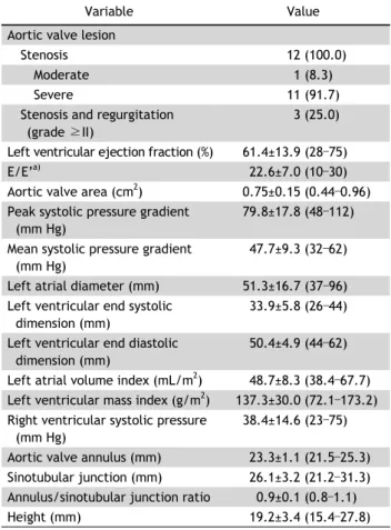
Table 2. Preoperative echocardiographic data (N=12)
Variable Value
Aortic valve lesion
Stenosis 12 (100.0)
Moderate 1 (8.3)
Severe 11 (91.7)
Stenosis and regurgitation (grade ≥II)
3 (25.0)
Left ventricular ejection fraction (%) 61.4±13.9 (28 –75)
E/E’
a)22.6±7.0 (10 –30)
Aortic valve area (cm
2) 0.75±0.15 (0.44 –0.96) Peak systolic pressure gradient
(mm Hg)
79.8±17.8 (48 –112) Mean systolic pressure gradient
(mm Hg)
47.7±9.3 (32 –62)
Left atrial diameter (mm) 51.3±16.7 (37 –96) Left ventricular end systolic
dimension (mm)
33.9±5.8 (26 –44) Left ventricular end diastolic
dimension (mm)
50.4±4.9 (44 –62)
Left atrial volume index (mL/m
2) 48.7±8.3 (38.4 –67.7) Left ventricular mass index (g/m
2) 137.3±30.0 (72.1 –173.2) Right ventricular systolic pressure
(mm Hg)
38.4±14.6 (23 –75) Aortic valve annulus (mm) 23.3±1.1 (21.5 –25.3) Sinotubular junction (mm) 26.1±3.2 (21.2 –31.3) Annulus/sinotubular junction ratio 0.9±0.1 (0.8 –1.1)
Height (mm) 19.2±3.4 (15.4 –27.8)
Values are presented as number (%) or mean±standard deviation (range).
a)
Ratio of early diastolic transmitral velocity to early diastolic tis- sue velocity.
the Fisher exact test. The long-term survival curve was evaluated using the Kaplan-Meier method. All p-values less than 0.05 were considered to indicate statistical significance, and rates are expressed with 95% confidence limits.
Results
1) Patient characteristics and preoperative echo- cardiographic data
The mean age of the patients was 77.1±5.8 years (range, 62.0 to 85.0 years) and 33.3% were at least 80 years old. The study population consisted of 7 women (58.3%) and 5 men (41.7%). The pre- operative characteristics of the patients and their cardiovascular comorbidities are summarized in Table 1. Eleven patients (91.7%) were classified as NYHA functional class III, and 1 (8.3%) was classified as NYHA functional class IV. The mean STS score was 9.2±17.7, the logistic Euroscore was 16.2±19.2, and the Euroscore II was 7.8±11.8. In 3 patients (25.0%), previous cardiac surgery was performed be-
fore the SU-AVR procedure, such as coronary artery bypass grafting or mitral valve replacement. As shown in Table 2, the preoperative left ventricular ejection fraction was 61.4%±13.9% (range, 28% to 75%) and the peak and mean systolic pressure gra- dients were 79.8±17.8 mm Hg and 47.7±9.3 mm Hg, respectively.
2) Perioperative data
The sutureless valve was successfully implanted in
all patients (Fig. 1). One patient required a second
attempt due to size mismatching. The valves were
sized medium (n=5, 41.7%), large (n=4, 33.3%), and
extra-large (n=3, 25.0%). Eleven patients underwent
isolated AVR, and concomitant coronary artery by-
pass grafting was performed in 1 patient with 3-ves-
sel disease. Minimally invasive surgery (upper ster-
Table 3. Perioperative data
Variable Value
Surgical approach
Median sternotomy 6 (50.0)
Minimal invasive approach 6 (50.0) Valve size
S (21 mm) 0
M (23 mm) 5 (41.7)
L (25 mm) 4 (33.3)
XL (27 mm) 3 (25.0)
Cardiopulmonary bypass time (min) 94.5±37.3 (55.0 –183.0) Aortic cross clamp time (min) 54.9±12.5 (39.0 –87.0) Concomitant cardiac surgery
Coronary artery bypass grafting 1 (8.3)
Values are presented as number (%) or mean±standard deviation (range).
Table 4. Clinical outcomes
Variable Value
Reoperation for bleeding 0
Newly required dialysis 0
Delayed ventilation 1 (8.3)
Early stroke 0
Gastrointestinal bleeding 0
Arrhythmia/pacemaker implantation 2 (16.7)/1 (8.3) Hospital stay (day) 16.8±14.8 (7 –59) Postoperative New York Heart Association class
I 5 (41.7)
II 7 (58.3)
In hospital mortality 0
Late mortality 3 (25.0)
Valve thrombosis 0
Endocarditis 0
Valve migration 0
Paravalvular leak 0
Values are presented as number (%) or mean±standard deviation (range).
Fig. 1. Computed tomography scan performed 1 month post- operatively, showing a well-functioning Perceval S sutureless bioprosthesis. A, anterior; P, posterior; H, head; F, foot.
Fig. 2. Kaplan-Meier curve showing the overall survival rate in patients who were implanted with a sutureless valve (SU-AVR).
SU-AVR, sutureless aortic valve replacement.
notomy) was performed in 6 patients (50.0%). Three patients underwent redo surgery. The mean CPB and ACC times were 94.5±37.3 minutes and 54.9±12.5 minutes, respectively (Table 3).
3) Clinical outcomes
The mean follow-up duration was 18.1±8.6 months (range, 3.3 to 28.9 months). The overall survival rate at 1 year was 83.3%±10.8%. The Kaplan-Meier risk curve for overall survival is shown in Fig. 2. There was no case of 30-day in-hospital mortality. Three
patients (25.0%) in the population died during the
follow-up period. The causes of death were cerebral
hemorrhage (n=1), fungal sepsis (n=1), and gastro-
intestinal bleeding due to the rupture of esophageal
varices (n=1). An 81-year-old woman who had
worked at a sanatorium as a physician died at 3.3
months postoperatively due to septic shock with cul-
tured Candida albicans, and another 81-year-old
woman with an uncontrolled international normal-
ized ratio died during follow-up due to anti-
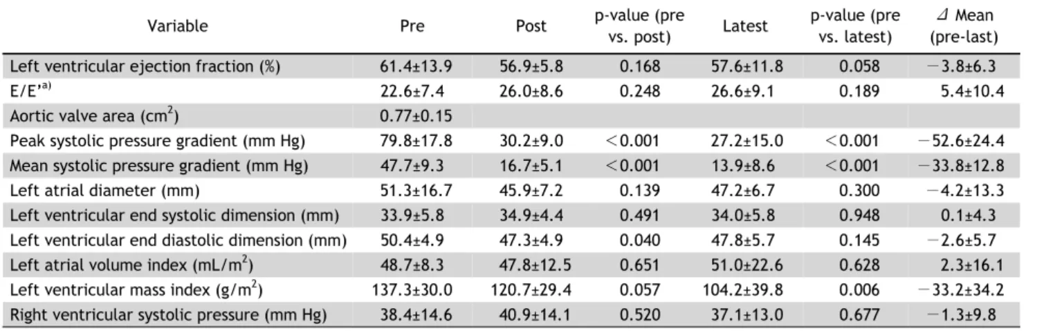
Table 5. Hemodynamic data from postoperative to latest follow-up
Variable Pre Post p-value (pre
vs. post) Latest p-value (pre vs. latest)
Δ Mean (pre-last) Left ventricular ejection fraction (%) 61.4±13.9 56.9±5.8 0.168 57.6±11.8 0.058 −3.8±6.3
E/E’
a)22.6±7.4 26.0±8.6 0.248 26.6±9.1 0.189 5.4±10.4
Aortic valve area (cm
2) 0.77±0.15
Peak systolic pressure gradient (mm Hg) 79.8±17.8 30.2±9.0 <0.001 27.2±15.0 <0.001 −52.6±24.4 Mean systolic pressure gradient (mm Hg) 47.7±9.3 16.7±5.1 <0.001 13.9±8.6 <0.001 −33.8±12.8
Left atrial diameter (mm) 51.3±16.7 45.9±7.2 0.139 47.2±6.7 0.300 −4.2±13.3
Left ventricular end systolic dimension (mm) 33.9±5.8 34.9±4.4 0.491 34.0±5.8 0.948 0.1±4.3 Left ventricular end diastolic dimension (mm) 50.4±4.9 47.3±4.9 0.040 47.8±5.7 0.145 −2.6±5.7 Left atrial volume index (mL/m
2) 48.7±8.3 47.8±12.5 0.651 51.0±22.6 0.628 2.3±16.1 Left ventricular mass index (g/m
2) 137.3±30.0 120.7±29.4 0.057 104.2±39.8 0.006 −33.2±34.2 Right ventricular systolic pressure (mm Hg) 38.4±14.6 40.9±14.1 0.520 37.1±13.0 0.677 −1.3±9.8 Values are presented as mean±standard deviation.
Pre, preoperative; Post, postoperative.
a)