CT Follow-Up of Postoperative Bronchopleural Fistula:
Risk Factors for Progression to Chronic Complicated Infection
수술 후 기관지 흉막루의 전산화단층촬영 추적 검사:
만성 복합성 감염 진행의 위험인자 분석
Ji-Yeon Han, MD1 , Ki-Nam Lee, MD2* , Yoo Sang Yoon, MD3 , Jihyun Lee, MD4 , Hongyeul Lee, MD5 , Seok Jin Choi, MD1 , Hye Jung Choo, MD1 , Jin Wook Baek, MD1 , Young Jin Heo, MD1 , Gi Won Shin, MD1 , Jinyoung Park, MD1 , Dasom Kim, MD1
Departments of 1Radiology, 3Thoracic Surgery, 5Internal Medicine, Respiratory Medicine, Inje University College of Medicine, Busan Paik Hospital, Busan, Korea
2Department of Radiology, Dong-A University College of Medicine, Busan, Korea
4Department of Radiology, Dongnam Institute of Radiological & Medical Sciences Cancer Center, Busan, Korea
Purpose We evaluated the risk factors for progression to chronic complicated bronchopleural fistula (BPF) after pulmonary resection using follow-up CT.
Materials and Methods We retrospectively reviewed 45 cases with BPF that had undergone pulmonary resection during 2010-2018. We compared the clinical and radiological characteris- tics of those with complicated BPF (n = 24) and those without complicated (sterilized) BPF (n = 21). The clinical and radiological risk factors for progression to chronic complicated BPF were examined by logistic regression analysis.
Results The thickness of the pleural cavity wall (p = 0.022), the size of the pleural cavity (p = 0.029), and the size increase of BPF on follow-up (p = 0.012) were significantly different be- tween the two groups. The risk factors for progression to chronic complicated BPF were age
> 70 years (odds ratio, 6.43; 95% confidence interval, 1.2–33.7), the thickness of the cavity wall
> 5 mm (odds ratio, 52.5; 95% confidence interval, 5.1–545.4), and an increase in the size of the pleural cavity on follow-up CT (odds ratio, 12.5; 95% confidence interval, 2.1–73.5), only in the univariate analysis.
Conclusion The risk factors for progression to chronic complicated BPF can be evaluated using follow-up CT.
Index terms Fistula; Lobectomy; Pneumonectomy
Received January 23, 2020 Revised April 21, 2020 Accepted June 11, 2020
*Corresponding author Ki-Nam Lee, MD Department of Radiology, Dong-A University Medical Center, 26 Daesingongwon-ro, Seo-gu, Busan 49201, Korea.
Tel 82-51-240-5367 Fax 82-51-253-4931 E-mail gnlee@dau.ac.kr This is an Open Access article distributed under the terms of the Creative Commons Attribu- tion Non-Commercial License (https://creativecommons.org/
licenses/by-nc/4.0) which permits unrestricted non-commercial use, distribution, and reproduc- tion in any medium, provided the original work is properly cited.
ORCID iDs Ji-Yeon Han https://
orcid.org/0000-0003-3780-358X Ki-Nam Lee
https://
orcid.org/0000-0003-0848-3935 Yoo Sang Yoon
https://
orcid.org/0000-0002-7458-661X Jihyun Lee
https://
orcid.org/0000-0001-8198-9241 Hongyeul Lee
https://
orcid.org/0000-0002-6121-3611 Seok Jin Choi
https://
orcid.org/0000-0002-2688-4006 Hye Jung Choo
https://
orcid.org/0000-0003-3941-6989 Jin Wook Baek
https://
orcid.org/0000-0003-4632-4951 Young Jin Heo
https://
orcid.org/0000-0002-4765-0727 Gi Won Shin
https://
orcid.org/0000-0002-6202-1945 Jinyoung Park
https://
orcid.org/0000-0003-2713-4490 Dasom Kim
https://
orcid.org/0000-0002-8501-9389
INTRODUCTION
Although the incidence of bronchopleural fistula (BPF) after pulmonary resection has de- creased to 1–2% in recent years, when it occurs, management and treatment remain a major challenge (1). The aim of management in this condition is to control active infection in the pleural cavity with closure of the fistula; however, there is still a considerable failure rate (2, 3). The incidence of initial treatment failure and progression to a refractory fistula with chronic empyema is known to be 8.3–25% (4, 5). Empyema-complicated BPF is particularly difficult to manage, despite various treatment options, including surgical and bronchoscopic maneuvers, has a long recovery period (6).
Early prediction of refractory fistula with chronic infection might reflect the response to current treatment and might facilitate deciding on changes in the treatment plan, which might reduce the overall duration and cost of treatment. The risk factors for developing BPF have been widely studied; however, limited data are available on the prediction of progres- sion to chronic disease. The purpose of this study was to identify factors that associated with the progression to chronic complicated BPF after pulmonary resection, based on CT follow-up.
MATERIALS AND METHODS STUDY POPULATION
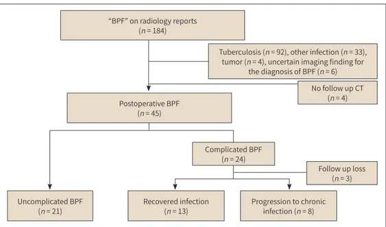
This retrospective study was approved by the Institutional Review Board of both participat- ing hospitals (D-1810-017-002, 19-0164) and the need to obtain informed patient consent was waived. We retrospectively identified 184 patients with “bronchopleural fistula” on radiology reports in the radiology information system databases of two hospitals, between July 2010 and June 2018. Patients with BPF caused by pulmonary infection (n = 33), tuberculosis (n = 92), tu- mor (n = 4), and uncertain imaging findings for the diagnosis of BPF (n = 6) were excluded. In addition, patients who had no follow-up CT (n = 4) were also excluded. Forty-five patients who had developed BPF after pulmonary resection were finally included (Fig. 1).
CT TECHNIQUES
CT scans were obtained with Brilliance iCT (Philips Medical Systems, Cleveland, OH, USA), Somatom Definition AS (Siemens Healthcare, Erlangen, Germany), Somatom Drive (Siemens Healthcare), Somatom Perspective (Siemens Healthcare), or Discovery CT 750HD (GE Health- care, Waukesha, WI, USA). The CT parameters were as follows: 0.9 or 0.625 mm collimation;
2–5 mm thickness and 2–5 mm increment without gap, and 1-mm thickness with 5-mm gap;
high spatial-frequency algorithm; 120 kVp, and 100 effective mAs, with dose modulation for chest CT with enhancement. Coronal multiplanar reformation images with a section thick- ness of 3 mm and an increment of 3 mm were routinely constructed.
CT ANALYSIS
All CT images were interpreted by two radiologists in consensus. An imaging diagnosis of BPF was made when the presence of a definite fistulous tract was detected between the pleu- ral space and airway (direct), or a suspicious air bubble, or a suspicious but not definite com-
munication between the pleural space and airway (indirect) was noted (7). Location of the BPF (central, more proximal than the lobar bronchus or peripheral, more distal than the lo- bar bronchus), onset of BPF (duration from operation to development of BPF), stump length, the size of the pleural cavity, and the thickness of the pleural cavity wall were evaluated on initial CT images when a BPF was diagnosed, or at the first follow-up CT when a complicated BPF developed. The thickness of the cavitary wall was measured at the thickest portion of the pleura in nondependent side and the size of the cavity was measured by the longest length of the aerated pleura either in axial or coronal scan (Fig. 2). Stump length was measured only in cases of central BPF. Measurements of continuous variables (stump length, size of pleural cavity, and thickness of pleural cavity wall) by each observer were averaged for statistical analysis. After the initial CT evaluation, all follow-up CTs were reviewed by 2 observers in consensus. Duration of BPF was defined by the period from the time of the first CT scan when BPF was detected until the time of the last follow-up CT, or the time when BPF was no longer visible. Development of complicated BPF was defined as the presence of empyema (air-fluid level with pleural thickening), recurrent aspiration pneumonia, and postoperative acute lung injury during the follow-up. We defined chronic infection when the complicated BPF persisted over 1 year from the development of the infection. The change in size of the aerated pleural cavity (increased size of pleural cavity/partially decreased size/total resolution of pleural cavity) and total duration of BPF were evaluated at follow-up CT.
CLINICAL ANALYSIS
Bronchosocpy was performed in 16 cases, and a direct fistulous tract was found in five cas- es. The presence of thoracic infection was confirmed either by positive microbial staining and/or culture of sputum, blood, pleural, bronchoscopic lavage, or by serum aspergillus anti- gen test. The patients with BPF who did not show evidence of thoracic infection in clinical and radiologic analysis were considered to have a sterilized pleural cavity. Three patients with complicated BPF were lost during the follow-up, before 1 year. Patients expired due to postoperative acute lung injury (n = 2), pneumonia (n = 2), or recurrence of cancer (n = 5) during the follow-up period. The median duration of radiographic follow-up was 10 months (range, 0.6–73 months).
STATISTICAL ANALYSIS
Patients with and without total resolution were compared by Student’s t test or the Mann–
Whitney U and chi-square tests. Clinical and radiological characteristics were compared be- tween the group who had complicated BPF and the group who had sterilized BPF, using ei- ther Student’s t test or the Mann–Whitney U and chi-square tests.
Logistic regression analysis was performed to identify the risk factors for progression to chronic complicated BPF. Variables used in the univariate analysis included clinical factors (age, sex, cause or extent of the operation, onset of BPF, postoperative chemoradiation thera- py) and CT findings (type and location of BPF, stump length, size of the pleural cavity, thick- ness of the pleural cavity wall, and course of BPF). Significant variables in the univariate analysis were further analyzed in a multivariate logistic regression analysis. Statistical analy- sis was performed using SPSS version 25.0 (IBM Corp., Armonk, NY, USA). p values less than
0.05 were considered significant.
RESULTS
Among the 184 patients with BPF, 26.6% (n = 49) of cases were related to pulmonary resec- tion. The causes of pulmonary resection were lung cancer (n = 26), metastasis (n = 11), asper- gilloma (n = 5), tuberculosis (n = 4) and other benign disease (n = 3). Patient selection and the number of those who progressed to chronic complications in postoperative BPF are summa- rized in Fig. 1.
In CT analysis, postoperative BPF was common in right side (32/45, 71.1%), and in upper lobe (24/45, 53.3%). Direct type was more common in peripheral BPF (15/22, 68.2%), than in central BPF (9/23, 39.1%). In follow up CT scans, 30.4% (7/23) of cases showed size increase in central BPF, while 13.6% (3/22) of cases showed size increase in peripheral BPF.
In clinical analysis of postoperative BPF, 46.7% (21/45) of cases did not have a thoracic in- fection, and were considered to have a sterilized pleural cavity. None of the patients showed meaningful evidence of infection in laboratory findings. Complicated BPF had developed in 53.3% (24/45) patients in the form of empyema (79.2%, n = 19), recurrent aspiration pneumo- nia (41.7%, n = 10), fungal infection (12.5%, n = 3), and postoperative lung injury (12.5%, n = 3).
BPF-associated deaths were caused by pneumonia (n = 3) and postoperative lung injury (n = 1), and the mortality rate of postoperative BPF was 8.9% (4/45).
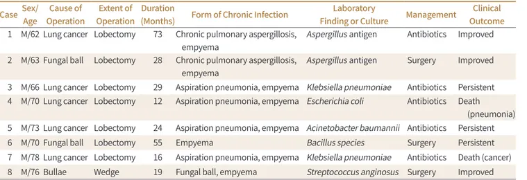
Among the cases of complicated BPF (n = 24), 3 patients were lost to follow-up before reach- ing 1 year of thoracic infection, while 8 patients had progressed to chronic infection (33.3%, 8/24). Clinical information of cases with chronic complicated BPF is reported in Table 1. Sur- gical management was required in 42.9% (3/7) of patients with chronic complicated BPF. In
“BPF” on radiology reports (n = 184)
Tuberculosis (n = 92), other infection (n = 33), tumor (n = 4), uncertain imaging finding for
the diagnosis of BPF (n = 6)
Postoperative BPF (n = 45)
Complicated BPF (n = 24)
No follow up CT (n = 4)
Follow up loss (n = 3) Uncomplicated BPF
(n = 21) Recovered infection
(n = 13) Progression to chronic infection (n = 8)
Fig. 1. Patient selection and the number of patients showing progression to chronic complicated postopera- tive BPF.
BPF = bronchopleural fistula
one patient, surgery failed to manage the chronic infection, and one patient died of pneumo- nia during the follow-up.
Approximately 44.4% (20/45) of patients showed total resolution of pleural cavity on longi- tudinal follow-up CT. These patients were significantly different from patients who did not, in terms of development of complicated BPF (p = 0.038), total duration of BPF (mean 9.5 months vs. 18.7 months, p = 0.039) and the thickness of the cavity wall (mean 3.3 mm vs. 4.4 mm, p = 0.044). Sex, cause of operation, peripheral BPF, indirect BPF, extent of operation, and a history of chemoradiation therapy did not increase the frequency of total resolution.
Patient characteristics and CT analysis of the groups with complicated BPF and sterilized BPF are reported in Table 2. Any of clinical factors were not significantly different between the two groups. In sterilized BPF group, the size of pleural cavity was smaller (p = 0.029), and the resolution of pleural cavity was common on follow-up CT (p = 0.014), compared with the complicated BPF group. The thickness of pleural cavity was also thinner in sterilized BPF with near statistical significance (p = 0.063).
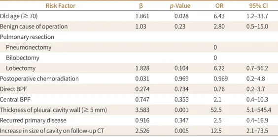
In logistic regression analysis, factors related to progression to chronic complicated BPF were age > 70 years (p = 0.028), thickness of the pleural cavity wall > 5 mm (p = 0.001), and in- crease in the size of the cavity on follow-up (p = 0.005) (Table 3). However, 95% confidence in- terval of the thickness of cavity wall was wide. Multivariate analysis failed to demonstrate a sig- nificant association of any of the factors above with progression to chronic complicated BPF. A representative case of progression to chronic complicated BPF is shown in Fig. 3.
DISCUSSION
Although a wide variety of bronchoscopic and surgical maneuvers have been developed, no gold-standard management for BPF has been established to date (8). Some recent studies have attributed importance to conservative management of BPF by infection control, using imaging follow-up or endoscopic applications, to avoid invasive surgical techniques (8-11). As conservative treatment requires a long period of time, determination of treatment failure and the time point at which the treatment option should be changed to avoid reaching re-
Table 1. Cases of Chronic Complicated Bronchopleural Fistula*
Case Sex/
Age
Cause of Operation
Extent of Operation
Duration
(Months) Form of Chronic Infection Laboratory
Finding or Culture Management Clinical Outcome 1 M/62 Lung cancer Lobectomy 73 Chronic pulmonary aspergillosis,
empyema
Aspergillus antigen Antibiotics Improved 2 M/63 Fungal ball Lobectomy 28 Chronic pulmonary aspergillosis,
empyema
Aspergillus antigen Surgery Improved 3 M/66 Lung cancer Lobectomy 29 Aspiration pneumonia, empyema Klebsiella pneumoniae Antibiotics Persistent 4 M/70 Lung cancer Lobectomy 12 Aspiration pneumonia, empyema Escherichia coli Antibiotics Death
(pneumonia) 5 M/73 Lung cancer Lobectomy 24 Aspiration pneumonia, empyema Acinetobacter baumannii Antibiotics Persistent
6 M/70 Fungal ball Lobectomy 55 Empyema Bacillus species Surgery Persistent
7 M/78 Lung cancer Lobectomy 16 Aspiration pneumonia, empyema Klebsiella pneumoniae Antibiotics Death (cancer) 8 M/76 Bullae Wedge 19 Fungal ball, empyema Streptococcus anginosus Surgery Improved
*Cases of complicated bronchopleural fistula to progress chronic infection.
fractory empyema is of importance during the conservative management follow-up period.
The risk factors for development of postoperative BPF, including clinical factors and opera- tive method, have been widely studied (6, 12-15). However, prognostic factors related to pro- gression of BPF are not well known. To our knowledge, no previous studies have assessed the risk factors associated with refractory complicated BPF.
Number of central (n = 23) and peripheral (n = 22) BPF were similar in postoperative BPF in our study. Postlobectomy or pneumonectomy BPF are suspected by clinical signs and symp- toms by thoracic surgeons initially (15). As clinical outcome of peripheral fistulas are known to be favorable even with conservative treatment (13), subclinical or peripheral BPF can be Table 2. Comparison of the Patient Characteristics with and without Complicated BPF
Sterilized BPF (n = 21)
Complicated BPF
(n = 24) p-Value
Age 60.9 63.5 0.392
Sex (M/F) 18/4 25/2 0.252
Diabetes mellitus 5 4 0.713
Cause of operation 0.354
Malignant 18 19
Benign 4 8
Extent of pulmonary resection 0.498
Pneumonectomy 3 1
Bilobectomy 1 3
Lobectomy 12 17
Segmentectomy or wedge resection 6 6
Postoperative management
Radiation therapy 2 8 0.152
Chemotherapy 7 7 0.650
Onset of BPF (months) 4.0 (0.5–300.0) 3.0 (0.2–121.0) 0.647*
Total duration of BPF (months) 8.0 (0.6–48.0) 13.0 (1.0-73.0) 0.214*
Management of BPF
Surgery 1 4 0.362
Chest tube insertion 3 9 0.182
CT findings
Direct/indirect 9/13 15/12 0.308
Central/peripheral 10/12 15/12 0.482
Stump length (mm) 11.7 6.1 0.193
Recurrence 2 7 0.159
Size of cavity (mm) 62.7 86.8 0.026
Thickness of pleural cavity wall (mm) 3.0 (1.0–5.0) 4.0 (1.0–9.0) 0.063*
Size change of cavity on follow-up CT 0.014
Increase in size of cavity 1 9 0.012
Partial decrease in size of cavity 7 8 1.0
Total resolution of cavity 13 7 0.038
*Mann-Whitney U test. Relevant data are medians with ranges in parentheses.
BPF = bronchopleural fistula
easily neglected by thoracic surgeons. We had selected the patients by CT image, which lead to increase the number of subclinical or sterilized BPF that thoracic surgeons could not have noticed. Radiologic features that are suggestive of the presence of a BPF are usually indirect (16), however direct type were more common in peripheral BPF in our study, similar with past study (7). Our study demonstrated that postoperative BPF does not always result in tho- racic infection. In our study, 46.7% (21/45) of postoperative BPF cases did not have a thoracic infection, and 59.1% (13/21) of sterilized BPF showed total resolution on follow-up CT. When Table 3. Risk Factors of Chronic Complicated BPF in the Logistic Regression Analysis
Risk Factor β p-Value OR 95% CI
Old age (≥ 70) 1.861 0.028 6.43 1.2–33.7
Benign cause of operation 1.03 0.23 2.80 0.5–15.0
Pulmonary resection
Pneumonectomy 0
Bilobectomy 0
Lobectomy 1.828 0.104 6.22 0.7–56.2
Postoperative chemoradiation 0.031 0.969 0.969 0.2–4.8
Direct BPF 0.274 0.734 0.76 0.2–3.7
Central BPF 0.747 0.355 2.1 0.4–10.3
Thickness of pleural cavity wall (≥ 5 mm) 3.583 0.001 52.5 5.1–545.4
Recurred primary disease 0.916 0.347 2.5 0.4–16.9
Increase in size of cavity on follow-up CT 2.526 0.005 12.5 2.1–73.5 BPF = bronchopleural fistula, CI = confidence interval, OR = odds ratio
Fig. 2. Measurement of the CT parameters of BPF in patients who had undergone a right upper lobectomy for lung cancer.
A. A central direct BPF distal to a long stump (arrow) is detected 4 months after the operation. The type, lo- cation, and stump length are evaluated on the initial CT.
B. After 9 months, a complicated BPF in the form of empyema is first detected. The thickness of the cavitary wall at the non-dependent portion of the pleura (black arrow) and the size of the cavity (white dotted ar- row) are measured. The size of the aerated pleural cavity is decreased compared with the cavity shown in (A).BPF = bronchopleural fistula
A B
thoracic infection was present, 33.3% (8/24) of patients with complicated BPF progressed to chronic infection in longitudinal follow-up of postoperative BPF. The incidence of progres- sion to chronic complicated BPF was 17.8% (8/45) among postoperative BPF cases in this study. In a study about the natural course of post-pneumonectomy BPF, Hollaus et al. (4) re- ported that 16.7% (16/96) patients were discharged with chronic empyema, with permanent drainage. Westcott and Volpe (5) reported that 20% (5/20) of nonoperative BPF progressed to chronic disease, as visualized in thin-section CT. However, the incidence of chronic disease after pulmonary resection had not been reported in previous studies. As slow progression of disease may not be apparent in clinical findings, CT analysis might be necessary in the fol- low-up of BPF.
Surgery was required in 37.5% (3/8) of patients with chronic complicated BPF in this study.
Fistula repair is not necessarily the first step in managing empyema-complicated BPF (10).
Many studies have described the safety of conservative management of BPF and minimizing surgery (9, 10, 17). In our three patients who underwent surgical correction, all cases re- ceived delayed medical treatment because of loss to follow-up and failure to notice the com- plications. Delayed diagnosis and treatment contribute to multiple operations and morbidity.
Late-developing fistulas are particularly difficult to manage, because of the delayed diagnosis (1). We emphasize the necessity of CT follow-up both in conservative and surgical manage- ment, particularly in cases of delayed BPF, to confirm diminution of the pleural cavity during treatment and to determine the appropriate time for surgical correction.
Patients who showed total resolution were younger, had fewer complications, and thinner cavitary walls than the patients who did not show resolution. The presence of complications at postoperative BPF might be important factors for patients’ prognosis. Mortality related to Fig. 3. A 63-year old male with chronic complicated BPF after a right upper lobectomy for aspergillosis.
A. Aerated pleural cavity and air bubbles beneath the surgical clip (arrow), suggesting an indirect BPF. These are first detected in the right up- per thorax on a non-contrast coronal CT scan performed 30 months after the operation.
B. Serial contrast-enhanced CT 10 months after the operation shows that the size of the pleural cavity had increased.
C. After another 7 months, an axial lung window scan demonstrates an air-fluid level and irregular internal debris inside the cavity with the development of consolidation around the cavity (arrows). Bronchoscopic washing demonstrates an elevated titer of aspergillus antigen (3.71).
Conservative management failed, and surgery was required.
BPF = bronchopleural fistula
A B C
BPF is related to the degree of contamination of the pleural space (1). In a sterilized cavity, formation of granuloma and scars may minimize the cavity and the fistula may gradually be occluded (10). Prevention of development of thoracic infection might be the initial step for successful management of postoperative BPF.
Larger size of pleural cavity, thicker pleural cavitary wall, and size increase of the pleural cavity were frequent findings in patients with complicated BPF in our study. Any clinical fac- tors were not helpful in predicting the development of thoracic infection. CT analysis and follow-up might be more important to predict disease progression.
The risk factors for progression to chronic disease were found to be older age (over 70 years), a cavitary wall thickness over 5mm, and an increase in cavity size on follow-up, in our study. The pleural thickness of empyema demonstrated on CT was in agreement with empy- ema stage in a previous report. Cavitary wall thickness over 5 mm correlated with late stage II and III empyema, requiring surgical decortication (18). In our patients with pleural thick- ness exceeding 5 mm, 58.3% (7/11) of patients progressed to chronic complicated BPF.
Mao et al. (14) suggested the significance of observation of gradual narrowing of cavities in follow-up of cases under conservative management of empyema-complicated BPF. Radiolog- ic improvement of pleural opacity was considered to be an important indicator for managing empyema without surgery (19). Although there have been several studies of the role of CT in management of BPF in prior studies, we suggest that CT follow-up as well as an initial CT evaluation is necessary for successful management (7, 17, 20). Evaluation of pleural cavity wall thickness and changes in pleural cavity size on follow-up CT may facilitate clinical deci- sion-making between surgical and conservative management.
This study had several limitations. First, this study was retrospective, which implies the possibility of selection bias when patients were initially selected by imaging diagnosis. BPF diagnosed by bronchoscopy or surgery only were not included. Determination of complica- tions was also based on imaging diagnosis. However, in recent clinical practice, CT is a more practical method to diagnose postoperative BPF, as bronchoscopy and surgical diagnosis is not appropriate for all patients. Second, the number of patients in each group was too small to generalize the results.
In conclusion, we here identified several findings on follow-up CT that can allow predic- tion of development of chronic BPF. Old age, thicker pleural cavitary wall, and size change of the pleural cavity on follow-up CT scans were factors associated with progression to chronic complicated BPF. Follow-up CT might be helpful for early detection of chronic complicated BPF and for the clinical decision-making about managing postoperative BPF.
Author Contributions
Conceptualization, H.J.; data curation, H.J.; formal analysis, H.J.; investiation, L.J.; methodology, H.J., Y.Y.S.; project administration, L.K.; resources, Y.Y.S., L.H.; software, H.J.; supervision, L.K.; vali- dation, Y.Y.S.; visualization, L.J., Y.Y.S.; writing—original draft, H.J.; and writing—review & editing, all authors.
Conflicts of Interest
The authors have no potential conflicts of interest to disclose.
REFERENCES
1. Khan JH, Rahman SB, McElhinney DB, Harmon AL, Anthony JP, Hall TS, et al. Management strategies for complex bronchopleural fistula. Asian Cardiovascular Thoracic Annals 2000;8:78-84
2. Uramoto H, Hanagiri T. The development of bronchopleural fistula in lung cancer patients after major sur- gery: 31 years of experience with 19 cases. Anticancer Res 2011;31:619-624
3. Varoli F, Roviaro G, Grignani F, Vergani C, Maciocco M, Rebuffat C. Endoscopic treatment of bronchopleural fistulas. Ann Thorac Surg 1998;65:807-809
4. Hollaus PH, Lax F, El-Nashef BB, Hauck HH, Lucciarini P, Pridun NS. Natural history of bronchopleural fistu- la after pneumonectomy: a review of 96 cases. Ann Thorac Surg 1997;63:1391-1396; discussion 1396-1397 5. Westcott JL, Volpe JP. Peripheral bronchopleural fistula: CT evaluation in 20 patients with pneumonia, em-
pyema, or postoperative air leak. Radiology 1995;196:175-181
6. Sonobe M, Nakagawa M, Ichinose M, Ikegami N, Nagasawa M, Shindo T. Analysis of risk factors in broncho- pleural fistula after pulmonary resection for primary lung cancer. Eur J Cardiothorac Surg 2000;18:519-523 7. Seo H, Kim TJ, Jin KN, Lee KW. Multi-detector row computed tomographic evaluation of bronchopleural
fistula: correlation with clinical, bronchoscopic, and surgical findings. J Comput Assist Tomogr 2010;34:
13-18
8. Okuda M, Go T, Yokomise H. Risk factor of bronchopleural fistula after general thoracic surgery: review arti- cle. Gen Thorac Cardiovasc Surg 2017;65:679-685
9. Pforr A, Pagès PB, Baste JM, Thomas P, Falcoz PE, Lepimpec Barthes F, et al. A predictive score for bron- chopleural fistula established using the french database Epithor. Ann Thorac Surg 2016;101:287-293 10. Deschamps C, Bernard A, Nichols FC 3rd, Allen MS, Miller DL, Trastek VF, et al. Empyema and bronchopleu-
ral fistula after pneumonectomy: factors affecting incidence. Ann Thorac Surg 2001;72:243-247
11. Li SJ, Fan J, Zhou J, Ren YT, Shen C, Che GW. Diabetes mellitus and risk of bronchopleural fistula after pul- monary resections: a meta-analysis. Ann Thorac Surg 2016;102:328-339
12. Boudaya MS, Smadhi H, Zribi H, Mohamed J, Ammar J, Mestiri T, et al. Conservative management of post- operative bronchopleural fistulas. J Thorac Cardiovasc Surg 2013;146:575-579
13. Tsubakimoto M, Murayama S, Iraha R, Kamiya H, Tsuchiya N, Yamashiro T. Can peripheral bronchopleural fistula demonstrated on computed tomography be treated conservatively? A retrospective analysis. J Comput Assist Tomogr 2016;40:86-90
14. Mao R, Ying PQ, Xie D, Dai CY, Zha JY, Chen T, et al. Conservative management of empyema-complicated post-lobectomy bronchopleural fistulas: experience of consecutive 13 cases in 9 years. J Thorac Dis 2016;
8:1577-1586
15. Fuso L, Varone F, Nachira D, Leli I, Salimbene I, Congedo MT, et al. Incidence and management of post-lo- bectomy and pneumonectomy bronchopleural fistula. Lung 2016;194:299-305
16. Sarkar P, Patel N, Chusid J, Shah R, Talwar A. The role of computed tomography bronchography in the management of bronchopleural fistulas. J Thorac Imaging 2010;25:W10-W13
17. Naranjo Gómez JM, Carbajo Carbajo M, Valdivia Concha D, Campo-Cañaveral de la Cruz JL. Conservative treatment of post-lobectomy bronchopleural fistula. Interact Cardiovasc Thorac Surg 2012;15:152-154 18. Waite RJ, Carbonneau RJ, Balikian JP, Umali CB, Pezzella AT, Nash G. Parietal pleural changes in empyema:
appearances at CT. Radiology 1990;175:145-150
19. Brims FJ, Lansley SM, Waterer GW, Lee YC. Empyema thoracis: new insights into an old disease. Eur Respir Rev 2010;19:220-228
20. Ricci ZJ, Haramati LB, Rosenbaum AT, Liebling MS. Role of computed tomography in guiding the manage- ment of peripheral bronchopleural fistula. J Thorac Imaging 2002;17:214-218
수술 후 기관지 흉막루의 전산화단층촬영 추적 검사:
만성 복합성 감염 진행의 위험인자 분석
한지연1 · 이기남2* · 윤유상3 · 이지현4 · 이홍열5 · 최석진1 추혜정1 · 백진욱1 · 허영진1 · 신기원1 · 박진영1 · 김다솜1
목적 수술 후 발생한 기관지 흉막루에서 만성 복합성 형태로 진행하는 위험인자를 추적 전산 화단층촬영 검사에서 평가하고자 한다.
대상과 방법 2010~2018년 동안 수술 후 발생한 기관지 흉막루 45예의 추적 전산화단층촬영 검사와 의무기록을 후향적으로 평가하였다. 합병된 기관지 흉막루(n = 24)와 합병되지 않은 기관지 흉막루(n = 21)의 임상적 영상의학적 특징을 비교하였다. 만성 복합성 기관지 흉막루 로의 진행 위험인자는 로지스틱 회귀분석으로 분석하였다.
결과 흉강 벽 두께(p = 0.022), 흉강 크기(p = 0.029) 그리고 추적검사에서 흉강의 크기 증가(p = 0.012)에서 두 집단 간의 차이를 보였다. 만성 복합성 기관지 흉막루로의 진행 위험인자는 단 일 변량 회귀 분석에서 70세 이상 고령(odds ratio, 6.43; 95% confidence interval, 1.2~
33.7), 5 mm 이상의 흉벽 두께(odds ratio, 52.5; 95% confidence interval, 5.1~545.4), 추적 전산화단층촬영에서 흉강의 크기 증가(odds ratio, 12.5; 95% confidence interval, 2.1~73.5) 로 나타났다.
결론 수술 후 발생한 기관지 흉막루에서 만성 복합성 형태로의 진행 위험인자를 추적 전산화 단층촬영검사에서 평가할 수 있다.
인제대학교 의과대학 부산백병원 1영상의학과, 3흉부외과, 5호흡기내과,
2동아대학교 의과대학 영상의학과, 4동남권원자력의학원 영상의학과