- 69 -
R eceived R e v i s e d A ccepted
: September 3, 2018 : October 12, 2018 : October 18, 2018
+Corresponding author: Dong Jin Lee, M.D. Ph.D.
1 Singil-ro Yeongdeungpo-gu, Seoul 150-950, Republic of Korea Tel: +82-2-829-5217, Fax: +82-2-842-5217
E-mail: [email protected]
대한두경부종양학회지, 제34권 제2호, 2018. pp.69-72 Korean Journal of Head & Neck Oncology, Vol.34, No.2
https://doi.org/10.21593/kjhno/2018.34.2.69 ISSN 1229-5183(Print) / ISSN 2586-2553(Online)
어깨 통증을 주증상으로 내원한 경부 해면상 림프관종 1례
박지훈1⋅이범상1⋅이종규1⋅장수경1⋅김진환1⋅김정원2⋅이동진1+
한림대학교 강남성심병원 이비인후과학교실1, 병리학교실2
A case of cavernous lymphangioma causing shoulder pain
Ji Hoon Park, MD1, Bum Sang Lee, MD1, Jong Kyu Lee, MD1, Soo Kyung Jang, MD1, Jin Hwan Kim, MD, PhD1, Jung Won Kim, MD2, Dong Jin Lee, MD, PhD1+
Department of Otolaryngology-Head and Neck surgery1, Department of Pathology2, Kangnam Sacred Heart Hospital, Hallym University College of Medicine, Seoul, Korea
= Abstract =
Cavernous lymphangioma is a rare congenital malformation that usually appears in the early childhood. The most common site is head and neck area, where approximately 75% of all lymphangiomas occur. We present a cavernous lymphangioma abutting brachial plexus and causing shoulder pain. A 28-year-old male patient pre- sented with right shoulder pain for 2 months. Neck MRI revealed a lobulated multiseptated cystic mass at the anterior superior aspect of the right neck. Inferior, medial aspect of the mass was abutting brachial plexus. Surgical excision was performed, and pathologic result with immunohistochemical analysis confirmed the diagnosis cav- ernous lymphangioma.
Key W ords : cavernous lymphangioma, brachial plexus, shoulder pain
Introduction
Lymphangiomas are lymphatic malformations which are characterized by abnormal proliferation of lymphatic vessels.
Lymphangiomas are comparatively rare diseases that most often occur in the head and neck area.1) Lymphangiomas are seen almost exclusively in children less than two years of age, and it is extremely rare in adults, with only about 100 cases reported in literature.2) Clinically, lymphangiomas tend to grow slowly and compress surrounding anatomical structures.3) Obstructive symptoms such as respiratory dis-
tress and dysphagia can occur with large masses especially those in the suprahyoid. Neural symptoms by nerve com- pression can occur with large lymphangiomas in the thoracic or axillary area. We present here a cavernous lymphangioma abutting brachial plexus and causing shoulder pain in 28- ear-old male patient.
Case report
A 28-year-old male patient, with a history of benign neck mass excision, when he was three years old, came to the Department of Orthopedic surgery with complaints of right shoulder pain for two months. Pain pattern was intermittent without any tender point. Physical examination revealed a 6cm sized rubbery mass in right supraclavicular area with a previous incision scar. (Fig. 1). Pathologic report or oper- ation record of previous mass excision was not available.
To evaluate the cause of right shoulder pain, we performed
- 70 -
Fig. 1. Preoperative clinical photographs showed a right neck mass with a previous incision scar (white arrow)
A
B
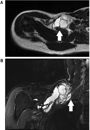
Fig. 2. Axial view of the T2-weighted image showed about 7 x 6 x 11 cm sized lobulated and multiseptated cystic mass in the anterior and superior aspect of the right neck (Fig.
2A)(white arrow). Coronal view showed that inferior and medical aspect of the cystic mass was abutting brachial plexus causing mild compression (Fig. 2B)(white arrow).
A
B
Fig. 3. The operative finding revealed cystic mass abutting scalene muscle (white arrow) and brachial plexus (blue ar- row) (Fig. 3A). With meticulous dissections, the mass was suc- cessfully dissected from scalene muscles (white arrow) and brachial plexus (blue arrow) (Fig. 3B)
Fig. 4. Excised surgical specimen showed about 7 (width) x 6 (thickness) x 11 (length) cm sized lobulated and multiseptated cystic mass
shoulder magnetic resonance image (MRI). Other tests in- cluding shoulder plain X-ray or electromyography (EMG) were not performed in this patient. Shoulder MRI revealed a 7 x 6 x 11cm sized, lobulated and multiseptated cystic mass at the superior anterior aspect of the right neck (Fig.
2A). An inferior and medial aspect of the mass was abutting brachial plexus (Fig. 2B). With these findings, initial im- pression was recurred lymphangioma arising from lym-
phatic structures around the brachial plexus. The patient was referred to the department of otolaryngology-head and neck surgery to get surgical excision. In the operative field, about 10 x 7 x 7cm sized cystic mass was found in front of scalene muscle and lateral to brachial plexus (Fig. 3A). With metic- ulous dissections using Harmonic scalpel, the mass was suc- cessfully dissected from scalene muscles and brachial plexus (Fig. 3B). Excised surgical specimen showed about 7 (width) x 6 (thickness) x 11 (length) cm sized lobulated and multiseptated cystic mass (Fig. 4). The final pathologic report was cavernous type lymphangioma. Microscopically, large, irregular vascular spaces (Fig. 5A, H&E x40) lined
- 71 -
A B C
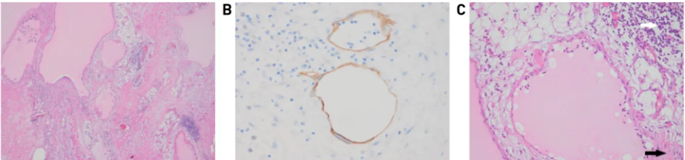
Fig. 5. Microscopically, large, irregular vascular spaces (Fig. 5A, H&E x40) lined by flattened endothelial cells, which were positively stained with D2-40 immunohistochemical stains (Fig. 5B, x400). The spaces were filled with proteinaceous fluid containing lympho- cytes (white arrow) and were surrounded by a fibrotic stroma with lymphocytes (black arrow) (Fig. 5C, H&E x200).
by flattened endothelial cells, which were positively stained with D2-40 immunohistochemical stains (Fig. 5B, x400).
The spaces were filled with proteinaceous fluid containing lymphocytes and were surrounded by a fibrotic stroma with lymphocytes (Fig. 5C, H&E x200). These histologic find- ings were consistent with cavernous lymphangioma. After surgical treatment, right shoulder pain was improved. Hemovac drain was removed on third day after surgical excision and the patient discharged without any complication. The patient followed-up outpatient clinic for one year without any re- currence of lymphangioma.
Discussion
Lymphangiomas are benign lymphatic malformations with a common predilection to the head and neck region.1) However other sites such as the axilla, mediastinum, retro- peritoneal area, and pelvis have been reported in the literature.4-6) They frequently occur in infants or children younger than two years of age.7) They may be developmental, hamartomatous or neoplastic in origin.8) The most widely accepted theory about the development of lymphangioma is that they arise from sequestrations of the primitive embry- onic lymph sacs.9) However, the etiology in the adult group is controversial. Some authors attribute adult lymphangioma to the delayed proliferation of the congenital or acquired lymphoid rests following trauma or preceding respiratory infection.10) Clinically, lymphangiomas tend to grow slowly and compress surrounding anatomical structures. In children cervical lesions can cause dysphagia and airway obstruction11) but these symptoms are rare in adults. Adults usually present with an asymptomatic, soft fluctuant and well-defined mass.12) In this case, large lymphangioma was abutting the brachial plexus and compressed it, causing right shoulder pain which
brought the patient to the department of orthopedic surgery.
Differential diagnosis of lymphangioma includes a number of cystic mass lesions such as hemangioma, teratoma, der- moid cyst, thyroglossal duct cyst, and brachial cleft cyst, all of which cause protruding neck mass. Histopathological features of lymphangioma consist of lymphatic vessels with marked dilatations. Vessels are often infiltrated into the ad- jacent soft tissue and demonstrated as lymphoid aggregate in their wall. Endothelial lining is thin, and the spaces con- sist of proteinaceous fluid and lymphocytes.13) Sometimes secondary hemorrhage may be noticed in the lymphatic vessels. The lymphatic space contains lymphatic fluid, red blood cells, lymphocytes, macrophages, and neutrophils.13) Although, various treatment modalities other than surgery such as radiation therapy, cryotherapy, electrocautery, scle- rotherapy, steroid injection, embolization, ligation, laser ablation using Nd-YAG or CO2, and radiofrequency tissue ablation, surgical excision is the treatment of choice as lym- phangiomas are encapsulated or partially circumscribed.14) For successful surgical treatment without recurrence, it is necessary to include sufficient surrounding normal tissue together in the specimen because vssels are often infiltrated into the adjacent soft tissue.15)
Here, we present a case of cavernous lymphangioma abut- ting brachial plexus and causing right shoulder pain. Irritation of brachial plexus by cavernous lymphangioma caused shoulder pain. Differential diagnosis of shoulder pain should include benign neck mass lesion around brachial plexus.
References
1) Mandel L. Parotid area lymphangioma in an adult: case report. J Oral Maxillofac Surg. 2004;62:1320-1323.
2) Shahi M, Bagga PK, Mahajan NC. Cervical cystic lymphangio- ma in an adult, diagnosed on FNAC. J Cytol. 2009;26:164-165.
- 72 -
3) Ozturk A, Yousem DM. Magnetic resonance imaging findings in diffuse lymphangiomatosis: neuroradiological manifestations.
Acta Radiol. 2007;48:560-564.
4) Izumi D, Toyama E, Shigaki H, Iwagami S, Baba Y, Hayashi N, et al. Laparoscopic excision of an adult retroperitoneal cystic lymphangioma coexisting with an esophageal hiatus hernia. Clin J Gastroenterol. 2015;8:130-133.
5) Chung JC, Song OP. Cystic lymphangioma of the jejunal mesen- tery presenting with acute abdomen in an adult. Can J Surg.
2009;52:E286-288.
6) Pannell TL, Jolles H. Adult cystic mediastinal lymphangioma simulating a thymic cyst. J Thorac Imaging. 1991;7:86-89.
7) Goodman J, Mc CJ, Denton GR, Stein A. Cystic hygromas in adults. Arch Surg. 1963;86:641-644.
8) Poyraz AS, Kilic D, Hatipoglu A, Ozulku M, Sar A, Bilezikci B.
Cystic lymphangioma confined to mediastinum in an adult. Jpn J Thorac Cardiovasc Surg. 2004;52:567-569.
9) Zanotti SD, LaRusso S, Coulson C. Prenatal sonographic diag-
nosis of axillary cystic lymphangiomas. J Clin Ultrasound. 2001;
29:112-115.
10) Aneeshkumar MK, Kale S, Kabbani M, David VC. Cystic lym- phangioma in adults: can trauma be the trigger? Eur Arch Otorhinolaryngol. 2005;262:335-337.
11) Damaskos C, Garmpis N, Manousi M, Garmpi A, Margonis GA, Spartalis E, et al. Cystic hygroma of the neck: single center expe- rience and literature review. Eur Rev Med Pharmacol Sci. 2017;
21:4918-4923.
12) Morley SE, Ramesar KC, Macleod DA. Cystic hygroma in an adult: a case report. J R Coll Surg Edinb. 1999;44:57-58.
13) Allen CM. Radiology and oral and maxillofacial pathology. Oral Surg Oral Med Oral Pathol Oral Radiol Endod. 1995;80:495.
14) Kennedy TL. Cystic hygroma-lymphangioma: a rare and still unclear entity. Laryngoscope. 1989;99:1-10.
15) Fung K, Poenaru D, Soboleski DA, Kamal IM. Impact of mag- netic resonance imaging on the surgical management of cystic hygromas. J Pediatr Surg. 1998;33:839-841.