Listeria innocua 유래 cyclomaltodextrinase의 유전자 클러스터 구조 및 효소 특성
장명운1†, 정창구2†, 강혜정1†, 김민정1, 이민재1, 손병삼1, 김태집1*
1충북대학교대학원축산·원예·식품공학부
2(주)에이피테크놀로지
Received: August 19, 2016 / Revised: August 26, 2016 / Accepted: August 26, 2016
서 론
Cyclodextrinase 계열의효소군에는 Glycoside Hydrolase (GH) family 13에속하는 cyclomaltodextrinases (CDases, EC 3.2.1.54), maltogenic amylases (MAases; EC 3.2.1.133), neopullulanases (NPases; EC 3.2.1.135), Thermoactinomyces vulgaris α-amylase II (TVAII) 등서로다른명칭의다양한 효소가포함된다[1, 14, 18, 19]. 지금까지 CDase 계열의효 소들은 Bacillus [2, 10], Paenibacillus [11], Clostridium
[21], Thermus [12], Lactobacillus [17], Lactococcus [8] 속 을포함한다양한미생물에서보고되었다. 특히이러한효소
유전자는대부분의 Bacillus 속미생물유전체내에폭넓게
분포하고있다[1, 18, 19]. 이들 CDase 계열효소는공통적 으로 glucose로구성된탄수화물중합체의α-(1,4)-결합부위 를가수분해하고, 다양한수용체분자에대한당전이활성을 통해 α-(1,4)-, α-(1,3)-, α-(1,6)-결합으로이루어진각종당전 이산물의생성이가능하기때문에이들효소를이용한신 규탄수화물소재생산등의응용분야에서많은연구가진 행되고있다[18].
그러나, 자연계에존재하는미생물유전체내에일반α-
amylase 계열 전분 분해효소에 비해 널리 분포하는이들
CDase 계열효소의역할에대해서는충분한연구가이루어
지지않았다. Klebsiella oxytoca 세포내에서 cyclodextrin을 Gene Cluster Analysis and Functional Characterization of Cyclomaltodextrinase from Listeria innocua
Myoung-Uoon Jang1†, Chang-Ku Jeong2†, Hye-Jeong Kang1†, Min-Jeong Kim1, Min-Jae Lee1, Byung Sam Son1, and Tae-Jip Kim1*
1Division of Animal, Horticultural, and Food Sciences, Graduate School of Chungbuk National University, Cheongju 28644, Republic of Korea
2Advanced Protein Technologies Co., Suwon 16229, Republic of Korea
A putative cyclomaltodextrinase gene (licd) was found from the genome of Listeria innocua ATCC 33090.
The licd gene is located in the gene cluster involved in maltose/maltodextrin utilization, which consists of various genes encoding maltose phosphorylase and sugar ABC transporters. The structural gene encodes 591 amino acids with a predicted molecular mass of 68.6 kDa, which shares less than 58% of amino acid sequence identity with other known CDase family enzymes. The licd gene was cloned, and the dimeric enzyme with C-terminal six-histidines was successfully produced and purified from recombinant Esche- richia coli. The enzyme showed the highest activity at pH 7.0 and 37℃. licd could hydrolyze β-cyclodextrin, starch, and maltotriose to mainly maltose, and it cleaved pullulan to panose. It could also catalyze the hydrolysis of acarbose to glucose and acarviosine-glucose. In particular, it showed significantly higher activity towards β-cyclodextrin and maltotriose than towards starch and acarbose. licd also showed trans- glycosylation activity, producing α-(1,6)- and/or α-(1,3)-linked transfer products from the acarbose donor and α-methyl glucopyranoside acceptor.
Keywords: Listeria innocua, cyclomaltodextrinase (CDase), gene cluster, enzymatic characterization
*Corresponding author
Tel: +82-43-261-3354, Fax: +82-43-271-4412 E-mail: [email protected]
†These authors contributed equally to this work.
© 2016, The Korean Society for Microbiology and Biotechnology
경유하는전분이용경로가제안되었고[3, 4], Bacillus subtilis 의 glycogen 대사에서 maltogenic amylase와 pullulanase 의역할이보고되었다[23]. 최근 Listeria monocytogenes의 maltose/maltodextrin 이용에관련된유전자클러스터와미 생물학적특성의상관관계에대한보고가이루어지면서[5], 병원성균주에속하는 Listeria 속미생물내독성유전자의 발현과탄소원이용시스템, 탄수화물대사경로에대한유전 체수준의연구가필요한시점이다[15, 22].
본연구에서는미생물유전체데이터베이스분석을통해 Listeria innocua ATCC 33090 균주로부터 cyclodextrinase (LICD)로예상되는유전자를발견하고, 클로닝, 발현하여효 소특성을확인하였다. 또한이유전자가포함된유전자클 러스터의구조를분석함으로써, 전분및 maltose 이용경로 에서 LICD의역할을제시하였다.
재료 및 방법
미생물 및 유전자
Listeria innocua ATCC 33090 (DSM 20649) 균주의 염 색체 DNA는한국생명공학연구원으로부터제공받았다. PCR
증폭 유전자의클로닝을위한 T-클로닝벡터는 pMD18-T
(Takara Biomedical Inc., Japan)를사용하였다. 대장균내 항시발현은 pHCEII/NdeI (BioLeaders Co., Korea)를변형 시킨 pHCXHD [10] 벡터를사용하였다. 유전자클로닝및 발현을위한숙주세포는 Escherichia coli MC1061 균주를 사용하였다.
시약 및 재료
일반 시약, 기질 및 미생물용 배지는 Sigma-Aldrich
(USA)와 Duchefa Biochemie (The Netherlands)에서구입 하였다. 유전자실험용제한효소및 DNA ligase 등은 Roche Applied Science (Germany)에서 구입하였으며, PCR 및 sequencing 프라이머는 Bioneer (Korea)에서합성하여사용 하였다.
유전자 증폭과 클로닝
Listeria innocua 염색체 DNA를주형으로하고, LICD-N (5'-TTTTGGATCCATGGAAAAAGCAGGGATTTATC-3') 및 LICD-C (5'-TTTTCTCGAGGCTGTTTTCTTTAATAA- CAAGAA-3')를 PCR 프라이머로사용하여 LICD 유전자를 증폭하였다. PCR 반응은 Taq DNA polymerase (Roche)와 C1000 thermal cycler (Bio-Rad Laboratories Inc., USA) 를사용하여 94℃에서 1분, 55℃에서 30초, 72℃에서 1분 30초로 30회반복하고, 최종적으로 72℃에서 5분간추가로 증폭하였다. 증폭된유전자는 pMD18-T 벡터에클로닝하여
pMDLICD를제조하고, SolGent (Korea)에의뢰하여염기서 열을확인하였다. 유전자발현을위해 pMDLICD를제한효 소 Nde I과 Xho I으로절단하고, 항시발현벡터인 pHCXHD 에삽입하여 pHCXLICD를얻었다.
재조합 효소 유전자의 발현 및 정제
pHCXLICD가 형질전환 된 재조합 E. coli MC1061를 LBA (1% bacto-tryptone, 0.5% yeast extract, 1% NaCl, 100 mg/ml ampicillin) 액체배지에접종하여 37℃에서 12시 간동안배양하였다. 원심분리로회수한균체를 ultrasonicator (VCX750, Sonics & Materials, Inc., USA)로 파쇄한 후, HisTrap-FF column (GE Healthcare, Sweden)과 AKTA PrimeTM system (GE Healthcare)을이용하여정제하였다. 최종적으로 elution buffer [20 mM Tris-HCl (pH 7.4), 500 mM NaCl, 500 mM imidazole] 1 ml/min의유속으로 효소를회수하였으며, 정제된효소는농축후최적 buffer로 투석하여실험에사용하였다.
단백질 정량 및 분자량 측정
재조합 단백질의 정제도는 Mini-protean II (Bio-Rad, USA)를이용한 12% SDS-PAGE로확인하였다. 전기영동후, Coomassie blue로 염색하였고, 단백질 표준시료(Sigma-
Aldrich)와비교하여단량체의크기를결정하였다. 단백질의
분자량과 4차 구조는 gel permeation chromatography (GPC; Superdex-200 column, 10 × 300 mm, GE Healthcare) 및 0.5 ml/min 유속의 50 mM sodium phosphate buffer
(pH 7.0)를이용하여분석하였다. 효소단백질의농도측정
은 BCATM protein assay kit (Pierce Biotechnology Inc., USA)를이용하였다.
효소 활성 측정
LICD의 활성을 측정하기 위해 1%의 β-CD, pullulan, soluble starch를 각각 50 mM sodium phosphate buffer (pH 7.0)에녹인후, 적당량의효소를첨가하여최종 100 μl 로반응하였다. 37℃에서 10분동안효소반응을통해생성 된 maltose의양을 dinitrosalicylic acid (DNS)법으로측정 하였다[16]. Maltotriose와 acarbose 기질의경우, 효소반응 으로 생성된 glucose의 양을 AceChem Glucose kit (YD Diagnostics Co., Korea)로측정하였다. LICD의활성 1 unit 는 1분당 1 μmol의 maltose 또는 glucose를생성하는효소 의양으로정의하였다.
반응 산물 분석
최적반응조건에서 1% 기질을 1시간동안효소반응하여 생성된가수분해산물을 thin layer chromatography (TLC)
로 분석하였다. Silica gel 60F254 TLC plate (Merck, Germany)에 1 μl의 시료를 spotting 하고, isopropanol, ethylacetate, 물을 3:1:1의부피비로혼합한전개용액을사 용하였다. TLC plate를 발색시약 (3 g N-(1-naphthyl)- ethylene-diamine, 50 ml H2SO4, 950 ml methanol)에 담 근후, 건조하고 110℃에서 10분간발색하여분석하였다.
당전이 산물 분석
5% acarbose (donor; Carbosynth, England)와 10% α- methyl glucopyranoside (acceptor; Sigma-Aldrich)를이용 하여최적반응조건에서 24시간반응하였다. 생성된당전 이 산물은 CarboPac PA1 column (0.4 × 25 cm, Thermo Fisher Scientific Co., USA)과 electrochemical detector (ED40, Thermo Fisher Scientific)를 사용하여 high performance anion exchange chromatography (HPAEC;
Bio-LC ICS-3000, Thermo Fisher Scientific)로분석하였 다. 시료의분석조건은이동상 A (150 mM NaOH, Thermo Fisher Scientific)를기본으로이동상 B (600 mM sodium acetate, Sigma-Aldrich)를분당 1%씩증가시키는방법으로 진행하였다. 이동상의속도는 1.0 ml/min으로일정하게유 지하였다.
결과 및 고찰
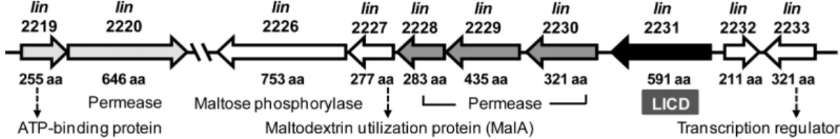
Listeria maltose/maltodextrin 이용 유전자 클러스터
CDase 계열효소유전자는다양한미생물유전체에폭넓
게분포하는것으로알려져있다[20]. 세포내에서발현되는
CDase 효소는 cyclodextrin (CD)의환형구조를절단하여α- glucosidase의최적기질인 maltose 단위로가수분해한다[18, 19]. 이미 Bacillus subtilis 168 세포내의탄수화물대사에 서 maltogenic amylase (MAase)와 debranching enzyme
(pullulanase)의생리적기능분석을위한다양한돌연변이
연구가 보고된 바 있다[23]. 따라서, 본 연구에서는 B.
subtilis MAase 유전자를 이용한 BLAST 검색을 통해 Listeria innocua ATCC 33090 유전체로부터 CDase로예상 되는 유전자(lin2231; GenBank accession No. NP471564) 를발굴하였고, 이를 LICD 유전자로명명하였다. LICD 유
전자를중심으로 L. innocua 유전체서열을광범위하게분
석하여, 이유전자가 maltose/maltodextrin 이용유전자클 러스터내에위치하는것을확인하였다(Fig. 1). LICD 유전 자는데이터베이스상에서초기에α-glycosidase로분류되어 있었으며, 이외에도다양한 sugar ABC transporter 유전자 군과 maltose phosphorylase 유전자가클러스터를구성함을 확인하였다. LICD의아미노산서열을 SignalP 서버[20]로 분석한결과, 특이적인 signal peptide 서열이발견되지않 았으며, 이는이효소가세포내의 cytoplasm 영역에서발현 됨을의미한다. 따라서, LICD와 maltose phosphorylase는 세포 내로운반된 cyclodextrin 또는 maltooligosaccharide 를서로다른방식으로저분자화하여대사경로에진입시키 는역할을담당하는것으로예측된다. 이러한유전자클러스 터 구조는 지금까지 알려진 B. subtilis 168 및 Listeria monocytogenes EGD-e의유전체분석결과와유사하였다[5, 23, 24].
LICD 유전자의 클로닝 및 발현
LICD 구조유전자(lin2231)는 1,773개의염기서열로이 루어지며, 총 591개의아미노산잔기를암호화하고있다. 목 적유전자를 LICD-N 및 LICD-C 프라이머를이용하여 PCR 로증폭하였다. 약 1.8 kb의증폭된 LICD 유전자단편을 T- 클로닝벡터에삽입하여 pMDLICD를얻었다. 이를제한효소 Nde I과 Xho I으로처리한후, pHCXHD 발현벡터에삽입 하여 재조합 플라스미드인 pHCXLICD를 제조하였다. pHCXLICD를 E. coli MC1061에형질전환하여재조합대 장균을얻었으며, 이를배양하여 C-말단에 6개의 histidine 잔기가결합된형태의재조합 LICD를얻었다. Ni-NTA 크 로마토그래피를이용하여정제한 LICD 효소를 SDS-PAGE 로분석한결과, 염기서열로부터예상한바와같이약 70 kDa 크기의단백질이발현되고성공적으로정제되었음을확인하 였다(Fig. 2).
LICD의 1차 및 4차 구조 분석
CAZy (Carbohydrate Active EnZymes) 데이터베이스에 서는아미노산의서열상동성을토대로하여, 전분등탄수 화물중합체를기질로하는가수분해효소를 GH 13 family
Fig. 1. Gene cluster for maltose/maltodextrin utilization in Listeria innocua. Each gene ID is shown above the arrow, and experi- mentally proven or presumptive protein functions below. The GenBank accession no. for the genome sequence is NC003212.
로분류하고있으며[1, 6], 넓은의미에서 CDase 계열의효 소역시 GH 13 계열에속한다[14]. 아미노산서열에따른상 관관계를분석한결과, LICD는다른미생물유래 CDase 계 열효소들과 39−57% 수준의서열상동성을나타내었다. 특 히, Bacillus 속미생물유래의효소들과높은상관관계를보 였으며, B. stearothermophilus MAase [2]와 57.2%, 호열성 균주인 Thermus MAase [12]와 51.8%, B. subtilis MAase [23]와 52.0%, B. halodurans CDase [10]와 50.7%, 또한 Thermoactinomyces vulgaris amylase II [9]와 38.5%의상 동성을나타내었다.
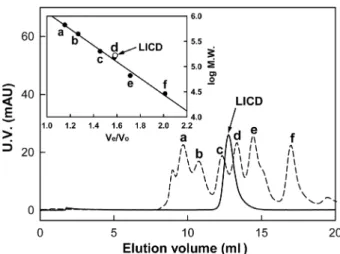
또한 LICD는 CDase 계열효소의구조적특징을공유하였 다. 예를들면, 일반적인α-amylase에존재하지않는 N-말단 부위를가지며, 상동부위 I (DAVFNH), II (GWRLDVANE), III (EIWH), IV (LLGSHD) 서열또한매우 높은유사도를 나타내었다. 상동부위 II, III, IV 내에위치하는주요활성 부위아미노산잔기인 Asp339, Glu368, Asp435 또한공유 하였다. 따라서, LICD는 CD 기질에 높은 활성을 보이는 CDase 계열효소들과 60% 미만의서열상동성을보이지만, 전체적인 1차구조및활성잔기의위치, 상동부위서열등 을비교할때유사한효소특성을가질것으로예상하였다. GPC 분석결과, 수용액상에서재조합 LICD의분자량은 약 166 kDa이었으며, 이는 LICD가 homo-dimer의형태로 존재함을의미한다(Fig. 3). 지금까지보고된 CDase 계열효
소들은대부분 homo-dimer의형태를가지는것으로알려져
있으며[2, 7, 9, 10, 12, 21], 일부효소에서 homo-dodecamer 의형태가보고되기도하였다[23]. 이들효소에공통으로존 재하는 N-말단 domain이 4차구조의형성에중요한역할을
하며, 이과정에서형성된좁고깊은형태의기질결합부위 구조가효소의기질특이성에영향을미치는것으로알려져 있다[13]. LICD도특유의 N-말단부위를가지므로, 이를통 해 homo-dimer 구조를형성할것으로예상하였다. LICD의 효소 특성
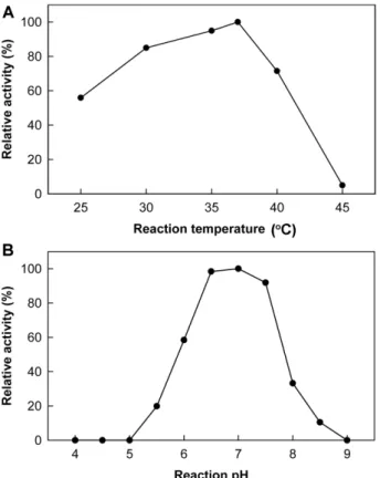
재조합 LICD는β-CD를기질로할때, 37℃에서최대활 성을나타내었으며, 25℃에서도 50% 이상의활성을보였지 만, 45℃에서는 매우 낮은 활성을 보였다(Fig. 4A). 한편, 50 mM sodium phosphate buffer (pH 7.0)에서 가장높은 β-CD 가수분해활성을보였으나, pH 6.0 이하또는 pH 8.0 이상의조건에서는최적 pH 대비 60% 이하의낮은활성을 나타내었다(Fig. 4B). 대부분의중온성미생물유래 CDase 계열효소들이 40−60℃, pH 5.5−8.0 범위에서최적의효소 활성을나타내는것을감안할때, L. innocua CDase는온도 및 pH 변화에민감한경향을나타내었으며, 이는병원성미 생물특성상숙주의생육적정온도와밀접한관련이있을 것으로예상하였다[5, 22].
LICD의 기질 특이성 및 가수분해 특성
CDase 계열의효소는β-CD, pullulan, starch 뿐아니라 maltooligosaccharide와 acarbose 등다양한기질에대해가 수분해및당전이활성을가지는것으로알려졌다. 따라서
다양한기질에대한 LICD의효소활성을측정하고, 이를기
존에연구된 CDase 계열효소들과직접비교하였다(Table
Fig. 2. Gene expression and purification of LICD. SDS-PAGE analysis showed the expression level and the purity of LICD from recombinant E. coli. Lane M, protein molecular weight marker;
lane 1, cell extract from E. coli harboring pHCXHD; lane 2, cell extract from E. coli harboring pHCXLICD; lane 3, LICD purified by Ni-NTA chromatography.
Fig. 3. Determination of quaternary structure of LICD.
Molecular mass of LICD was estimated by gel permeation chro- matography (Superdex-200). The purified LICD was drawn as a solid line and the molecular weight markers (a dashed line) were used as the mixture of six proteins: (a) thyroglobulin (669 kDa), (b) apoferritin (443 kDa), (c) α-amylase (200 kDa), (d) alcohol dehydrogenase (150 kDa), (e) bovine serum albumin (66 kDa), (f) carbonic anhydrase (29 kDa).
1). LICD를포함한모든 CDase 계열 효소는β-CD에대해 가장높은활성을나타내었으나, 다른기질에대한활성은 효소에따라다소차이를보였다. 예를들어 LICD의β-CD 및 pullulan 가수분해활성은 BHCD와 ThMA의중간수준 이었으며, starch와 acarbose에대한활성은다른두효소보 다낮았다. 그러나, glucose 3개분자로구성된 maltotriose 에대한가수분해활성은 BHCD, ThMA에비해각각 1.5배,
4.4배높은활성을보였다. 특히, starch 분해활성에비해 maltotriose에대한활성이 26배이상으로매우높았으며, 이
는 LICD가고분자중합체보다저분자소당류에대해높은
활성을가지는효소임을의미한다. 따라서중합체기질에대 해높은선호도를나타낸 BHCD와비교할때, LICD의기질 특이성이매우상이함을알수있었다. 또한, α-glucosidase
저해제로당뇨병 치료에널리 사용되는 acarbose의경우,
ThMA와 BHCD 등의 CDase 계열 효소에 의해 glucose와 acarviosine-glucose로분해된다[10, 12]. LICD의경우에도 acarbose를분해하지만, 그활성은 ThMA와 BHCD에비해 약 0.6% 및 53% 수준으로크게 낮았다. 비교적유사한형 태의 저분자 기질인 maltotriose와 acarbose 중, 특히 maltotriose에대해매우높은활성을가지는반면 acarbose 에 대해낮은 활성을 나타내는것은 각각의 기질에 대한
LICD의구조적친화도가다른효소와크게다르기때문으
로추측하였다.
각기질로부터생성된반응산물을 TLC로분석하여 LICD
Fig. 4. Effects of reaction temperature (A) and pH (B) on LICD activity. Optimal reaction temperature and pH of LICD were determined on the basis of its hydrolyzing activity towards β-CD.
Sodium acetate (pH 4.0−6.0); sodium phosphate (pH 6.0−7.5);
Tris-HCl (pH 7.5−8.0); borate-NaOH (pH 8.0−9.0).
Table 1. Comparison of the multi-substrate specificity between LICD and other known CDase-family enzymes.
Enzymea Specific activity (U/mg)b Activity ratioc
β-CD Pullulan Starch Maltotriose Acarbose C/S P/S M/S M/A
LICD 56.3 ± 0.7 13.0 ± 0.2 3.0 0.0 77.9 ± 0.5 1.6 ± 0.0 18.8 4.3 26.0 48.7 BHCD 52.9 ± 0.3 22.8 ± 0.5 11.2 ± 0.1 17.9 ± 0.1 3.0 ± 0.1 4.7 2.0 1.6 6.0 ThMA 65.2 ± 0.9 5.0 ± 0.1 4.4 ± 0.1 48.9 ± 0.3 27.3 ± 0.3 14.8 1.1 11.1 1.8
aLICD, L. innocua CDase; BHCD, B. halodurans CDase; ThMA, Thermus sp. MAase
bEach hydrolyzing activity on β-CD, pullulan, or starch was determined by DNS reducing sugar assay, whereas the activity on maltotriose or acarbose was measured by glucose oxidase-peroxidase method.
cThe abbreviations for the activity ratios between substrates were used as follows: C, β-CD; P, pullulan; S, soluble starch; M, maltotri- ose; A, acarbose.
Fig. 5. TLC analysis of hydrolysis patterns of LICD on various substrates. LICD was reacted with 1% of each substrate: CD, β- CD; PL, pullulan; SS, soluble starch; G3, maltotriose; AG, acarvios- ine-glucose; AC, acarbose; PN, panose; S, maltooligosaccharide standards, and the reaction products with (+) or without (-) enzyme.
의기질가수분해패턴을확인하였다(Fig. 5). LICD는β-CD 의환형구조를 endo-형으로절단하여 7개의 glucose로구 성된직쇄형의 maltoheptaose로전환하며, 이어지는가수분 해작용을통해 maltose를최종산물로, glucose를부산물로 생성하였다. Pullulan 및 starch에대한활성은상대적으로 낮았지만, 장시간반응을통해얻어진산물은예상과같이 각각 maltose와 panose임을확인하였다. Maltotriose의분해 산물은최종적으로 glucose와 maltose였으며, maltose는더 이상분해되지않고축적되었다. 이상의결과를통해 LICD 가α-(1,4)-결합을분해하여주로 maltose를생성하는전형 적인 CDase 계열의효소임을알수있었다. 한편 acarbose 의경우에는극히낮은효소활성으로인해기질이불완전하 게분해되었으나, 소량의 glucose와 acarviosine-glucose가
생성된것으로판단할때, 역시다른 CDase 효소들과유사
한가수분해특성을가지는것으로판단하였다.
결과적으로 LICD는전형적인 CDase 계열의효소이나, pullulan 및 starch와같은큰분자량의중합체기질보다 β- CD, maltotriose와같은저분자소당류의기질에더잘작용 하며, acarbose에대한분해활성이상대적으로매우낮은특 징을보였다.
LICD의 당전이 특성
CDase 계열효소의일종인 ThMA 및 BHCD에대한연구
에서, 당전이반응으로얻어진α-(1,6)- 및α-(1,3)-결합의전 이생성물은다시분해되지않아축적되며, 반대로초기에
생성된α-(1,4)-전이생성물은생성과동시에빠르게재분해
되는것으로보고되었다[10, 12]. 선행연구를토대로 LICD 의당전이활성을확인하고자, 공여체로 acarbose, 수용체로 α-methyl glucopyranoside (α-MG)를이용한당전이반응을 실시하고, 반응산물을 HPAEC로분석하였다. 당전이반응 을통해 BHCD와유사하게α-(1,6)- 및α-(1,3)-전이생성물 이생성되었다(Fig. 6).
LICD와기존 CDase 계열효소간상동부위잔기들의차 별성에주목하여효소구조및기능의상관관계에대한지 속적인연구가이루어진다면, 고유의기질특이성및당전이 활성에관여하는핵심아미노산잔기를발굴할수있을것 이다. 이를상호치환하여효소의기질특이성을변화시키는 단백질공학적연구에중요한정보를제공할수있을것이 다. 또한향후보다다양한미생물에서유래한 CDase 계열 효소를비교·연구함으로써, 이들유전자의자연계내다양 성및탄수화물대사경로에서의역할에대한과학적이해를 향상시킬것으로기대한다.
요 약
Listeria innocua ATCC 33090 유전체로부터 maltose/
maltodextrin 이용과관련한유전자클러스터를발견하였으
며, 그로부터 cyclomaltodextrinase (LICD)로예상되는유전 자를클로닝하고, 대장균내에서발현하였다. LICD는총 591 개의아미노산으로이루어진 68.6 kDa 크기의효소이며, 일 반적인 CDase 계열효소들과 39−58%의아미노산서열상 동성을나타내었다. 재조합 LICD는 37℃, pH 7.0의조건에 서최대활성을나타내었으며, cyclodextrin, starch, maltotriose 에작용하여주로 maltose를생성하였다. 또한 pullulan을분 해하여 panose를, 그리고 acarbose를 분해하여 glucose와 acarviosine-glucose를생성하는전형적인 CDase 계열효소 임을확인하였다. 그러나, starch 및 pullulan과같은고분자 기질대비 cyclodextrin 및 maltotriose의저분자소당류에 대해상대적으로높은활성을나타내며, acarbose 분해활성
이매우낮아다른효소들과차별성을가진다. 또한 LICD는
acarbose 공여체를가수분해하여수용체에전이하는당전이
활성을보였다.
Acknowledgments
This work was supported by the research grant of Chungbuk National University in 2013.
Fig. 6. HPAEC analysis of acarbose transglycosylation prod- ucts. LICD was reacted with 5% acarbose (donor) and 10% α- methyl glucopyranoside (acceptor) at 37oC for 2 h. At each time interval of (A) 0 min, (B) 120 min, the reaction mixture was taken and analyzed by HPAEC. Abbreviations used here: α-MG, α- methyl glucopyranoside; G1, glucose; AG, acarviosine-glucose, AC, acarbose; 3, α-(1,3)-transglycosylation product; 6, α-(1,6)- transglycosylation product.
References
1. Cantarel BL, Coutinho PM, Rancurel C, Bernard T, Lombard V, Henrissat B. 2009. The Carbohydrate-Active EnZymes database (CAZy): an expert resource for glycogenomics. Nucl. Acids Res.
37: D233-238.
2. Cha HJ, Yoon HG, Kim YW, Lee HS, Kim JW, Kweon KS, et al.
1998. Molecular and enzymatic characterization of a malto- genic amylase that hydrolyzes and transglycosylates acarbose.
Eur. J. Biochem. 253: 251-262.
3. Feederle R, Pajatsch M, Kremmer E, Bock A. 1996. Metabolism of cyclodextrins by Klebsiella oxytoca M5a1: purification and characterisation of a cytoplasmically located cyclodextrinase.
Arch. Microbiol. 165: 206-212.
4. Fiedler G, Pajatsch M, Bock A. 1996. Genetics of a novel starch utilisation pathway present in Klebsiella oxytoca. J. Mol. Biol.
256: 279-291.
5. Gopal S, Berg D, Hagen N, Schriefer EM, Stoll R, Goebel W, et al.
2010. Maltose and maltodextrin utilization by Listeria monocy- togenes depend on an inducible ABC transporter which is repressed by glucose. PLoS One 5: e10349.
6. Henrissat B, Bairoch A. 1996. Updating the sequence-based classification of glycosyl hydrolases. Biochem. J. 316: 695-696.
7. Hondoh H, Kuriki T, Matsuura Y. 2003. Three-dimensional struc- ture and substrate binding of Bacillus stearothermophilus neopullulanase. J. Mol. Biol. 326: 177-188.
8. Jang MU, Kang HJ, Jeong CK, Park JM, Yi AR, Kang JH, et al.
2013. Enzymatic characterization of Lactococcus lactis subsp.
lactis cyclomaltodextrinase expressed in E. coli. Korean J. Micro- biol. Biotechnol. 41: 391-397.
9. Kamitori S, Abe A, Ohtaki A, Kaji A, Tonozuka T, Sakano Y. 2002.
Crystal structures and structural comparison of Thermoactino- myces vulgaris R-47 alpha-amylase 1 (TVAI) at 1.6 Å resolution and alpha-amylase 2 (TVAII) at 2.3 Å resolution. J. Mol. Biol. 318:
443-453.
10. Kang HJ, Jeong CK, Jang MU, Choi SH, Kim TJ, Kim MH, et al.
2009. Expression of cyclomaltodextrinase gene from Bacillus halodurans C-125 and characterization of its multisubstrate specificity. Food Sci. Biotechnol. 18: 776-781.
11. Kaulpiboon J, Pongsawasdi P. 2004. Expression of cyclodextri- nase gene from Paenibacillus sp. A11 in Escherichia coli and characterization of the purified cyclodextrinase. J. Biochem.
Mol. Biol. 37: 408-415.
12. Kim TJ, Kim MJ, Kim BC, Kim JC, Cheong TK, Kim JW, et al. 1999.
Modes of action of acarbose hydrolysis and transglycosylation catalyzed by a thermostable maltogenic amylase, the gene for
which was cloned from a Thermus strain. Appl. Environ. Micro- biol. 65: 1644-1651.
13. Kim TJ, Nguyen VD, Lee HS, Kim MJ, Cho HY, Kim YW, et al. 2001.
Modulation of the multisubstrate specificity of Thermus malto- genic amylase by truncation of the N-terminal domain and by a salt-induced shift of the monomer/dimer equilibrium. Bio- chemistry 40: 14182-14190.
14. Lee HS, Kim MS, Cho HS, Kim JI, Kim TJ, Choi JH, et al. 2002.
Cyclomaltodextrinase, neopullulanase, and maltogenic amy- lase are nearly indistinguishable from each other. J. Biol. Chem.
277: 21891-21897.
15. Milenbachs AA, Brown DP, Moors M, Youngman P. 1997. Car- bon-source regulation of virulence gene expression in Listeria monocytogenes. Mol. Microbiol. 23: 1075-1085.
16. Miller GL. 1959. Use of dinitrosalicylic acid reagent for determi- nation of reducing sugar. Anal. Chem. 31: 426-428.
17. Oh KW, Kim MJ, Kim HY, Kim BY, Baik MY, Auh JH et al. 2005.
Enzymatic characterization of a maltogenic amylase from Lac- tobacillus gasseri ATCC 33323 expressed in Escherichia coli.
FEMS Microbiol. Lett. 252: 175-181.
18. Park KH. 2006. Function and tertiary- and quaternary-structure of cyclodextrin-hydrolyzing enzymes (CDase), a group of mul- tisubstrate specific enzymes belonging to the α-amylase fam- ily. J. Appl. Glycosci. 53: 35-44.
19. Park KH, Kim TJ, Cheong TK, Kim JW, Oh BH, Svensson B. 2000.
Structure, specificity and function of cyclomaltodextrinase, a multispecific enzyme of the alpha-amylase family. Biochim.
Biophys. Acta 1478: 165-185.
20. Petersen TN, Brunak S, von Heijne G, Nielsen H. 2011. SignalP 4.0: discriminating signal peptides from transmembrane regions. Nat. Methods 8: 785-786.
21. Podkovyrov SM, Zeikus JG. 1992. Structure of the gene encod- ing cyclomaltodextrinase from Clostridium thermohydrosulfuri- cum 39E and characterization of the enzyme purified from Escherichia coli. J. Bacteriol. 174: 5400-5405.
22. Poncet S, Milohanic E, Maze A, Nait Abdallah J, Ake F, Larribe M, et al. 2009. Correlations between carbon metabolism and viru- lence in bacteria. Contrib. Microbiol. 16: 88-102.
23. Shim JH, Park JT, Hong JS, Kim KW, Kim MJ, Auh JH, et al. 2009.
Role of maltogenic amylase and pullulanase in maltodextrin and glycogen metabolism of Bacillus subtilis 168. J. Bacteriol.
191: 4835-4844.
24. Takami H, Nakasone K, Takaki Y, Maeno G, Sasaki R, Masui N, et al. 2000. Complete genome sequence of the alkaliphilic bacte- rium Bacillus halodurans and genomic sequence comparison with Bacillus subtilis. Nucl. Acids Res. 28: 4317-4331.