Tuberc Respir Dis 2011;70:132-138
CopyrightⒸ2011. The Korean Academy of Tuberculosis and Respiratory Diseases. All rights reserved.
비소세포폐암에서 Maspin의 발현과 임상적 의의
부산대학교 의학전문대학원
1내과학교실,
2병리학교실
윤성훈1, 김원진1, 신경화1, 김미현1, 조우현1, 김기욱1, 박혜경1, 전두수1, 김윤성1, 이창훈2, 이민기1, 박순규1
Maspin Expression and Its Clinical Significance in Non-Small Cell Lung Cancer
Seong Hoon Yoon, M.D.
1, Won Jin Kim, M.D.
1, Kyung Hwa Shin, M.D.
1, Mi Hyun Kim, M.D.
1, Woo Hyun Cho, M.D.
1, Ki Uk Kim, M.D.
1, Hye-Kyung Park, M.D.
1, Doo Soo Jeon, M.D.
1, Yun Seong Kim, M.D.
1, Chang Hun Lee, M.D.
2, Min Ki Lee, M.D.
1, Soon Kew Park, M.D.
1Departments of
1Internal Medicine,
2Pathology, Pusan National University School of Medicine, Busan, Korea
Background: Maspin (mammary serine protease inhibitor) is a member of the serpin superfamily. A few studies have examined the role of maspin in tumor suppression of non-small cell lung cancer (NSCLC); however, its role in the development and progression of NSCLC still remains controversial. We evaluated the immunohistochemical expression of maspin in order to elucidate its clinical significance in NSCLC.
Methods: We analyzed 145 patients with pathologically confirmed NSCLC, including 66 cases of squamous cell carcinomas (SCCs) and 79 cases of adenocarcinomas (ADCs). We performed a immuno-histochemical stain with maspin and PCNA (proliferating cell nuclear antigen) using tissue microarray blocks.
Results: There were 108 men and 37 women in the study population. The mean age of patients in the study was 63.7 years (range, 40.0∼82.0; median, 65.0). The proportion of maspin expression was significantly higher in SCCs (52/66, 78.8%; p<0.01) than in ADCs (17/79, 21.5%; p<0.01). Maspin expression was not associated with PCNA (p=0.828), lymph node involvement (p=0.483), or tumor stage (p=0.216), but showed correlation with well-to-moderate tumor differentiation (p=0.012). There was no observed correlation between maspin expression and survival with NSCLC (p=0.218).
Conclusion: The present study suggests that maspin expression was significantly higher in SCCs than in ADCs and was associated with low histological grade. However, maspin expression was not an independent factor to predict a prognosis in NSCLC.
Key Words: Maspin; Non-Small Cell Lung Cancer; Carcinoma, Squamous Cell; Pathology; Prognosis
Address for correspondence: Soon Kew Park, M.D.
Department of Internal Medicine, Pusan National University School of Medicine, 1-10, Ami-dong, Seo-gu, Busan 602-739, Korea
Phone: 82-51-240-7222, Fax: 82-51-254-3127 E-mail: [email protected]
Received: Feb. 24, 2010 Accepted: Feb. 5, 2011
서 론
Mammary serine protease inhibitor (maspin) 단백은 serine protease inhibitor (serpin)의 일종으로, 1994년 정
상 유선조직에서 처음 분리되었다1. 이후 전립선, 흉선, 고환, 소장, 대장 등의 정상 상피조직에서도 발견되었다2. Maspin과 종양과의 관계에서 일부 연구들은 maspin이 유 방암에서 세포운동성, 침습 및 전이의 억제를 통해 종양억 제기능을 가진다고 했지만3,4, 다른 연구들에서는 유방암, 췌장암 및 난소암에서 maspin의 발현이 나쁜 예후 및 종 양의 진행과 연관이 있다고 하였다5,6. 이처럼 아직까지 종 양에서 maspin의 정확한 기능은 정립되어 있지 않은 실정 이다.
원발성 폐암은 전 세계적으로 암사망률의 가장 흔한 원 인이며, 우리나라에서도 2007년 기준, 암으로 인한 사망
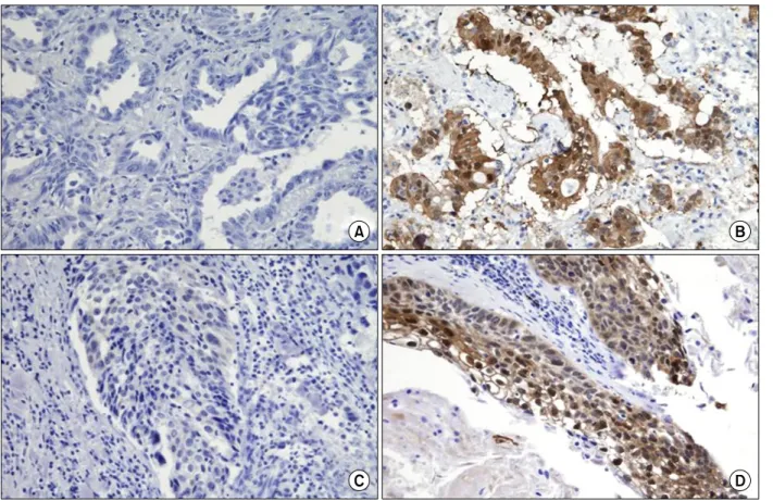
Figure 1. Immunohistochemical staining for maspin using the tissue microarray (×200). (A) ADC without maspin expression. (B) ADC with maspin expression. (C) SCC without maspin expression. (D) SCC with maspin expression.
원인 중 1위이다7,8. 비소세포폐암은 원발성 폐암의 80%를 차지하며9, 비소세포폐암의 임상적인 예후지표에 대한 필 요성이 제기되었으나 그 간의 연구에서는 특정한 지표를 정립하지 못하였다. 최근 여러 연구에서 비소세포폐암에 서 maspin의 발현을 보고하고 있지만, 아직 이 단백의 정 확한 기능 및 역할에 대해서는 보고마다 차이가 있고, maspin의 세포 내 발현위치에 따라서도 기능이 다르다고 알려져 있다. Smith 등10은, 세포핵 내에서 발현되는 mas- pin은 좋은 예후와 관련된다고 보고했지만, Hirai 등11은 세포질에서 발현되는 maspin은 종양의 진행 및 나쁜 예후 와 연관이 있다고 보고하였다.
이상에서 보듯이 비소세포폐암에서 아직 maspin의 정 확한 기능은 잘 정립되어 있지 않다. 따라서 본 연구에서 는 비소세포폐암의 주요 조직학적 유형에 따른 maspin의 발현에 대해 분석하였고, 비소세포폐암의 예후에 대한 maspin의 역할에 대해 알아보고자 하였다. 또한 병기, 조 직학적 분화도, 종양의 증식능과 maspin 발현과의 연관성 에 대해서도 평가를 하였으며, 기존의 예후인자로 알려진
나이, 종양의 증식, TNM 병기, 림프절 병기 및 조직학적 분화도와 예후와의 연관성에 대한 분석도 시행하였다.
대상 및 방법
1. 대상 환자
1998년 4월 1일부터 2008년 4월 30일까지 부산대학교 병원에서 조직학적으로 비소세포폐암으로 확진된 145명 의 환자를 대상으로 하였고, 이들의 조직학적 분류, 병기, 증식 정도 및 임상적 소견을 후향적으로 분석하였다. 전 체 환자 중 편평상피세포암은 66명이었고, 선암은 79명이 었다. 평균 나이는 63.7세였고 평균 추적관찰 기간은 44 개월이었으며, 145명 중 남자가 109명, 여자가 36명이었 다. 모든 환자에서 의무기록 및 영상학적 사진(엑스레이, 흉부 전산화 단층촬영 등)을 검토하였고 maspin의 발현과 종양의 증식 정도는 면역조직화학염색을 이용하여 분석 하였으며, 병리학적 병기는 기존의 TNM 병기분류법12을 따랐다.
Table 1. Data sheet for maspin and PCNA
Antigen Antibody Clone Source Dilution Detection Antigen retrieval (min)
Maspin Gpab C-20 Santa Cruz, USA 1:400 Envision Kit, DAKO Room temperature (60)
PCNA Mmab PC10 SIGMA, USA 1:2,000 Envision Kit, DAKO Overnight
Gpab: polyclonal gout antibody; PCNA: proliferating cell nuclear antigen; Mmab: monoclonal mouse antibody.
Table 2. Characteristics of patients and maspin expression
No. of patients
Maspin expression
p-value Negative (<5) Positive (≥5)
All patients 145 (100) 76 (52.4) 69 (47.6)
Age, yr 63.7±9.5 62.22±8.93 65.33±8.2 0.043
<65 68 (46.9) 39 (57.4) 29 (42.6) 0.263
≥65 77 (53.1) 37 (48.1) 40 (51.9)
Sex
Male 108 (74.5) 45 (41.7) 63 (58.3) <0.01
Female 37 (25.5) 31 (83.8) 6 (16.2)
Histological type
Squamous cell carcinoma 66 (45.5) 14 (21.2) 52 (78.8) <0.01
Adenocarcinoma 79 (54.5) 62 (78.5) 17 (21.5)
Values are presented as number (%) unless otherwise indicated.
2. 면역조직화학염색
Maspin의 종양세포 내 발현 정도를 보기 위해 조직 미 세배열 블록(tissue microarray block)을 사용하여 maspin 에 대한 면역조직화학염색을 하였다(Figure 1). 각 증례에 서 종양세포핵의 5% 이상에서 암갈색의 염색반응을 보일 경우 양성으로 하였고 세포질에서의 발현은 무시하였다.
종양의 증식 정도를 평가하기 위해서는 proliferating cell nuclear antigen (PCNA)에 대한 면역조직화학염색을 실 시하였다(Table 1). PCNA의 양성 소견은 종양세포의 핵 에서 암갈색의 염색 소견을 보이는 경우로 하였고, 각 증 례에서 1,000개의 종양세포 중 양성 소견을 보이는 종양 세포를 계수하여 백분율(PCNA index)로 표시하였다.
3. 통계학적 분석
Maspin의 발현과 여러 임상병리학적 특징들(나이, 성 별, 조직학적 유형, 종양의 증식 정도, TNM 병기 등)과의 연관성은 Chi-square검사를 이용하였고, 생존분석은 Kaplan-Meier법 및 Cox's proportional hazards model을 이용하였다. 모든 통계학적 분석은 SPSS version 15.0 (SPSS Inc., Chicago, IL, USA)를 이용하였고 p값이 0.05
미만인 경우 유의한 것으로 판정하였다.
결 과
1. 대상환자의 특성 및 maspin의 발현율
전체 환자 145명 중, 69명(47.6%)에서 maspin 단백은 양성 발현을 보였다. 환자군의 중간 연령인 65세를 기준 으로 하였을 때, 65세 이상인 환자에서 maspin의 발현율 이 51.9% (40/77)였고 65세 이하에서는 42.6% (29/68)를 보였으나 의미있는 차이를 보이지는 않았다(p=0.263). 전 체 환자 중 남성에서는 maspin의 발현율이 58.3% (63/
108)였고, 여성에서는 16.2% (6/37)로 남성에서 maspin 의 발현율이 유의하게 더 높았다(p<0.01). 조직학적 유 형에 따른 maspin의 발현율은 편평상피세포암은 78.8%
(52/66)였고, 선암은 21.5% (17/79)로 편평상피세포암에 서 maspin의 발현율이 더 높았다(p<0.01, Table 2).
2. 임상병리학적 특성과의 관련성
Maspin의 발현과 종양의 조직학적 분화도(differenti- ation)와의 관련성을 살펴보면, 조직의 분화도 등급이 낮 을 때(well or moderate differentiation), maspin의 발현
Table 3. Relationship between maspin expression and clinicopathologic features
Patients
Maspin expression
p-value Negative (<5) Positive (≥5)
All patients 145 (100) 76 (52.4) 69 (47.6)
Differentiation
Well, moderate 117 (82.9) 55 (47) 62 (53) 0.012*
Poor 24 (17.1) 18 (75) 6 (25)
T classification
T1 48 (33.1) 26 (54.1) 22 (45.9) 0.216
T2∼4 97 (66.9) 47 (48.5) 50 (51.5)
Lymph node involvement
N0 101 (69.7) 51 (50.5) 50 (49.5) 0.483
N1∼3 44 (30.3) 25 (56.8) 19 (43.2)
Pathologic stage
I 93 (64.1) 50 (53.8) 43 (46.2) 0.284
†II 19 (13.1) 7 (36.8) 12 (63.2)
IIIA ↑ 33 (22.8) 19 (57.6) 14 (42.4)
Values are presented as number (%).
*Regardless of histologic type, maspin expression was significantly associated with low histological grade (p-value: SCC, <0.01;
ADC, 0.04);
†I, II vs. IIIA ↑.
Figure 2. Comparison of cumulative survival according to maspin expression in non-small cell lung cancer.
Table 4. Relationship between maspin expression and PCNA
Tumors Maspin expression
No. of
patients PCNA (%) p-value
NSCLC Positive 68 52.8±2.5 0.828
Negative 76 53.2±2.8
SCC Positive 52 52.7±2.8 0.21
Negative 14 60.7±7.2
ADC Positive 16 53.4±6.4 0.77
Negative 62 51.5±3.0
NSCLC: non-small cell lung cancer; SCC: squamous cell carci- noma; ADC: adenocarcinoma; PCNA: pro-liferating cell nuclear antigen.
율은 전체 117명 중 62명으로 53%를 나타냈고 등급이 높 을 때는(poor differentiation) 발현율이 전체 24명 중 6명 으로 25%를 나타내었다. 분화도의 등급이 낮을 때, mas- pin의 발현율이 의미 있게 더 높았으며(p=0.012), 이는 조 직학적 유형별 분화도에 따른 비교에서도 똑같은 결과를 보였다. 반면, maspin의 발현은 다른 임상병리학적 특징 들(T병기, 림프절의 침범, TNM 병기)에 따른 유의한 차이 를 보이지 않았다(Table 3).
3. Maspin의 발현과 종양의 증식정도
종양의 증식을 나타내는 PCNA의 경우, maspin이 양성 인 군에서 52.8%였고 음성인 군에서 53.2%를 보였으나 의미 있는 차이는 보이지 않았다(p=0.828). 조직학적 유 형을 나누어 살펴보면, 편평상피세포암의 경우 maspin이 양성인 군에서 PCNA가 52.7%였고 maspin이 음성인 군에 서는 60.7%를 나타내었다. 선암의 경우는 각각 53.4%와 51.5%였다. 조직학적 유형에 따라 살펴보았지만 maspin
Table 5. Multivariate analysis of the prognostic factors in non-small cell lung cancer
Factors Beta p-value Hazard ratio (95% CI)
PCNA, % (≥50 vs. <50) −1.178 0.01 0.31 (0.18∼0.49)
Age, yr (≥65 vs. <65) −1.438 <0.001 0.24 (0.15∼0.31)
N-stage (+ vs. −) −0.815 0.033 0.44 (0.17∼0.89)
PCNA: proliferating cell nuclear antigen; CI: confidence interval.
의 발현과 종양의 증식은 유의한 연관성이 없었다(Table 4).
4. Maspin의 발현과 비소세포폐암의 예후
Maspin이 양성인 군에서는 비소세포폐암의 5년 생존율 이 52%인 반면, maspin이 음성인 군에서는 5년 생존율이 65%를 나타냈다. 하지만, 전체 추적기간 중에는 maspin 의 발현과 생존율은 유의한 연관성이 없었다(Figure 2).
조직학적 유형에 따라서도 maspin의 발현과 생존율과는 유의성이 없었다(ADC, p=0.677; SCC, p=0.563).
5. 임상병리인자들의 예후적 의의
종양의 증식능, TNM 병기, 나이, 림프절 병기, 조직학 적 분화도와 생존과의 연관성을 살펴보면, PCNA, 나이 및 림프절 병기가 생존율과 유의한 연관성을 보였고(p=0.029 for PCNA; p=0.009 for age; p=0.029 for N stage), 이러한 인자를 다변량 분석을 통해 위험비를 산출하면 종양의 증 식이 심할수록, 나이가 많을수록, 림프절 병기가 진행될수 록 나쁜 예후를 나타내는 것을 알 수 있었다(Table 5).
고 찰
Maspin은 18번 염색체에 위치하는 유전자에 의해 발현 되며, 9개의 α-나선(helices), 3개의 β-시트(sheets) 및 reative site loop로 구성된다13. Maspin은 1994년 sub- tractive hybridization방법에 의해 유선 상피조직에서 처 음 분리가 되었고, 이후 전립선, 흉선, 고환 등의 상피세포 뿐만 아니라 정상 폐조직에서도 발현되었다14. 기존의 몇 몇 연구들은 정상 폐조직에서 세포의 종류 및 위치에 따라 서 maspin의 발현율이 차이가 난다고 보고하였는데, Yatabe 등15에 의하면, 기관지 상피의 기저세포와 샘꽈리 (glandular acinus)에는 maspin이 잘 발현되지만 말초 폐 세포에는 전반적으로 발현율이 떨어진다고 하였다. 이러 한 결과는 여러 연구에서 다른 조직학적 유형보다 말초
폐에서 주로 기인하는 선암의 maspin 발현율이 떨어지는 것으로 설명할 수 있다. 본 연구에서도 maspin의 발현율 이 선암에서 편평상피세포암보다 유의하게 더 낮음을 알 수 있었다.
종양조직에서 maspin의 발현은 주로 유방암, 전립선암, 췌장암, 난소암 등에서 기능 및 임상적 중요성에 대한 연 구가 많이 진행되었는데, Hendrix16는 유방암 및 전립선암 에서 maspin은 종양세포의 운동, 침범 및 전이 등을 억제 하여 종양억제기능을 한다고 하였다. 또한 Zou 등17은 maspin이 p53을 통하여 종양의 진행을 억제하고 좋은 예 후와 연관된다고 하였다. 이와는 대조적으로 다른 몇몇 연구에서는 유방암 및 췌장암에서 maspin의 발현이 종양 의 진행 및 나쁜 예후와 연관된다고 보고하였다18,19. 따라 서 아직 종양의 진행 및 예후에 있어서 maspin의 정확한 역할은 정립되어 있지 않다. 비소세포폐암에서도 maspin 의 기능 및 임상적 중요성에 대한 여러 보고가 있었으나 아직 정확한 역할은 잘 모르는 실정이다. 특히 생존과 관 련된 예후측면에서 여러 보고가 있었는데, Nakagawa 등20 은 편평상피세포암에서 maspin의 발현은 좋은 예후를 나 타내는 지표가 된다고 하였고, 다른 한 연구에서는 mas- pin의 발현이 기존에 잘 알려진 p53이 아닌 다른 전사인 자(transcription factor)인 p63에 의해 나타나고 폐암의 침 범을 억제하는 역할을 한다고 하였다21. 이와 대조적으로 Hirai 등11은 비소세포폐암에서 maspin의 발현은 종양의 진행 및 나쁜 예후와 관련됨을 보고하였다. 이렇게 예후 인자로서 maspin의 역할이 다른 결과를 보이는 이유 중 일부는 면역조직화학염색시 maspin 발현에 대한 정확한 정량화가 어렵기 때문으로 생각된다22. 다른 연구에서는 세포 내 maspin의 발현위치(핵/세포질)에 따라 다른 예후 를 보고하기도 하였는데, 핵 내의 maspin 발현은 좋은 예 후와 관련되고 세포질 내 발현은 나쁜 예후와 연관된다고 보고하였다10,11. 본 연구에서는 비소세포폐암의 핵 내의 maspin 발현만을 양성으로 판정해서 연구하였고 예후와 는 의미 있는 결과를 보이지 못하였다.
기존의 한 연구에서 maspin의 발현은 림프절의 침범이 나 높은 T병기와 연관된다고 하였는데21, 본 연구에서는 maspin의 발현과 조직학적 분화도, T병기, N병기 및 TNM병기와의 관련성에 대해 평가하였으나 조직학적 분 화도만이 유의한 연관성을 가졌으며 분화도의 등급이 낮 을 때 maspin의 발현율이 더 높았다(p=0.012). 또한 종양 의 증식능과의 상관성을 보기 위해 PCNA를 이용하여 면 역조직화학적 염색방법을 통해 정량적으로 분석을 하였 으나 maspin과 종양의 증식능과는 유의한 연관성이 없었 다(p=0.828). 조직학적 유형을 나누어서 평가를 하였지만 모두 의미가 없었다(SCC, p=0.21; ADC, p=0.77). 한편, 지금까지 널리 알려진 비소세포폐암의 예후인자와 생존 과의 상관성을 살펴보기 위해 다변량 분석을 하였을 때, 나이가 많을수록, 종양의 증식이 심할수록, 림프절 침범이 있을수록 나쁜 예후를 나타냈다.
결론적으로 maspin은 선암보다는 편평상피세포암에서 더 많이 발현되고, 남자에서 더 흔히 발현됨을 알 수 있었 다. 또한, 여러 임상병리학적 특징 중 조직학적 분화도와 연관이 되며, 비소세포폐암의 예후와는 유의한 연관성이 없음을 알 수 있었다. 본 연구는 대상 인원의 수가 적고 후향적으로 연구되었기 때문에 환자의 선택 및 생존분석 에 있어 제한점이 있었다. 따라서, 비소세포폐암에서 maspin의 발현과 예후를 정확하게 평가하기 위해서는 더 많은 환자를 대상으로 객관적인 방법으로 수치를 정량화 할 수 있는 전향적인 연구가 필요할 것으로 생각된다.
감사의 글
This work was supported for two years by Pusan National University Research Grant.
참 고 문 헌
1. Zou Z, Anisowicz A, Hendrix MJ, Thor A, Neveu M, Sheng S, et al. Maspin, a serpin with tumor-suppress- ing activity in human mammary epithelial cells. Science 1994;263:526-9.
2. Pemberton PA, Tipton AR, Pavloff N, Smith J, Erickson JR, Mouchabeck ZM, et al. Maspin is an intracellular serpin that partitions into secretory vesicles and is pres- ent at the cell surface. J Histochem Cytochem 1997;
45:1697-706.
3. Sheng S, Carey J, Seftor EA, Dias L, Hendrix MJ, Sager
R. Maspin acts at the cell membrane to inhibit invasion and motility of mammary and prostatic cancer cells.
Proc Natl Acad Sci USA 1996;93:11669-74.
4. Sheng S, Truong B, Fredrickson D, Wu R, Pardee AB, Sager R. Tissue-type plasminogen activator is a target of the tumor suppressor gene maspin. Proc Natl Acad Sci USA 1998;95:499-504.
5. Umekita Y, Ohi Y, Sagara Y, Yoshida H. Expression of maspin predicts poor prognosis in breast-cancer patients. Int J Cancer 2002;100:452-5.
6. Bièche I, Girault I, Sabourin JC, Tozlu S, Driouch K, Vidaud M, et al. Prognostic value of maspin mRNA ex- pression in ER alpha-positive postmenopausal breast carcinomas. Br J Cancer 2003;88:863-70.
7. Jemal A, Siegel R, Ward E, Murray T, Xu J, Thun MJ.
Cancer statistics, 2007. CA Cancer J Clin 2007;57:43-66.
8. Korea National Statistical Office. Deaths and death rate by cause. Daejeon: Korea National Statistical Office;
2007.
9. Buccheri G, Ferrigno D. Prognostic value of stage grouping and TNM descriptors in lung cancer. Chest 2000;117:1247-55.
10. Smith SL, Watson SG, Ratschiller D, Gugger M, Betticher DC, Heighway J. Maspin - the most com- monly-expressed gene of the 18q21.3 serpin cluster in lung cancer - is strongly expressed in preneoplastic bronchial lesions. Oncogene 2003;22:8677-87.
11. Hirai K, Koizumi K, Haraguchi S, Hirata T, Mikami I, Fukushima M, et al. Prognostic significance of the tu- mor suppressor gene maspin in non-small cell lung cancer. Ann Thorac Surg 2005;79:248-53.
12. Mountain CF. Revisions in the international system for staging lung cancer. Chest 1997;111:1710-7.
13. Ngamkitidechakul C, Warejcka DJ, Burke JM, O'Brien WJ, Twining SS. Sufficiency of the reactive site loop of maspin for induction of cell-matrix adhesion and in- hibition of cell invasion. Conversion of ovalbumin to a maspin-like molecule. J Biol Chem 2003;278:31796- 806.
14. Futscher BW, Oshiro MM, Wozniak RJ, Holtan N, Hanigan CL, Duan H, et al. Role for DNA methylation in the control of cell type specific maspin expression.
Nat Genet 2002;31:175-9.
15. Yatabe Y, Mitsudomi T, Takahashi T. Maspin ex- pression in normal lung and non-small-cell lung can- cers: cellular property-associated expression under the control of promoter DNA methylation. Oncogene 2004;23:4041-9.
16. Hendrix MJ. De-mystifying the mechanism(s) of mas-
pin. Nat Med 2000;6:374-6.
17. Zou Z, Gao C, Nagaich AK, Connell T, Saito S, Moul JW, et al. p53 regulates the expression of the tumor suppressor gene maspin. J Biol Chem 2000;275:6051-4.
18. Maass N, Hojo T, Ueding M, Lüttges J, Klöppel G, Jonat W, et al. Expression of the tumor suppressor gene Maspin in human pancreatic cancers. Clin Cancer Res 2001;7:812-7.
19. Maass N, Teffner M, Rösel F, Pawaresch R, Jonat W, Nagasaki K, et al. Decline in the expression of the ser- ine proteinase inhibitor maspin is associated with tu- mour progression in ductal carcinomas of the breast.
J Pathol 2001;195:321-6.
20. Nakagawa M, Katakura H, Adachi M, Takenaka K, Yanagihara K, Otake Y, et al. Maspin expression and its clinical significance in non-small cell lung cancer.
Ann Surg Oncol 2006;13:1517-23.
21. Kim S, Han J, Kim J, Park C. Maspin expression is transactivated by p63 and is critical for the modulation of lung cancer progression. Cancer Res 2004;64:6900-5.
22. Katakura H, Takenaka K, Nakagawa M, Sonobe M, Adachi M, Ito S, et al. Maspin gene expression is a significant prognostic factor in resected non-small cell lung cancer (NSCLC). Maspin in NSCLC. Lung Cancer 2006;51:323-8.