─ 188 ─
대 한 외 상 학 회 지
Vol. 23, No. 2, December, 2010 � 증 례 �
급성 경막하 혈종에 대한 감압술 후 발생한 반대편의 천막 상, 하 급성 경막외 혈종
국립중앙의료원 신경외과 이정식∙좌철수∙심숙영∙김강현
─ Abstract ─
Postoperative Contralateral Supra- and Infratentorial Acute Epidural Hematoma after Decompressive Surgery for an Acute Subdural Hematoma
- A Case Report -
Jeong Shik Lee, M.D., Cheol Su Jwa, M.D., Sook Young Sim, M.D., Gang Hyun Kim, M.D.
Department of Neurosurgery, National Medical Center, Seoul, Korea
A postoperative contralateral supra- and infratentorial epidural hematoma after decompressive surgery is an extremely rare event. We describe a 38-year-old male with a contralateral supra- and infratentorial acute epidural hematoma just after decompressive surgery for an acute subdural hematoma. A contralateral skull fracture involving a lambdoidal suture and an intraoperative brain protrusion may be warning signs. The mech- anisms, along with relevant literature, are discussed. (J Korean Soc Traumatol 2010;23:188-191)
Key Words: Acute epidural hematoma, Acute subdural hematoma, Surgery
� Address for Correspondence : Cheol Su Jwa, M.D.
Department of Neurosurgery, National Medical Center 243 Euljiro, Jung-gu, Seoul 100-799, Korea
Tel : 82-2-2260-7279, Fax : 82-2-2271-2708, E-mail : [email protected]
접수일: 2010년 6월 9일, 심사일: 2010년 6월 23일, 수정일: 2010년 8월 23일, 승인일: 2010년 9월 8일
I. Introduction
A postoperative contralateral epidural hematoma (EDH) after decompressive surgery for an acute subdural hematoma is not common. Clinical progress is slow and indolent, but the deterioration is sudden and quick to become fatal if not promptly treated. We present a rare case of a postoperative contralateral supra- and infratentorial epidural hematoma after decompressive surgery of an acute
subdural hematoma.
II. Case report
A 38-year-old man presented with mental change after a severe head trauma following an accidental fall. Past his- tory was unremarkable. At the emergency room, blood pressure was 131/82 mmHg, pulse rate was 73 beats/minute and breathing was slow and superficial.
Physical examinations revealed a scalp hematoma on right
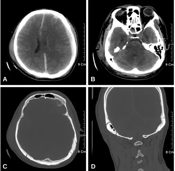
temporo-parietal areas. Multiple contusion of trunk and limbs was also noticed. On neurological examination, he was stuporous and responded by withdrawal of his extremities in response to painful stimuli. His right pupil was dilated and fixed. He had an initial Glasgow Coma Scale Score of 6. Plain x-rays of the skull showed a diastatic fracture of right lambdoidal suture along the course of the distal por- tion of the transverse sinus. Initial computed tomography (CT) scan showed left-sided dominant, bilateral acute sub- dural hematoma, multiple hemorrhagic contusions, traumatic subarachnoid hemorrhages. Severe brain swelling and marked midline shift from left to right side were also observed. CT scan on bone window demonstrated a diasta- tic fracture of right lambdoidal suture near the right trans- verse sinus (Fig. 1). He underwent emergent operation for left-sided subdural hematoma after decompressive craniecto-
my. A large subdural hematoma was evacuated and multi- ple cerebral contusions were scattered. The bleeding source was a lacerated, small cortical vein. Meticulous hemostasis was carried out. Just before the closure of the operative wound, the brain was markedly swollen and bulging.
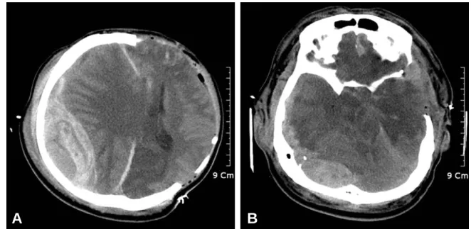
Endotracheal tube was patent and arterial blood gas analy- sis was within normal limit. Bone flap was not replaced and the dura was left open. Postoperatively, he was comatose and the both pupils were fully dilated to 5 mm without response to light. Immediate postoperative CT scan revealed massive acute EDH in the right hemisphere with significant midline shift and he coexisting acute EDH in the right-sided posterior fossa simultaneously (Fig. 2). Second operation was immediately performed to evacuate the con- tralateral supratentorial hematoma, After massive contralat- eral supratentorial EDH was removed, then the dura was
─ 189 ─
— 이정식 외: 급성 경막하 혈종에 대한 감압술 후 발생한 반대편의 천막 상, 하 급성 경막외 혈종 —
Fig. 1. Preoperative computed tomography (CT) scans. a, b: Preoperative CT scans show left dominant, bilateral subdural hematoma with midline shift to the right. c, d: Bone windows of CT show a linear skull fracture extended to the right lambdoidal suture overlying the right tentorium.
A B
C D
open. Scanty acute subdural hematoma and multiple corti- cal contusions were observed. However, the infratentorial EDH could be not removed because of postoperative circu- latory collapse. After 2nd operation, he showed no improve- ment and died 4 days after surgery.
III. Discussion
A postoperative contralateral EDH after an acute subdur- al hematoma evacuation has occasionally reported in the lit- erature.(1-10) A traumatic infratentorial EDH is also a rare condition estimated to complicate about 0.3% of all head injuries, and represents 4% to 12.9% of the entire group of EDHs.(11) An infratentorial epidural hematoma is known for the indistinct signs and symptoms and a disreputable finish that varies from recovery to sudden death. Because of the small volume of the posterior fossa, included critical structures, and usually without prior warning signs, It may cause sudden and lethal deterioration of the patient by compression of the brain stem, if not diagnosed early and treated promptly.(1-10) To our knowledge, this is the first case of a postoperative contralateral simultaneous supra- and infratentorial acute EDH after decompressive surgery of an acute subdural hematoma. A postoperative contralateral EDH after acute SDH evacuation is typically manifested as an intraoperative brain protrusion(1-10) and is potentially fatal if unrecognized early. An overlying skull fracture is a typical preoperative warning sign of a contralateral delayed
EDH.(1-10)
The mechanism of postoperative contralateral EDH devel- opment is not clear, but has been explained as loss of tam- ponade effect. The initial impact causes contrecoup injury as well as coup injury with a skull fracture and hemor- rhage from the dura or fracture site. However, mass effect from the contrecoup hematoma probably increases the intracranial pressure and produces a tamponade effect on the contralateral EDH source, delaying the formation of an EDH.(12) Therefore, the measures, such as decompressive surgery and use of hyperosmolar agents undertaken to reduce the increased intracranial pressure, may release the tamponade effect and contribute to the formation of a postoperative contralateral EDH.(1,9) The usual bleeding source in delayed contralateral EDH is a skull fracture, stripped dura or venous sinus injury, rather than a torn middle meningeal artery. In the present case, bleeding source of a supra- and infratentorial EDH may be a detached dura or lambodoidal skull fracture near the con- tralateral transverse sinus.
Decompressive craniectomy and removal of hematoma may make patients more at risk for developing a fatal con- tralateral EDH when compared with craniotomy as our present case. However, in facing those patients with an intraoperative brain protrusion, immediate decompressive craniectomy should be performed to release mass effect. In these patients, a CT scan should be performed immediately to evaluate the cause of the brain displacement and detect
─ 190 ─
— 대한외상학회지 제 23 권 제 2 호 —
Fig. 2. Computed tomography (CT) scans just after decompressive surgery. a, b: Immediate postoperative CT scans show a con- tralateral supra- and infratentorial epidural hematoma with significant mass effect, resulting in severe brain swelling.
A B
─ 191 ─
— 이정식 외: 급성 경막하 혈종에 대한 감압술 후 발생한 반대편의 천막 상, 하 급성 경막외 혈종 —
this potential complication. In recent years, the values of intraoperative imaging have been reevaluated. It can be used for immediate checks for preventing intraoperative unexpected events, for example, contralateral hemorrhage, ipsilateral parenchymal hemorrhage. Intraoperative imaging techniques can be divided into several systems, such as magnetic resonance imaging, computed tomography and ultrasound. The benefit of CT-based technique compared with MR imaging is its simplicity combined with sufficient image quality. Another benefit of CT is that it can dimin- ish uncertainty during operations.(13) Anytime, intraopera- tive imagings are necessary, an immediate imaging check can be performed to check on unexpected events. The confirmation of an unexpected event provides an opportuni- ty for immediate diagnosis and prevent to avoid serious complications.(14) If the swelling is massive intraoperatively and there is evidence of a contralateral skull fracture, intra- operative hemorrhage in the contralateral hemisphere should be considered.
IV. Conclusion
A postoperative contralateral supra- and infratentorial EDH after acute SDH evacuation is an extremely rare event. An intraoperative brain protrusion in a patient with a skull fracture involving the lambdoidal suture may be a warning sign. A high clinical suspicion or awareness of this entity is necessary to diagnose this dangerous disorder early.
REFERENCES
01) Cohen JE, Rajz G, Itshayek E, Umansky F: Bilateral acute epidural hematoma after evacuation of acute subdural hematoma : brain shift and the dynamics of extraaxial collections. Neurol Res 2004;26:763-6.
02) Feuerman T, Wackym PA, Gade GF, Lanman T, Becker D: Intraoperative development of contralateral epidural hematoma during evacuation of traumatic extraaxial hematoma. Neurosurgery 1988;23:480-4.
03) Matsuno A, Katayama H, Wada H, Morikawa K, Tanaka K, Tanaka H, et al: Significance of consecu- tive bilateral surgeries for patients with acute subdural hematoma who develop contralateral acute epi- or sub- dural hematoma. Surg Neurol 2003;60:23-30.
04) Meguro K, Kobayashi E, Maki Y: Acute brain swelling during evacuation of subdural hematoma caused by delayed contralateral extradural hematoma : report of two cases. Neurosurgery 1987;20:326-8.
05) Mohindra S, Mukherjee KK, Gupta R, Chhabra R, Gupta SK, Khosla VK: Decompressive surgery for acute subdural haematoma leading to contralateral extradural hematoma : a report of two cases and review of literature. Br J Neurosurg 2005;19:490-4.
06) Piepmeier JM, Wagner FC Jr: Delayed post-traumatic extracerebral hematomas. J Trauma 1982;22:455-60.
07) Servadei F, Nanni A, Nasi MT, Zappi D, Vergoni G, Giuliani G, et al: Evolving brain lesions in the first 12 hours after head injury: analysis of 37 comatose patients. Neurosurgery 1995;37:899-906.
08) Su TM, Lee TH, Chen WF, Lee TC, Cheng CH:
Contralateral acute epidural hematoma after decompres- sive surgery of acute subdural hematoma: clinical fea- tures and outcome. J Trauma 2008;65:298-302.
09) Thibodeau M, Melanson D, Ethier R: Acute epidural hematoma following decompressive surgery of a sub- dural hematoma. Can Assoc Radiol J 1987;38:52-3 10) Yagu¨e LG, Rodr´guez-Sa´nchez J, Polaina M, Porras LF,l
Lorenzana L, Cabezudo JM: Contralateral extradural hematoma following craniotomy for traumatic intracra- nial lesion. Case report. J Neurosurg Sci 1991;35:107-9 11) Garza-Mercado R: Extradural hematoma of the poste-
rior cranial fossa. Report of seven cases with survival. J Neurosurg 1983;59:664-72.
12) Servadei F, Nasi MT, Giuliani G, Cremonini AM, Cenni P, Zappi D, et al: CT prognostic factors in acute subdural hematomas: the value of the ‘worst’ CT scan. Br J Neurosurg 2000;14:110-6
13) Kim JW, Lee SH, Son YJ, Yang HJ, Chung YS, Jung HW: Mobile computed tomography : early experience in Korea. J Korean Neurosurg Soc 2010;48:31-6 14) Foroglou N, Zamani A, Black P: Intra-operative MRI
(iop-MR) for brain tumour surgery. Br J Neurosurg 2009;23:14-22