A Comprehensive Prognostic Stratification for Patients with Metastatic Renal Clear Cell Carcinoma
Kang Su Cho,1 Young Deuk Choi,1 Se Joong Kim,2 Chun Il Kim,3 Byung Ha Chung,1 Do Hwan Seong,4 Dong Hyeon Lee,5 Jin Seon Cho,6 In Rae Cho,7 and Sung Joon Hong1
Department of Urology, 1Urological Science Institute, Yonsei University, Seoul; 2Ajou University, Suwon; 3Keimyung University, Daegu; 4Inha University, Incheon; 5Ewha Womans University, Seoul; 6Hallym University, Chuncheon; 7Inje University, Busan, Korea.
Received September 28, 2007 Accepted November 27, 2007
Reprint address: requests to Dr. Sung Joon Hong, Department of Urology, Urological Science Institute, Yonsei University College of Medicine, 250 Seongsanno, Seodaemun-gu, Seoul 120-752, Korea. Tel: 82-2-2228-2315, Fax: 82-2-312-2538, E-mail: sjhong [email protected]
Purpose: To develop a reliable prognostic model for patients with metastatic renal cell carcinoma (RCC) based on features readily available in common clinical settings. Patients and Methods: A total of 197 patients with RCC who underwent nephrectomy and immunotherapy from 1995 to 2004 were retrospectively reviewed. Their mean age was 55.1 ± 11.8 yrs (24 - 83 yrs) and mean survival time from metastasis was 22.6
± 20.2 mos (3 - 120 mos). The impact of 24 clinicopathological features on disease specific survival was investigated. Results:
On univariate analysis, constitutional symptoms, sarcomatoid differentiation, tumor necrosis, multiple primary lesions, liver metastasis, Eastern Cooperative Oncology Group Performance Status (ECOG-PS), thrombocytosis, alkaline phosphatase, hematocrit, T stage, N stage, and nuclear grade had significant influence on survival (p < 0.05). Multivariate analysis revealed the following features associated with survival: sarcomatoid differentiation [hazard ratio (HR) = 2.99, p < 0.001], liver metastasis (HR = 2.09, p = 0.002), ECOG-PS (HR = 1.95, p = 0.005), N stage (HR = 1.94, p = 0.002), and number of metastatic sites (HR = 1.76, p = 0.003). An individual prognostic score was defined as the sum of the weight of these features.
According to prognostic scores, patients could be subdivided into 3 groups: low risk (score 0), intermediate risk (score 1 or 2), and high risk (score ≥ 3). Conclusion: A comprehensive prognostic stratification model was developed to predict survival and stratify patients for prospective clinical trials.
Key Words: Carcinoma, renal cell, neoplasm metastasis, nephrectomy, immunotherapy, prognosis
INTRODUCTION
If untreated, the prognosis for patients with metastatic renal cell carcinoma (RCC) is generally poor, with an overall median survival time of no more than 12 mos and a 5-yr survival rate of less than 10%.1 However, it is not easy to predict the individual prognosis of these patients since the natural history of RCC is complex and influenced by various patient- and tumor-related factors.2
Combination therapies of nephrectomy and immunotherapy for patients with metastatic RCC demonstrate only limited benefits.3,4 Recently, novel molecular-targeted agents showed a significant benefit on progression-free survival in patients for whom cytokine therapy had failed.5 A variety of important prognostic indicators in metastatic RCC have previously been identified, and several prognosis prediction models have been suggested.6-12 These models can be used to counsel patients, determine the need for adjuvant therapy, stratify patients for clinical trials, and develop appropriate postoperative surveillance programs to monitor the risk of cancer progression.
In this study, a readily available comprehen- sive model that is capable of predicting survival of patients with metastatic RCC who underwent radical nephrectomy and immunotherapy in common clinical settings was developed.
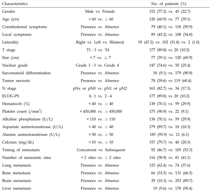
Table 1. Patient Characteristics
Characteristics No. of patients (%)
Gender Male vs. Female 152 (77.2) vs. 45 (22.7)
Age (yrs) < 60 vs. ≥ 60 120 (60.9) vs. 77 (39.1)
Constitutional symptoms Presence vs. Absence 79 (40.1) vs. 118 (59.9)
Local symptoms Presence vs. Absence 89 (45.2) vs. 108 (54.8)
Laterality Right vs. Left vs. Bilateral 93 (47.2) vs. 102 (51.8) vs. 2 (1.0)
T stage T1 - 3 vs. T4 177 (89.8) vs 20 (10.2)
Size (cm) < 7 vs. ≧ 7 77 (39.1) vs. 120 (60.9)
Nuclear grade Grade 1 - 3 vs. Grade 4 147 (74.6) vs. 50 (25.4)
Sarcomatoid differentiation Presence vs. Absence 18 (9.1) vs. 179 (90.9)
Tumor necrosis Presence vs. Absence 78 (39.6) vs 119 (60.4)
N stage pNx or pN0 vs. pN1 or pN2 163 (82.7) vs. 34 (17.3)
ECOG-PS 0, 1 vs. 2 - 4 177 (89.8) vs 20 (10.2)
Hematocrit (%) < 40 vs. ≥ 40 138 (70.1) vs. 59 (29.9)
Platelet count (/mm3) < 450,000 vs. ≥ 450,000 175 (90.9) vs. 22 (9.1) Alkaline phosphatase (U/L) < 110 vs. ≥ 110 138 (70.1) vs. 59 (29.9) Aspartate aminotransferase (U/L) < 40 vs. ≥ 40 179 (89.7) vs. 18 (10.3) Alanine aminotransferase (U/L) < 50 vs. ≥ 50 185 (93.9) vs. 12 (6.1)
Calcium (mg/dL) < 10 vs. ≥ 10 157 (79.7) vs. 40 (20.3)
Timing of metastasis Concurrent vs. Subsequent 92 (46.7) vs. 105 (53.3) Number of metastatic sites < 2 sites vs. ≥ 2 sites 116 (58.9) vs. 81 (41.1)
Lung metastasis Presence vs. Absence 123 (62.4) vs. 74 (37.6)
Bone metastasis Presence vs. Absence 66 (33.5) vs. 131 (66.5)
Brain metastasis Presence vs. Absence 29 (10.3) vs. 253 (89.7)
Liver metastasis Presence vs. Absence 19 (9.6) vs. 178 (90.4)
ECOG-PS, eastern cooperative oncology group performance status.
PATIENTS AND METHODS
The medical records of 368 patients with histologically proven metastatic RCC from 8 university hospitals were retrospectively reviewed.
The cohort was limited to patients who underwent radical nephrectomy and treatment between 1995 and 2004 with at least 1 cycle of immunotherapy [interferon-α, interleukin-2 (IL-2), or a combination thereof with or without 5-flourouracil]. Patients who received other biologic response modifiers
and chemotherapeutic regimens were excluded.
Exclusion criteria also included non-clear cell histology, von Hippel-Lindau disease, other malignant disease, and followup duration of less than 3 mos. A total of 197 patients were eligible for this study, with 152 males (77.2%) and 45 females (22.8%). Interferon-α monotherapy was performed in 72 patients, IL-2 monotherapy in 9, and combination therapy in 116 patients. The mean age was 55.1 ± 11.8 yrs (range, 24 to 83 yrs) and the mean survival time was 22.6 ± 20.2 mos
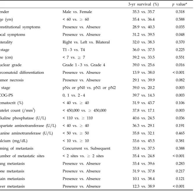
Table 2. Univariate Analysis for Disease-Specific Survival
3-yr survival (%) p value*
Gender Male vs. Female 35.3 vs. 35.7 0.318
Age (yrs) < 60 vs. ≥ 60 35.4 vs. 36.4 0.588
Constitutional symptoms Presence vs. Absence 28.9 vs. 40.3 0.035
Local symptoms Presence vs. Absence 31.2 vs. 39.5 0.048
Laterality Right vs. Left vs. Bilateral 32.0 vs. 38.3 0.370
T stage T1 - 3 vs. T4 36.0 vs. 37.5 0.225
Size (cm) < 7 vs. ≧ 7 39.2 vs. 33.5 0.551
Nuclear grade Grade 1 - 3 vs. Grade 4 39.0 vs. 25.6 0.016
Sarcomatoid differentiation Presence vs. Absence 13.9 vs. 38.0 < 0.001
Tumor necrosis Presence vs. Absence 29.1 vs. 39.9 0.082
N stage pNx or pN0 vs. pN1 or pN2 39.0 vs. 20.2 0.003
ECOG-PS 0, 1 vs. 2 - 4 39.7 vs. 14.3 0.003
Hematocrit (%) < 40 vs. ≥ 40 31.9 vs. 43.7 0.106
Platelet count (/mm3) < 450,000 vs. ≥ 450,000 37.8 vs. 17.1 0.003
Alkaline phosphatase (U/L) < 110 vs. ≥ 110 40.6 vs. 24.5 0.036
Aspartate aminotransferase (U/L) < 40 vs. ≥ 40 36.3 vs. 29.1 0.191
Alanine aminotransferase (U/L) < 50 vs. ≥ 50 35.8 vs. 32.1 0.465
Calcium (mg/dL) < 10 vs. ≥ 10 33.6 vs. 45.5 0.381
Timing of metastasis Concurrent vs. Subsequent 33.8 vs. 37.5 0.388
Number of metastatic sites < 2 sites vs. ≥ 2 sites 35.4 vs. 24.8 < 0.001
Lung metastasis Presence vs. Absence 33.4 vs. 39.6 0.283
Bone metastasis Presence vs. Absence 31.9 vs. 37.8 0.217
Brain metastasis Presence vs. Absence 10.1 vs. 38.4 0.121
Liver metastasis Presence vs. Absence 12.3 vs. 38.9 < 0.001
ECOG-PS, eastern cooperative oncology group performance status.
*p value by log-rank test.
(range, 3 to 120 mos). Survival time was defined as the time from diagnosis of metastatic disease to the date of death or last followup.
The impact of various clinicopathological factors on disease-specific survival was investigated. The following patient-related, laboratory, tumor-related, and metastasis-related features were assessed:
gender, age, constitutional symptoms at pre- sentation, local symptoms at presentation, Eastern Cooperative Oncology Group Performance Status
(ECOG-PS) score, commonly used laboratory tests (hematocrit, platelet, alkaline phosphatase, aspartate aminotransferase, alanine aminotransferase, and calcium level), T stage, tumor size, nuclear grade, sarcomatoid differentiation, tumor necrosis, N stage, timing of metastasis, site of metastatic disease (lung, bone, brain, and liver), and number of metastatic sites. The clinicopathological infor- mation of this study cohort is summarized in Table 1.
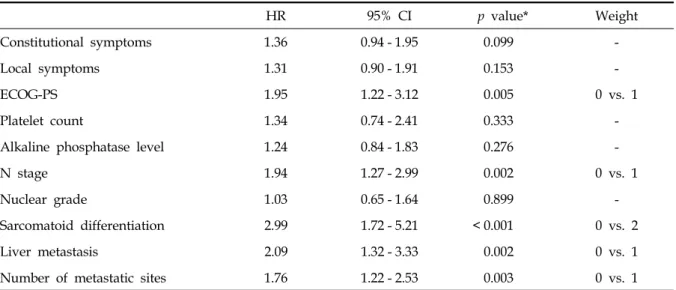
Table 3. Multivariate Analysis for Disease-Specific Survival
HR 95% CI p value* Weight
Constitutional symptoms 1.36 0.94 - 1.95 0.099 -
Local symptoms 1.31 0.90 - 1.91 0.153 -
ECOG-PS 1.95 1.22 - 3.12 0.005 0 vs. 1
Platelet count 1.34 0.74 - 2.41 0.333 -
Alkaline phosphatase level 1.24 0.84 - 1.83 0.276 -
N stage 1.94 1.27 - 2.99 0.002 0 vs. 1
Nuclear grade 1.03 0.65 - 1.64 0.899 -
Sarcomatoid differentiation 2.99 1.72 - 5.21 < 0.001 0 vs. 2
Liver metastasis 2.09 1.32 - 3.33 0.002 0 vs. 1
Number of metastatic sites 1.76 1.22 - 2.53 0.003 0 vs. 1
HR, hazard ratio; CI, confidence interval; ECOG-PS, eastern cooperative oncology group performance status score.
*p value by multivariate Cox proportional hazard model.
Kaplan-Meier curves were generated and compared by using log-rank test for univariate survival analyses. To assess the independent impact of clinicopathological factors on disease- specific survival, Cox proportional hazards regression was used for multivariate survival analyses. Based on rounded regression coefficients [log hazard ratios (HR) in the final Cox model] of variables, the weights of prognostic features were determined. A prognostic score was defined as the sum of the weights of the independent prognostic factors. SPSS for Windows version 12.0 was used for statistical analyses. All p values were 2-sided, and a p value of less than 0.05 was considered to be significant.
RESULTS
Univariate analysis
Of 197 patients, 127 (64.5%) died of cancer at last followup, while 2 (1.0%) died of other causes.
The disease-specific survival rate was 65.4% at 1 yr, 35.7% at 3 yrs, and 21.8% at 5 yrs. For univariate analysis, there was no apparent association between survival and gender, age, laterality, T stage, tumor size, tumor necrosis, hematocrit level, liver enzyme level, serum calcium level, timing of
metastasis, lung metastasis, bone metastasis or brain metastasis (p > 0.05). However, the presence of constitutional symptoms (p = 0.035), presence of local symptoms (p = 0.048), nuclear grade 4 (p = 0.016), presence of sarcomatoid differentiation (p <
0.001), nodal involvement (p = 0.003), ECOG-PS score of 2 or greater (p = 0.003), thrombocytosis (p
= 0.003), increased alkaline phosphatase (p = 0.036), multiple metastatic sites (p < 0.001), and presence of liver metastasis (p < 0.001) appeared to have a significant influence on survival (Table 2).
Multivariate analysis
Multivariate analysis revealed that the following 5 features were associated with disease-specific survival: sarcomatoid differentiation (presence vs.
absence, HR = 2.99, p < 0.001), liver metastasis (presence vs. absence, HR = 2.09, p = 0.002), ECOG- PS (≥ 2 vs < 2, HR = 1.95, p = 0.005), N stage (≥
1 vs 0, HR = 1.94, p = 0.002), and number of metastatic sites (≥ 2 vs 1, HR = 2.024, p = 0.003) (Table 3).
Prognostic stratification model
The weights of independent prognostic factors were defined as follows: sarcomatoid differen- tiation was given a weight of 2, and the remaining
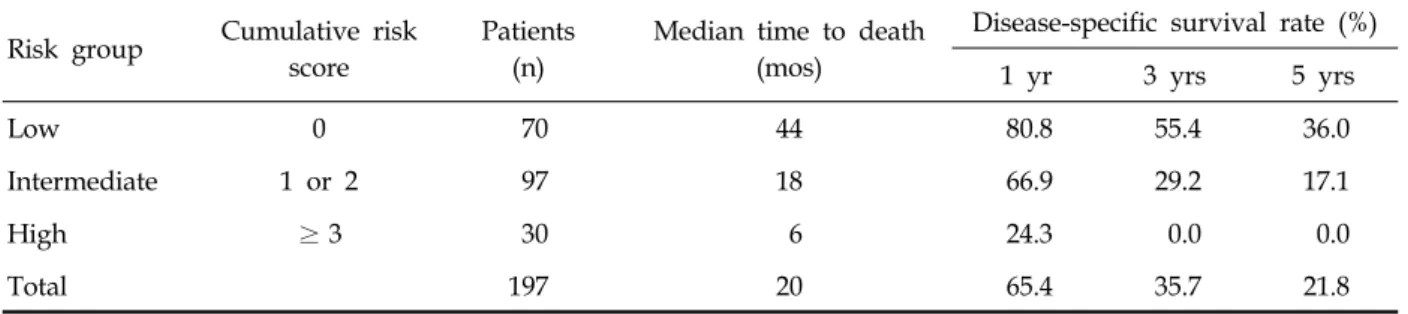
Table 4. Disease-Specific Survival Rates by Risk Group Risk group Cumulative risk
score
Patients (n)
Median time to death (mos)
Disease-specific survival rate (%)
1 yr 3 yrs 5 yrs
Low 0 70 44 80.8 55.4 36.0
Intermediate 1 or 2 97 18 66.9 29.2 17.1
High ≥3 30 6 24.3 0.0 0.0
Total 197 20 65.4 35.7 21.8
Fig. 1. Disease-specific survival curves according to risk scores (A) and risk groups (B). p value = 0.001 between low risk and intermediate risk, p value < 0.001 between intermediate risk and high risk.
4 factors were assigned a weight of 1 (Table 4).
An individual prognostic score was defined as the sum of the weights of these factors. The prognostic scores were 0 in 70 patients, 1 in 55, 2 in 42, 3 in 17, 4 in 9, and 5 in 4. Survival curves showed a statistically significant difference between those with scores 0 and 1 (p = 0.038) and scores 2 and 3 (p = 0.039). However, there was no differ- ence between those with scores 1 and 2 (p = 0.052), 3 and 4 (p = 0.061), and 4 and 5 (p = 0.618) (Fig. 1).
As a consequence, the cohort was subdivided into 3 groups as follows: a low risk group (score 0), intermediate risk group (score 1 or 2), and high risk group (score 3 or greater) (Fig. 1), each of which had disease-specific survival rates that are shown in Table 4.
DISCUSSION
Investigators have concentrated on the prognostic
stratification of patients with advanced or metastatic RCC for years, and a number of RCC outcome prediction models have previously been reported.6-12 Zisman et al. reported a single predictive system of patients with and without metastatic RCC, the University of California Los Angeles Integrated Staging System (UISS), which is based on 661 patients and incorporates TNM pathologic stage, ECOG-PS, and Fuhrman grade to predict overall survival after patients undergo radical nephrectomy. Frank et al.13 proposed the stage, size, nuclear grade, and necrosis (SSIGN) score, which is based on 1801 patients with clear cell RCC treated by radical nephrectomy. The SSIGN score stratifies the risk of death from RCC based on these features.
Recently, prognostic prediction systems have focused only on metastatic RCC.6-10 Motzer et al.10 suggested the prognostic stratification of 670 patients with advanced RCC. In their study, 5 prognostic factors (Karnofsky performance status,
A B
serum lactate dehydrogenase, hemoglobin, corrected serum calcium, and prior nephrectomy) were identified and used to categorize patients with metastatic RCC into 3 risk groups. They also reviewed 463 patients who were treated with interferon-α for metastatic RCC and developed another algorithm that consists of Karnofsky performance status, lactate dehydrogenase, hemoglobin, corrected serum calcium, and time from diagnosis to immunotherapy.9 Leivobich et al.8 proposed the first predictive algorithm in patients with metastatic RCC after nephrectomy and IL-2-based immunotherapy. In their model, regional lymph node status, constitutional symptoms, location of metastases, sarcomatoid histology, and thyroid stimulating hormone (TSH) levels were associated with survival. Atzpodien et al.6 demonstrated a comprehensive prognostic system of pretreatment clinical parameters in patients with metastatic RCC treated with different subcutaneous recombinant cytokine-based home therapies in consecutive trials. Six parameters (neutrophil counts, lactate dehydrogenase, C- reactive protein, time from diagnosis of tumor to metastatic disease, number of metastatic sites, and bone metastasis) were identified as independent prognostic factors.
In our study, 24 clinicopathological features were evaluated for their impacts on survival of patients with metastatic RCC who were treated with nephrectomy and immunotherapy. Sub- sequently, a new comprehensive prognostic model was devised that consisted of 3 risk groups.
The study data showed various median survival times according to each risk group ranging from 6 mos in the high-risk group to 44 mos in the low- risk group. This scoring system was determined by the weighted sum of 5 features (sarcomatoid differentiation, liver metastasis, ECOG-PS, N stage, and number of metastatic sites). The weight of each variable was considered and only sarcomatoid differentiation was given a weight of 2 because of its strong prognostic impact (HR = 2.99). The relatively strong impact of sarcomatoid differentiation has also been identified in previous reports.8,11
Generally, extrapulmonary metastasis has been regarded as an independent prognostic factor in metastatic RCC patients.7,8,14,15 In this study, only
hepatic involvement showed a significant influence on survival whereas brain and bone involvement did not. Although the cut-off value to define multiplicity of metastatic sites has been inconsistent, many authors have emphasized the significance of the number of metastatic sites in these patients.
6,7,16-18 Performance status was established as an
important prognostic factor without any uncer- tainty,9,10,16-19 suggesting a prognostic significance for regional node involvement similar to the work of Leibovich et al. However, it is not certain whether regional nodal involvement is a feasible prognostic factor in any metastatic RCC patients since both this study and those of Leibovich et al.
are restricted to patients who underwent nephrectomy and immunotherapy.
The present study was limited to patients with clear cell RCC since the histological subtype is associated with the biologic aggressiveness of RCC.20 To eliminate possible confounding factors, the study cohort was limited to patients with metastatic RCC who were treated with nephrectomy and immunotherapy. Nevertheless, the study still showed some limitations as well as the general limitations of a retrospective study, where immunotherapy protocol and number of cycles were not identical, and this difference was not considered in the data analysis. However, systemic immunotherapy demonstrated a minimal impact on outcome in advanced RCC, suggesting that the difference did not cause a significant bias in developing this prognostic model.
The objectives of this study were to develop a reliable prognostic model based on features readily available to clinicians and pathologists.
There are no other ancillary tests such as erythrocyte sedimentation rate, thyroid stimulating hormone, C-reactive protein, and lactate dehydrogenase that have previously been studied and identified as useful in the management of patients with RCC.6,
8-10,14,15 For this reason, the clinical feasibility of this model is expected to be greater in common clinical settings. Recently, race has been shown to be a significant predictor of overall survival within a clinical trial patient population with RCC, even in those with metastatic RCC.21,22 However, there was no literature in English about a predictive model for patients treated with nephrectomy and immunotherapy based on Asian
populations including Koreans. Therefore, the prognostic model described herein can be used to predict survival and stratify patients for prospective clinical trials although further validation of the model through prospectively designed clinical trials is needed.
In summary, sarcomatoid differentiation, liver metastasis, ECOG-PS, N stage, and number of metastatic sites were found to be independently associated with survival of patients with metastatic RCC who were treated with nephrectomy and immunotherapy. Based on these features, a comprehensive prognostic stratification model was developed to predict survival and stratify patients for prospective clinical trials.
ACKNOWLEDGEMENTS
We wish to thank the other members of the Severance Urologic Oncology Group: Yun Seob Song, and Won Jae Yang, Soonchunhyang University; Hong Sup Kim, Konkuk University;
Young-Sig Kim, National Health Insurance Corporation Ilsan Hospital; Sun Il Kim, Ajou University; Sang Hyeon Cheon, Ulsan University;
Joong Shik Lee, Sungkyunkwan University; and Ki Hak Song, Konyang University.
REFERENCES
1. Motzer RJ, Bander NH, Nanus DM. Renal-cell carcinoma. N Engl J Med 1996;335:865-75.
2. Pantuck AJ, Zisman A, Belldegrun AS. The changing natural history of renal cell carcinoma. J Urol 2001;166:
1611-23.
3. Flanigan RC, Salmon SE, Blumenstein BA, Bearman SI, Roy V, McGrath PC, et al. Nephrectomy followed by interferon alfa-2b compared with interferon alfa-2b alone for metastatic renal-cell cancer. N Engl J Med 2001;345:1655-9.
4. Mickisch GH, Garin A, van Poppel H, de Prijck L, Sylvester R; European Organisation for Research and Treatment of Cancer (EORTC) Genitourinary Group.
Radical nephrectomy plus interferon-alfa-based im- munotherapy compared with interferon alfa alone in metastatic renal-cell carcinoma: a randomised trial.
Lancet 2001;358:966-70.
5. Bellmunt J, Montagut C, Albiol S, Carles J, Maroto P, Orsola A. Present strategies in the treatment of metastatic renal cell carcinoma: an update on molecular targeting
agents. BJU Int 2007;99:274-80.
6. Atzpodien J, Royston P, Wandert T, Reitz M; DGCIN- German Cooperative Renal Carcinoma Chemo- Immunotherap Trial Group. Metastatic renal carcinoma comprehensive prognostic system. Br J Cancer 2003;88:
348-53.
7. Leibovich BC, Cheville JC, Lohse CM, Zincke H, Frank I, Kwon ED, et al. A scoring algorithm to predict survival for patients with metastatic clear cell renal cell carcinoma: a stratification tool for prospective clinical trials. J Urol 2005;174:1759-63; discussion 1763.
8. Leibovich BC, Han KR, Bui MH, Pantuck AJ, Dorey FJ, Figlin RA, et al. Scoring algorithm to predict survival after nephrectomy and immunotherapy in patients with metastatic renal cell carcinoma: a stratification tool for prospective clinical trials. Cancer 2003;98:2566-75.
9. Motzer RJ, Bacik J, Murphy BA, Russo P, Mazumdar M. Interferon-alfa as a comparative treatment for clinical trials of new therapies against advanced renal cell carcinoma. J Clin Oncol 2002;20:289-96.
10. Motzer RJ, Mazumdar M, Bacik J, Berg W, Amsterdam A, Ferrara J. Survival and prognostic stratification of 670 patients with advanced renal cell carcinoma. J Clin Oncol 1999;17:2530-40.
11. Zisman A, Pantuck AJ, Dorey F, Said JW, Shvarts O, Quintana D, et al. Improved prognostication of renal cell carcinoma using an integrated staging system. J Clin Oncol 2001;19:1649-57.
12. Zisman A, Pantuck AJ, Wieder J, Chao DH, Dorey F, Said JW, et al. Risk group assessment and clinical outcome algorithm to predict the natural history of patients with surgically resected renal cell carcinoma.
J Clin Oncol 2002;20:4559-66.
13. Frank I, Blute ML, Cheville JC, Lohse CM, Weaver AL, Zincke H. An outcome prediction model for patients with clear cell renal cell carcinoma treated with radical nephrectomy based on tumor stage, size, grade and necrosis: the SSIGN score. J Urol 2002;168:2395-400.
14. de Forges A, Rey A, Klink M, Ghosn M, Kramar A, Droz JP. Prognostic factors of adult metastatic renal carcinoma: a multivariate analysis. Semin Surg Oncol 1988;4:149-54.
15. Lopez Hänninen E, Kirchner H, Atzpodien J.
Interleukin-2 based home therapy of metastatic renal cell carcinoma: risks and benefits in 215 consecutive single institution patients. J Urol 1996;155:19-25.
16. Jones M, Philip T, Palmer P, von der Maase H, Vinke J, Elson P, et al. The impact of interleukin-2 on survival in renal cancer: a multivariate analysis. Cancer Biother 1993;8:275-88.
17. Palmer PA, Vinke J, Philip T, Negrier S, Atzpodien J, Kirchner H, et al. Prognostic factors for survival in patients with advanced renal cell carcinoma treated with recombinant interleukin-2. Ann Oncol 1992;3:475- 80.
18. Elson PJ, Witte RS, Trump DL. Prognostic factors for survival in patients with recurrent or metastatic renal cell carcinoma. Cancer Res 1988;48:7310-3.
19. Fosså SD, Kramar A, Droz JP. Prognostic factors and survival in patients with metastatic renal cell carcinoma treated with chemotherapy or interferon-alpha. Eur J Cancer 1994;30A:1310-4.
20. Amin MB, Amin MB, Tamboli P, Javidan J, Stricker H, de-Peralta Venturina M, et al. Prognostic impact of histologic subtyping of adult renal epithelial neoplasms:
an experience of 405 cases. Am J Surg Pathol 2002;26:
281-91.
21. Berndt SI, Carter HB, Schoenberg MP, Newschaffer CJ.
Disparities in treatment and outcome for renal cell cancer among older black and white patients. J Clin Oncol 2007;25:3589-95.
22. Tripathi RT, Heilbrun LK, Jain V, Vaishampayan UN.
Racial disparity in outcomes of a clinical trial population with metastatic renal cell carcinoma. Urology 2006;68:
296-301.