- 62 -
서 론
신세포암은 25~57%에서 진단과 동시에 폐, 뼈, 뇌 등의 다 른 장기로의 전이를 동반하고 있다. 또한 원발암을 제거한 수 년 뒤에도 림프절, 폐, 간 등 신체 다른 부위에서 전이가 발견 되는 예측하기 힘든 종양이다.1)
신세포암이 두경부로 전이되는 경우는 비교적 드물다. 두경 부로의 전이 부위로는 치은(63%), 설부(17%), 구개(6%), 이하 선(6%), 구개수(3%), 하악골(3%), 구순(3%) 등으로 Smith 등 이 보고되나 갑상선으로의 전이는 매우 드물다.2) 반면에, 갑상 선암으로 전이되는 다른 장기의 원발암 중에서는 신세포암이 차지하는 비율이 42%로 가장 높으며, 갑상선 종물을 첫 증상 으로 내원한 환자가 갑상선으로 전이된 신세포암으로 밝혀지 는 경우도 있다.3) 신세포암이 갑상선에 전이된 경우는 외국 문
헌에서 드물게 보고되며, 국내 문헌에서도 2예가 보고된 상태 이다.4)
저자들은 처음 진단 당시 갑상선으로 전이된 신세포암을 경 험하였기에 이를 문헌고찰과 함께 보고하는 바이다.
증 례
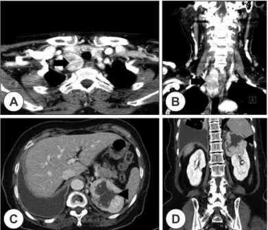
58세 여자환자가 흉막 삼출증으로 타 병원에서 치료 중 우 측 갑상선 및 좌측 신장에 종괴가 발견되어 본원 내분비내과 로 전원되었다. 전산화단층촬영검사상 갑상선 우엽에 9.8 cm 크기의 종물이 흉곽 내까지 위치하고 좌엽에도 다발성 결절이 관찰되었다. 복부에서는 좌측 신장의 상부에 조영 증강이 되 는 7 cm 크기의 종괴가 관찰되었다(Fig. 1). 우측 갑상선 결절 에 대한 세침흡인검사상 결절성 과증식으로 진단되었고 BRAF 변이검사는 음성의 결과를 보였다. 좌측 신장 종괴는 복부 전 산화단층촬영검사상 신세포암으로 의심되어 추가적인 검사를 진행하였다. 마취과를 포함한 관련된 과들 간의 협진을 통해 신장 종괴에 대한 수술적 치료를 먼저 계획하고 환자의 상태에 따라 갑상선 수술을 시행하기로 하였다. 수술 전 좌측 신동맥 에 혈관색전술을 시행한 후에 비뇨기과에서 복강경하 좌측 부
Received : August 20, 2013 / Revised : September 13, 2013
Accepted : September 17, 2013
교신저자 : 박기철, 630-522 경남 창원시 마산회원구 팔용로 158 성균관대학교 의과대학 삼성창원병원 이비인후과학교실 전화 : (055) 290-6068 ・ 전송 : (055) 290-6465 E-mail : [email protected]
대한 두경부 종양 학회지 제 29 권 제 2 호 2013
갑상선에 전이된 신세포암 1예
성균관대학교 의과대학 삼성창원병원 이비인후과학교실
고 영 범·박 기 철
=
Abstract
=A Case of Metastatic Renal Cell Carcinoma to Thyroid Gland
Young-Bum Ko, MD, Gi Cheol Park, MD
Department of Otolaryngology, Samsung Changwon Hospital, Sungkyunkwan University School of Medicine, Changwon, Korea
The distant metastasis is found out in about 25-57% of the patients with renal cell carcinoma at the time of diagnosis. But, the incidence of metastases to the head and neck region, especially to the thyroid gland, is rare.
Most of patients with metastatic renal cell carcinoma to the thyroid gland are asymptomatic at presentation as pa- tients with primary thyroid carcinoma. In the presence of clear cell tumor of the thyroid gland, the diagnostic con- siderations must include metastatic renal cell carcinoma. We report a case of thyroid metastasis from renal cell carcinoma at the time of diagnosis.
KEY WORDS
:Thyroid cancerㆍMetastasisㆍRenal cell carcinoma.online©MLComm
- 63 -
분적 신적출술 및 좌측 부신적출술을 시행하였다. 수술 후 병 리조직 검사상 피막 침범을 보이지 않고 림프혈관계로의 전이 가 없는 신장에 국한된 투명세포암으로 진단되었으며 좌측 부 신의 조직검사상에서도 특이소견을 보이지 않았다. 신적출술 2주 후 본과에서 갑상선 결절에 대한 수술을 진행하였다. 우 측 갑상선 결절은 8.0×4.0×2.5 cm 크기로 종격동을 열지 않 고 절제가 가능하였다. 술 중 우측 갑상선 결절에 대한 동결절 편 조직 검사 결과 결절성 과증식으로 나왔으나 보호자와 상 의 후 좌측 갑상선 절제술도 함께 시행하였다. 수술 후 병리조 직 검사 결과 양측 갑상선에서 결절성 과증식의 결과 보였으나 우측 갑상선에서는 면역염색검사상 CD10, epithelial mem- brane antigen(EMA)에 양성, 갑상선 글로불린, 크로모그라 닌에 음성을 보이는 0.2 cm 크기의 전이성 신세포암이 진단되 었다(Fig. 2). 수술 후 환자는 항암치료를 치료를 시행하였고 이 후 14개월이 지난 현재 재발 소견 없이 외래 추적관찰 중이다.고 찰
신세포암은 신장에 발생하는 원발암의 80~90%를 차지하 며, 20~30%는 진단 당시에 다른 부위에 전이된 상태로 발견 된다. 국소 신세포암의 약 50%에서 타 장기로 전이가 되는데,5) 흔히 폐(50~60%), 림프절(30%), 뼈(30%), 간(30%), 뇌(5~10%) 순으로 발생하며 두경부는 8% 정도인 것으로 알려져 있다.6,7) 신세포암의 전이 경로는 동맥을 통한 혈행성 전이, 정맥이나 림 프절을 통한 역행성 전이, 수술 창상을 통한 전이, 요로를 통 한 전이로 나누어지며, 이중 주로 혈행성으로 전이되는 것으 로 알려져 있다.8,9)
신세포암의 임상적 양상은 다양하나 전형적으로 통증, 종 물, 혈뇨 등이 있다. 타 장기로의 전이가 있는 경우에는 쿠싱증 후군, 고칼슘혈증, 다혈구혈증 등 전이된 부위에 따른 특이 증 상이 나타날 수 있으며, 전이된 부위의 조직학적 검사에서 대 부분 투명세포 형태로 나타난다.10) 갑상선으로 전이된 신세포 암의 경우에는 연하곤란, 천명, 애성 및 경부 종물 등의 증상 이 있을 수 있으나 대부분은 무증상인 경우가 많다. 본 증례에 서도 흉막 삼출증으로 인한 활동성 호흡곤란 외에 다른 증상 은 보이지 않았다. 갑상선에 고립된 종괴로 나타나는 전이된 신세포암의 경우에는 원발성 갑상선암과 임상적으로는 구별 이 되지 않으며, 진단시 세침흡인세포검사 후 PAS 염색이 도움 이 된다.11) 특히 갑상선 투명세포암의 경우에는 전이된 갑상선 암과 조직학적 차이를 구별하기가 매우 힘들기 때문에 갑상선 글로불린 면역화학염색을 통해 구별해야 한다.12) 본 증례의 경 우에도 H-E 염색상에서는 갑상선 투명세포암에 적합한 소견 을 보였으나 갑상선 글로불린 염색에서는 음성소견으로 원발 병소가 갑상선이 아닌 타 장기에서 기원한 전이암으로 확인할 수 있었다.
신세포암의 5년 생존률은 45% 정도로 종양이 신장에 국한 된 경우에는 60~75% 정도이고 피막을 넘어선 국소 임파절 전 이가 있는 경우에는 12~25% 정도이며 원격전이가 있는 경우 의 5년 생존률은 5% 이하로 알려져 있다.13,14) 전이성 신세포암 의 예후는 원발암이 발견된 후 신체 다른 부위에 전이가 발견 되기까지의 기간과 밀접한 관계가 있다. 원발암과 원격전이가 진단 동시에 발견되면 평균생존기간이 1년 전후로 대부분 2년 이내에 사망하지만, 2년 이상 경과 후에 원격전이가 발견된 경 우에는 대부분 5년 이상 생존한다.15) 특히 골, 간 및 뇌 전이가 있는 경우에는 예후가 불량하며 단일 전이보다 다발성의 전이 가 있는 경우에 훨씬 더 예후가 불량하다.16,17) 단일 전이가 있
A
C D
B
Fig. 3. Preoperative contrast enhanced neck CT(A : axial scan.
B : coronal scan) show large hyperdense mass on the right thy- roid gland. The thyroid nodule(arrow) extended to intrathoracic space. Preoperative enhanced abomen CT(C : axial scan. D : coronal scan) show a solid hypodense mass(arrowhead) in the upper pole of the left kidney.
A
C D
B
Fig. 2. Histology of thyroid tumor shows nests of cells with clear cy-
toplasm(A), and renal tumor was surrounded by abundant thin
walled blood vessels(B)(H&E stain). The C and D show immu-
noreativity for CD 10 stain(C), and negativity for thyroglobulin
immmunostain(D) of the thyroid tumor(original magnification ×40).
- 64 -
는 신세포암 환자는 광범위 근치적 신적출술과 전이부위의 수 술적 제거로 생존율의 향상을 기대할 수 있다.18)이번 증례는 신세포암 환자에서 신적출술과 동시에 시행된 갑상선 종물에 대한 수술적 치료 후 갑상선 조직에서 전이성 신세포암이 우연히 발견된 경우이다. 일반적으로 타 장기로의 전이가 있는 신세포암인 경우에는 원발부위와 전이 부위에 대 한 수술적 치료, 면역요법, 항암화학요법, 호르몬요법 등이 고 려될 수 있으며, 본 증례의 경우에는 결과적으로 원발부위와 전이부위를 동시에 수술적 절제를 하게 된 상태로 술 후 항암 화학요법을 추가로 시행하였다. 향후 정기적인 병력 청취 및 신 체검사와 더불어 혈액검사 및 영상학적 검사를 통한 철저한 추적 관찰이 필요하다.
중심 단어 : 갑상선암 ・전이 ・신세포암.