INTRODUCTION
Renal cell carcinoma is characterized by a lack of early warning signs, resulting in a high proportion of metastasis at diagnosis. Relapse occurs in 30% to 50% of patients with completely resected renal cell carcinoma after a radical neph- rectomy. Metastatic renal cell carcinoma (mRCC) is a disease with a poor prognosis and a 5-yr survival rate of less than 10% and is resistant to chemotherapy or radiotherapy (1).
Many immunotherapy protocols have been investigated since Rosenberg and colleagues discovered the clinical efficacy of high-dose bolus interleukin-2 (IL-2) in the treatment of patients with mRCC (2). With an overall response rate of approximately 20% and a durable complete response, the use of high-dose bolus IL-2 has been the best treatment for mRCC. Thus, it remained as the only U.S. Food and Drug Administration-approved drug for the treatment of meta- static renal cancer for more than a decade before the intro- duction of new drugs (3). However, some investigators have encountered significant multi-system toxicities resulting in treatment-related mortality, and consequently, its applica- tion has been limited to the highly selected patients treated
at specialized centers (4).
The pronounced toxicities of high-dose bolus IL-2 treat- ment prompted the development of regimens with subcu- taneous injections of IL-2. In addition, attempts were also made to improve treatment efficacy by adding interferon- (IFN- ) and combinations of low-dose IL-2 and other che- motherapeutic agents (5-8).
Given the toxicity and expense, treatment should be lim- ited to patients most likely to benefit from immunotherapy.
Therefore, many groups have attempted to determine the immunologic prognostic factors as well as to establish clini- cal prognostic factors for patients with mRCC who receive im- munotherapy (9-12).
It is believed that antitumor effects of IL-2 are due to sev- eral mechanisms: it stimulates the generation of natural killer (NK) cells; it enhances not only the cytotoxic activities of T cells but also the T-helper cells and eosinophils (13-15). IL-2 based immunotherapy results in varying degrees of lympho- cytosis and eosinophilia in each patient.
The aims of the present study were to evaluate the clinical effectiveness of an IL-2, IFN- , and 5-fluorouracil (5-FU) combination immunotherapy regimen and to correlate the
In Gab Jeong, Kyung Seok Han, Jae Young Joung, Woo Suk Choi, Seung-Sik Hwang*, Seung Ok Yang, Ho Kyung Seo, Jinsoo Chung, Kang Hyun Lee
Urologic Oncology Clinic, National Cancer Center, and National Cancer Control Research Institute*, National Cancer Center, Goyang, Korea
Address for correspondence Jinsoo Chung, M.D.
Urologic Oncology Clinic, National Cancer Center, 809 Madu 1-dong, Ilsandong-gu, Goyang 410-769, Korea
Tel : +82.31-920-1677, Fax : +82.31-920-0998 E-mail : [email protected]
S122
Analysis of Changes in the Total Lymphocyte and Eosinophil Count during Immunotherapy for Metastatic Renal Cell Carcinoma:
Correlation with Response and Survival
The aims of this study were to analyze lymphocyte and eosinophil counts in con- secutive peripheral blood samples taken during immunotherapy for metastatic renal cell carcinoma (mRCC) and to correlate the findings with objective response and survival. A total of 40 patients with mRCC who received immunotherapy with inter- leukin-2, interferon- , and 5-fluorouracil were analyzed. Objective responses were observed in 14 patients, including 2 (5%) who showed a complete response (CR) and 12 (30%) who showed a partial response (PR). Eleven patients (27%) achieved stable disease (SD), and 15 patients (38%) had progressive disease (PD). Changes from baseline in the total lymphocyte counts were significantly higher in the respond- ing patients (CR+PR+SD) than in the non-responding patients (PD) (p=0.017), but no difference was seen in the total eosinophil counts (p=0.275). Univariate analy- sis identified the Eastern Cooperative Oncology Group (ECOG) performance sta- tus (p=0.017), the presence of a primary renal tumor (p<0.001) and the peripheral lymphocyte counts at week 4 (p=0.034) as prognostic factors, but a low ECOG performance status (p=0.003) and the presence of a primary renal tumor (p=0.001) were identified as independent poor prognostic factors by multivariate analysis. This study provides further evidence that changes in blood lymphocyte counts may serve as an objective indicator of objective responses.
Key Words : Renal Cell Carcinoma; Interleukin-2; Interferon-alpha; Lymphocytes; Eosinophils
Received : 13 February 2007 Accepted : 1 May 2007
objective response and survival with the changes in the blood lymphocyte and eosinophil counts during treatment.
MATERIALS AND METHODS Patient selection
From August 2001 to July 2006, 40 patients with histolo- gically confirmed and measurable progressive mRCC were recruited for this study. Patient assessment at entry into the study consisted of a clinical evaluation, a complete blood cell count, blood chemistry studies, urinary status, radionuclide bone scan, abdominal, thoracic and cranial computerized tomography (CT), and electrocardiography. Of these patients, nephrectomy was performed in 37 patients before treatment with immunotherapy. Three patients did not wish to under- go surgery and embolization was performed following the biopsy.
The eligibility criteria included an Eastern Cooperative Oncology Group (ECOG) performance status of 0 or 1, a life expectancy of at least 3 months, adequate blood counts (hemoglobin greater than 10 g/dL, a white blood cell count greater than 4,000/mL and a platelet count greater than 100,000/mL), adequate renal and hepatic functions (serum creatinine 1.4 mg/dL or less, serum total bilirubin 1.2 mg/
dL or less, and serum alanine aminotransferase 40 IU/L or less), and adequate cardiac and pulmonary function. Exclu- sion criteria included cardiovascular disease, hematopoietic, pulmonary, hepatic or renal dysfunction, ECOG performance status >1, active infection, autoimmune disease, HIV and hepatitis, concomitant therapy with drugs influencing im- munity, and prior malignancies or brain metastases. All pa- tients provided written, informed consent before study entry.
Treatment plan
Immunotherapy was given with the initial 4 weeks con- sisting of treatment with subcutaneous IL-2 (weeks 1 and 4:
20×106U/m2on day 1, 3 and 5; weeks 2 and 3: 5×10⁶ U/m2on days 1, 3 and 5) and treatment with subcutaneous IFN- (weeks 1 and 4: 6×10⁶U/m2on day 1; weeks 2 and 3: 6×10⁶U/m2on days 1, 3 and 5) injections, followed by 4 weeks of injections of subcutaneous IFN- (weeks 5 to 8:
9×10⁶U/m2on days 1, 3 and 5) and intravenous bolus in- jections of 5-FU (weeks 5 to 8: 750 mg/m2on day 1) (16).
The doses in weeks 1 and 4 were administered on an inpa- tient basis, with the rest of the treatment administered on an outpatient basis, except in cases of excessive toxicity.
Collection of blood samples
Blood samples were obtained at baseline and on day 6 of every week throughout the treatment. Total and differential
white blood cell counts were determined for all patients.
Response assessment
Patients were assessed by radiological evaluation or by phy- sical measurement of all sites of disease following the com- pletion of the immunotherapy regimen. The clinical response was evaluated according to the World Health Organization (WHO) criteria (17). After immunotherapy, metastasecto- my was considered for patients with residual disease and who presented with solitary or a few respectable metastases with an acceptable performance status. Response duration was measured from the first observation, and time to survival was counted from the beginning of treatment.
Treatment toxicity
Systemic toxicity of the treatment regimen was determined every week using a grading system according to the WHO classification, namely blood cell count and serum biochemi- cal tests of hepatic and renal function (17). Thyroid stimulat- ing hormone (TSH), triiodothyronine (T3), thyroxine (T4), and human anti-thyroid antibodies were assayed in all pati- ents before and after immunotherapy to quantify possible adverse effects on thyroid function.
Statistical analysis
The primary end-points of the study were therapeutic res- ponse and overall survival after immunotherapy. The abso- lute number of blood lymphocyte and eosinophil counts were analyzed as the dependent variables in a general linear model repeated measures methods to evaluate the associations of these variables and the clinical response following immu- notherapy. To evaluate the predictive value of measured blood lymphocyte counts at each time (baseline and weeks 1 to 4) for therapeutic response, receiver operating characteristic (ROC) curves were generated and areas under the curves (AUC) were calculated for the measured lymphocyte counts at each time. Kaplan-Meier survival curves for overall sur- vival were generated, and the log-rank test was used to com- pare survival according to the variable. The Cox proportion- al hazards regression model was used to estimate the relative importance of the variables. For all statistical analyses p<
0.05 was considered significant.
RESULTS
Patient characteristics and response to treatment
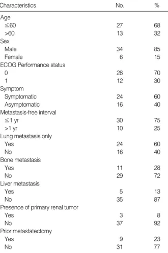
Demographic and clinical characteristics are summarized in Table 1. The median age of the 40 patients was 56 yr (range, 36-72). All patients included in this study showed a clear
cell histology, and sarcomatoid differentiation was seen in 2 patients. The overall response rate was 35% (2 complete res- ponses and 12 partial responses), stable disease was achieved in 11 (27%) patients, and progressive disease occurred in 15 (38%). Complete responses were found only in patients with a single lung metastasis, and the duration of complete remis- sion was 33 and 36 months, respectively.
Changes in the blood total lymphocyte and eosinophil count
Total lymphocyte counts were significantly higher in res- ponding patients (CR+PR+SD) than in non-responding patients (PD) (Fig. 1). No difference was seen in the total eosinophil counts (Fig. 2). Among the serial blood lympho- cyte counts at baseline and for each week, the total lympho- cyte count at week 4 was the most predictive of therapeutic response. The ROC curve for the lymphocyte count at week 4 demonstrated the most significant predictive ability (AUC 0.84, 95% confidence interval [CI]0.76 to 0.91). The AUC for the baseline, and weeks 1, 2, and 3 were 0.68 (95% CI 0.60 to 0.77), 0.49 (95% CI 0.40 to 0.59), 0.74 (95% CI 0.66 to 0.82), and 0.68 (95% CI 0.60 to 0.77), respectively.
Survival
The median follow-up period after the start of the immu- notherapy was 16 months. The overall survival rate was 61%
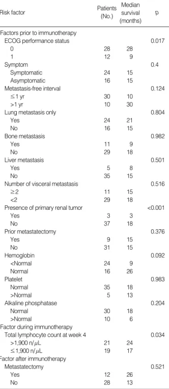
(95% CI 53% to 70%), 39% (95% CI 31% to 49%), and 27% (95% CI 18% to 36%) at 1, 2, and 3 yr, respectively, and median survival was 15 months (Fig. 3). On univariate analysis unfavorable prognostic variables for overall survival were low ECOG performance status, the presence of a pri- mary renal tumor, and a low peripheral lymphocyte count at week 4. In patients with a lymphocyte count higher than 1,900 n/ L, the 3-yr survival rate was 36%, compared to 15% when the lymphocyte count was 1,900 n/ L or less (Table 2 and Fig. 4). On multivariate analysis low ECOG performance status and the presence of a primary renal tumor were associated with decreased overall survival (Table 3).
ECOG, Eastern Cooperative Oncology Group.
Characteristics No. %
Age
≤60 27 68
>60 13 32
Sex
Male 34 85
Female 6 15
ECOG Performance status
0 28 70
1 12 30
Symptom
Symptomatic 24 60
Asymptomatic 16 40
Metastasis-free interval
≤1 yr 30 75
>1 yr 10 25
Lung metastasis only
Yes 24 60
No 16 40
Bone metastasis
Yes 11 28
No 29 72
Liver metastasis
Yes 5 13
No 35 87
Presence of primary renal tumor
Yes 3 8
No 37 92
Prior metastatectomy
Yes 9 23
No 31 77
Table 1.Baseline characteristics of patients (n=40)
Lymphocyte count
3,000
2,500
2,000
1,500
0 1 2 3 4
Week
Responder Non-responder
p=0.017
Fig. 1.Changes in the blood total lymphocyte counts in the first month during immunotherapy.
Eosinophil count
5,000
4,000
3,000
2,000
1,000
0
0 1 2 3 4
Week
Responder Non-responder
p=0.275
Fig. 2.Changes in the blood total eosinophil counts in the first month during immunotherapy.
Toxicity
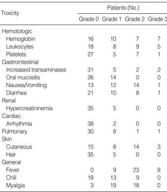
The most common side effects were flu-like symptoms, fatigue, mucositis, diarrhea, vesicle, changes in leukocyte, hemoglobin, and platelet levels, and nausea during the treat- ment. Table 4 shows a detailed description of systemic toxi- cities and their severity. In all patients treatment-related sys- temic toxicity resolved after the end of the therapeutic regi- men. No patient had grade 4 toxicity, and there were no treat- ment-related deaths.
DISCUSSION
Because of the significant toxicity and its high cost, it is very important to select patients with mRCC who may ben- efit mostly from immunotherapy prior to treatment. With
regard to the histologic subtype, responses to immunother- apy are most frequently seen in patients with renal cell car- cinoma of clear cell histology. Among the clear cell subtypes, the response to IL-2 was associated with the presence of al- veolar features and the absence of granular or papillary fea- tures (11). Gez and colleagues identified good performance status, absence of bone metastases, and no other concomitant
Risk factor Patients
(No.)
Median survival (months)
p
Factors prior to immunotherapy
ECOG performance status 0.017
0 28 28
1 12 9
Symptom 0.4
Symptomatic 24 15
Asymptomatic 16 15
Metastasis-free interval 0.124
≤1 yr 30 10
>1 yr 10 30
Lung metastasis only 0.804
Yes 24 21
No 16 15
Bone metastasis 0.982
Yes 11 9
No 29 18
Liver metastasis 0.501
Yes 5 8
No 35 15
Number of visceral metastasis 0.516
≥2 11 15
<2 29 18
Presence of primary renal tumor <0.001
Yes 3 3
No 37 18
Prior metastatectomy 0.376
Yes 9 15
No 31 15
Hemoglobin 0.092
<Normal 24 9
Normal 16 26
Platelet 0.983
Normal 35 18
>Normal 5 13
Alkaline phosphatase 0.204
Normal 30 18
>Normal 10 6
Factor during immunotherapy
Total lymphocyte count at week 4 0.034
>1,900 n/ L 21 24
≤1,900 n/ L 19 17
Factor after immunotherapy
Metastatectomy 0.521
Yes 12 26
No 28 13
Table 2.Univariate survival analysis of clinical variables before and after immunotherapy
ECOG, Eastern Cooperative Oncology Group.
Overall survival
1.0
0.8
0.6
0.4
0.2
0.0
0 10 20 30 40 50 60
Months
Fig. 3.Overall survival of the 40 patients with metastatic renal cell carcinoma treated with immunotherapy.
Overall survival
1.0
0.8
0.6
0.4
0.2
0.0
0 10 20 30 40 50 60
Months
Fig. 4.Survival curves by blood total lymphocyte counts at week 4.
>1,900 n/ L
<1,900 n/ L
p=0.034
comorbidity as important predictors of response in patients receiving immunochemotherapy. A prior nephrectomy had no influence on response to treatment in their study (18).
Recent data from the Cytokine Working Group phase III trial have suggested that patients with bone or liver metas- tasis or a primary tumor in place might little benefit mini- mally from a lower-dose IL-2 regimen. Based on their study, it was believed that high-dose IL-2 therapy was superior to a low-dose IL-2 regimen in selected patients with an access to such treatment (19). In our study, ECOG performance status and a prior nephrectomy were independent predictors of survival in patients receiving immunotherapy.
The results from the present study demonstrated a higher response rate of 35% and three-year survival rate of 27% for this regimen, which are similar to those described by other investigators (18, 20). There may be a possible explanation for the satisfactory results of this study. First of all, the majori- ty of patients had an excellent ECOG performance status (0 to 1). In addition, all of the patients had a clear cell histolo- gy, which was regarded as a good predictor of response to IL-2 therapy, and a large number of patients had undergone a prior nephrectomy. Even though a higher response rate was observed in the present study, the long-term survival of patients was poor. Possible explanation for poor survival is that in this study the response to treatment was assessed fol- lowing the completion of the first cycle. Subsequently, many of responding patients to the first cycle of immunotherapy progressed eventually.
Considering the anti-tumor activity of host immune cells, it has been speculated that the host immune system plays an important role in immunotherapy for the treatment of patients with mRCC. Most recently, Donskov and colleagues identified clinical and immunologic independent predictors of survival for patients with mRCC receiving IL-2. In this study, lactate dehydrogenase, lymph node metastases, hemo- globin, Karnofsky performance status, and bone metastases were identified as clinical independent prognostic factors.
Furthermore, a high blood neutrophil count, the presence of intratumoral neutrophils, and a low intratumoral CD57+NK cell count were also identified as independent poor prognos- tic immunologic factors (12).
After the administration of IL-2, the number of lympho-
cytes in the peripheral blood increases following transient lymphocytopenia for about 36 to 48 hr. The degree of lym- phocytosis is variable (21). It is known that a greater lym- phocyte increase has been observed after IL-2 therapy in pa- tients with mRCC who achieve an objective response, as seen in several studies. Furthermore, elevated baseline inflamma- tory markers prevent IL-2-induced lymphocytosis and results in tumor progression (22). With regard to the role of lym- phocytosis in predicting the response to IL-2, a significant positive correlation between the absolute number of periph- eral blood lymphocytes after 2 weeks of treatment and an objective response was demonstrated (9). Similarly, Fuma- galli and colleagues also found that the baseline and the ma- ximum lymphocyte count on therapy were associated with overall survival (10).
Our understanding of the mechanism by which IL-2 me- diates its antineoplastic actions is incomplete. Published studies have demonstrated an increased number of CD3+T cells and CD57+ NK cells in responding patients during immunotherapy. Moreover, these studies have found that intratumoral CD3+, CD8+T cells and CD57+NK cells were associated with an objective response (9, 23).
In our study, ROC analysis showed that the total lympho- cyte counts at weeks 2 and 4 were significantly more predic- tive of a therapeutic response. Because a relatively high-dose of IL-2 (20×106U/m2) was administrated at weeks 1 and 4, it is possible that the difference in the predictive value is related to differences in the administrated dose of IL-2. Tran- sient lymphocytopenia may be considered as a possible cause of the low predictive value of the lymphocyte count at week
CI, confidence interval; ECOG, Eastern Cooperative Oncology Group.
Risk factor
(categories compared) Hazard ratio 95% CI p
ECOG performance status 0.033
1 vs. 0 3.4 1.1-10.2
Presence of primary renal tumor 0.001
Yes vs. No 16.3 3.1-85.3
Total lymphocyte count at week 4 0.576 1,900 n/ L vs. >1,900 n/ L 1.3 0.5-3.5
Table 3.Multivariate survival analysis of clinical variables before and after immunotherapy
Toxicity Patients (No.)
Grade 0 Grade 1 Grade 2 Grade 3 Hemotologic
Hemoglobin 16 10 7 7
Leukocytes 18 8 9 5
Platelets 27 5 7 1
Gastrointestinal
Increased transaminases 31 5 2 2
Oral mucositis 26 14 0 0
Nausea/Vomiting 13 12 14 1
Diarrhea 21 10 8 1
Renal
Hypercreatininemia 35 5 0 0
Cardiac
Arrhythmia 38 2 0 0
Pulmonary 30 8 1 1
Skin
Cutaneous 15 8 14 3
Hair 35 5 0 0
General
Fever 0 9 23 8
Chill 18 13 9 0
Myalgia 3 19 18 0
Table 4.Systemic toxicities
1. However, the multivariate Cox model using a cutoff for a total lymphocyte count at week 4 of 1,900 n/ L failed to pre- dict the survival.
It has been reported that blood eosinophil counts as well as lymphocyte counts significantly increased after immuno- therapy with low-dose IL-2 and IFN- for mRCC (15). So far, an exact role of eosinophil in immunotherapy using IL- 2 remains unclear. Rodgers and colleagues studied the prop- erties of eosinophils from 16 patients with renal cell carci- noma who received low-dose IL-2 therapy, and in their study, the maximum eosinophil count achieved during IL-2 thera- py is of prognostic significance (24). Moroni and colleagues also showed that a large eosinophil number predicts the fail- ure of IL-2 treatment (25). In our study, the absolute num- ber of eosinophils in peripheral blood increased following immunotherapy in both responding and non-responding patients. Although the degree of increase showed a trend that it was higher in the responding patients, the difference was not statistically significant.
Several investigators have attempted to confirm the rela- tionship between changes in blood lymphocyte count and response to immunotherapy. However, the limitations of previous studies were the use of heterogeneous immunother- apy protocols and static analysis on the count at a single point of time (10, 22). In agreement with previous reports, our study showed that the blood total lymphocyte count had an important role in predicting the responses to low-dose IL-2 therapy. On the other hand, in contrast to their studies, all patients included in our study were homogeneous with regard to the immunotherapy protocol, and we analyzed the dyna- mic change of blood lymphocyte and eosinophil counts.
A lymphocyte subset was not determined in our study.
Thus, it is difficult to know which of the lymphocyte sub- sets correlates with the response to immunotherapy. How- ever, if we can easily predict the responding patients from the changes in their blood lymphocyte counts, this will be a useful way to sub-classify patients treated with immunother- apy because of its non-invasiveness. In addition, we verified that the degree of increase in the blood eosinophil counts did not differ between the responding and non-responding patients during immunotherapy. Further studies are needed to confirm whether the changes in the blood lymphocyte count during immunotherapy are associated with long-term survival.
In summary, we observed a significant increase in blood lymphocyte and eosinophil counts during immunotherapy for mRCC. Total lymphocyte counts were significantly higher in responding patients than in non-responding patients, but no difference was observed in the total eosinophil counts.
This study provides further evidence that changes in blood lymphocyte counts may serve as an objective indicator of the objective responses when treating patients with mRCC using immunotherapy.
REFERENCES
1. Bukowski RM. Natural history and therapy of metastatic renal cell carcinoma: the role of interleukin-2. Cancer1997; 80: 1198-220.
2. Rosenberg SA, Lotze MT, Muul LM, Leitman S, Chang AE, Etting- hausen SE, Matory YL, Skibber JM, Shiloni E, Vetto JT. Observa- tions on the systemic administration of autologous lymphokine-acti- vated killer cells and recombinant interleukin-2 to patients with me- tastatic cancer. N Engl J Med 1985; 313: 1485-92.
3. Rosenberg SA, Yang JC, White DE, Steinberg SM. Durability of complete responses in patients with metastatic cancer treated with high-dose interleukin-2: identification of the antigens mediating res- ponse. Ann Surg 1998; 228: 307-19.
4. Parkinson DR, Abrams JS, Wiernik PH, Rayner AA, Margolin KA, Van Echo DA, Sznol M, Dutcher JP, Aronson FR, Doroshow JH.
Interleukin-2 therapy in patients with metastatic malignant melano- ma: a phase II study. J Clin Oncol 1990; 8: 1650-6.
5. Atzpodien J, Lopez Hanninen E, Kirchner H, Bodenstein H, Pfre- undschuh M, Rebmann U, Metzner B, Illiger HJ, Jakse G, Niesel T.
Multiinstitutional home-therapy trial of recombinant human inter- leukin-2 and interferon alfa-2 in progressive metastatic renal cell carcinoma. J Clin Oncol 1995; 13: 497-501.
6. Negrier S, Escudier B, Lasset C, Douillard JY, Savary J, Chevreau C, Ravaud A, Mercatello A, Peny J, Mousseau M, Philip T, Tursz T. Recombinant human interleukin-2, recombinant human interfer- on alfa-2a, or both in metastatic renal-cell carcinoma. Groupe Fran- cais d’Immunotherapie. N Engl J Med 1998; 338: 1272-8.
7. Atzpodien J, Hoffmann R, Franzke M, Stief C, Wandert T, Reitz M.
Thirteen-year, long-term efficacy of interferon 2alpha and interleu- kin 2-based home therapy in patients with advanced renal cell car- cinoma. Cancer 2002; 95: 1045-50.
8. Yang JC, Sherry RM, Steinberg SM, Topalian SL, Schwartzentru- ber DJ, Hwu P, Seipp CA, Rogers-Freezer L, Morton KE, White DE, Liewehr DJ, Merino MJ, Rosenberg SA. Randomized study of high-dose and low-dose interleukin-2 in patients with metastatic renal cancer. J Clin Oncol 2003; 21: 3127-32.
9. Donskov F, Bennedsgaard KM, Von Der Maase H, Marcussen N, Fisker R, Jensen JJ, Naredi P, Hokland M. Intratumoural and peri- pheral blood lymphocyte subsets in patients with metastatic renal cell carcinoma undergoing interleukin-2 based immunotherapy:
association to objective response and survival. Br J Cancer 2002;
87: 194-201.
10. Fumagalli LA, Vinke J, Hoff W, Ypma E, Brivio F, Nespoli A. Lym- phocyte counts independently predict overall survival in advanced cancer patients: a biomarker for IL-2 immunotherapy. J Immunother 2003; 26: 394-402.
11. Upton MP, Parker RA, Youmans A, McDermott DF, Atkins MB.
Histologic predictors of renal cell carcinoma response to interleu- kin-2-based therapy. J Immunother (1997) 2005; 28: 488-95.
12. Donskov F, von der Maase H. Impact of immune parameters on long- term survival in metastatic renal cell carcinoma. J Clin Oncol 2006;
24: 1997-2005.
13. Parmiani G. An explanation of the variable clinical response to inter- leukin 2 and LAK cells. Immunol Today 1990; 11: 113-5.
14. Forni G, Giovarelli M, Santoni A, Modesti A, Forni M. Interleukin 2 activated tumor inhibition in vivo depends on the systemic involve- ment of host immunoreactivity. J Immunol 1987; 138: 4033-41.
15. Buzio C, Andrulli S, Santi R, Pavone L, Passalacqua R, Potenzoni D, Ferrozzi F, Giacosa R, Vaglio A. Long-term immunotherapy with low-dose interleukin-2 and interferon-alpha in the treatment of pa- tients with advanced renal cell carcinoma. Cancer 2001; 92: 2286-96.
16. Hofmockel G, Langer W, Theiss M, Gruss A, Frohmuller HG. Im- munochemotherapy for metastatic renal cell carcinoma using a regi- men of interleukin-2, interferon-alpha and 5-fluorouracil. J Urol 1996; 156: 18-21.
17. Miller AB, Hoogstraten B, Staquet M, Winkler A. Reporting results of cancer treatment. Cancer 1981; 47: 207-14.
18. Gez E, Rubinov R, Gaitini D, Meretyk S, Best LA, Native O, Stein A, Erlich N, Beny A, Zidan J, Haim N, Kuten A. Interleukin-2, inter- feron-alpha, 5-fluorouracil, and vinblastine in the treatment of meta- static renal cell carcinoma: a prospective phase II study: the experi- ence of Rambam and Lin Medical Centers 1996-2000. Cancer 2002;
95: 1644-9.
19. McDermott DF, Regan MM, Clark JI, Flaherty LE, Weiss GR, Lo- gan TF, Kirkwood JM, Gordon MS, Sosman JA, Ernstoff MS, Tret- ter CP, Urba WJ, Smith JW, Margolin KA, Mier JW, Gollob JA, Dutcher JP, Atkins MB. Randomized phase III trial of high-dose interleukin-2 versus subcutaneous interleukin-2 and interferon in patients with metastatic renal cell carcinoma. J Clin Oncol 2005;
23: 133-41.
20. Atzpodien J, Kirchner H, Jonas U, Bergmann L, Schott H, Heyne- mann H, Fornara P, Loening SA, Roigas J, Muller SC, Bodenstein
H, Pomer S, Metzner B, Rebmann U, Oberneder R, Siebels M, Wan- dert T, Puchberger T, Reitz M; Prospectively Randomized Trial of the German Cooperative Renal Carcinoma Chemoimmunotherapy Group (DGCIN). Interleukin-2- and interferon alfa-2a-based immu- nochemotherapy in advanced renal cell carcinoma: a Prospectively Randomized Trial of the German Cooperative Renal Carcinoma Chemoimmunotherapy Group (DGCIN). J Clin Oncol 2004; 22:
1188-94.
21. Konrad MW, Hemstreet G, Hersh EM, Mansell PW, Mertelsmann R, Kolitz JE, Bradley EC. Pharmacokinetics of recombinant inter- leukin 2 in humans. Cancer Res 1990; 50: 2009-17.
22. Fumagalli L, Lissoni P, Di Felice G, Meregalli S, Valsuani G, Mengo S, Rovelli F. Pretreatment serum markers and lymphocyte response to interleukin-2 therapy. Br J Cancer 1999; 80: 407-11.
23. Donskov F, Bennedsgaard KM, Hokland M, Marcussen N, Fisker R, Madsen HH, Fode K, von der Maase H. Leukocyte orchestration in blood and tumour tissue following interleukin-2 based immuno- therapy in metastatic renal cell carcinoma. Cancer Immunol Immu- nother 2004; 53: 729-39.
24. Rodgers S, Rees RC, Hancock BW. Changes in the phenotypic char- acteristics of eosinophils from patients receiving recombinant human interleukin-2 (rhIL-2) therapy. Br J Haematol 1994; 86: 746-53.
25. Moroni M, Porta C, De Amici M, Quaglini S, Cattabiani MA, Buzio C. Eosinophils and C4 predict clinical failure of combination immu- notherapy with very low dose subcutaneous interleukin-2 and inter- feron in renal cell carcinoma patients. Haematologica 2000; 85:
298-303.