Yonsei Medical Journal
Vol. 46, No. 5, pp. 733 - 736, 2005
Yonsei Med J Vol. 46, No. 5, 2005 Pulmonary embolism (PE) is a common disease with a high
mortality rate due to right ventricular dysfunction and under- filling of the left ventricle. We present a case of a 33-year-old man with hemodynamically compromised massive PE. His left atrium was collapsed with marked dilatation of the right atrium and ventricle on multi-detector-row CT scans. The patient was treated with an intracatheter injection of a mutant tissue-type plasminogen activator and subsequently showed clinical and radiological improvements. The small left atrial size in com- bination with a right ventricular pressure overload was consi- dered to be an adjunctive sign of hemodynamically compro- mised massive PE.
Key Words: Diagnostic imaging, cardiac output, computed tomography, echocardiography, thrombolytic therapy
Pulmonary embolism (PE) is frequently encoun- tered not only in the hospital setting, but as well in private clinical practice. Because untreated PE is associated with an increased mortality risk, accurate and prompt diagnosis is of critical impor- tance.
1,2A multi-detector-row CT (MDCT) scan provides information about pulmonary arteries, the morphology of heart chambers, and the vol- ume assessment obtained during a single breath
hold.
3-5We present MDCT findings of hemo- dynamically compromised massive pulmonary embolism and emphasize the clinical significance of a small left atrium.
A 33-year-old man was referred to our institu- tion with a 2-day history of dyspnea at rest and syncope. His past history and family history were not remarkable except that he had made two long-distance trips 1 month previously. His physi- cal examination was remarkable for tachycardia (114 beats per minute) and tachypnea (30 breaths per minute). He was obese with a body mass index of 30.4 (body weight: 90 kg, height: 172 cm).
The blood pressure was 116/60 mmHg, and the oxygen saturation decreased to 85% on room air.
A laboratory examination showed a D-dimer level of 8.7 g/mL (normal, < μ 1.0 g/mL), a fibrinogen μ degradation product of 41 g/mL (normal, < μ 5.0 g/mL), and a C-reactive protein level of 2.2 mg/
μ
dL (normal, < 0.3 mg/dL). The electrocardio- gram (ECG) demonstrated sinus tachycardia and an SI, QIII, TIII pattern (Fig. 1) suggestive of pulmonary embolism. Chest radiography demon- strated prominent bilateral central pulmonary arteries. Echocardiography showed a small left atrium (left atrial diameter, 26 mm) with marked dilatation of the right atrium (Fig. 2). The left ventricular end- diastolic volume was 85 mL and the end-systolic volume was 22 mL. That is, the left ventricular stroke volume was 63 mL and the cardiac index was 3.5 L/min/m
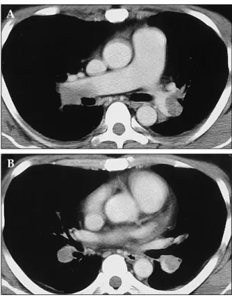
2. Contrast-en- hanced CT scans using a 4-slice MDCT scanner (Aquillion, Toshiba, Tokyo, Japan) demonstrated large clots in the pulmonary arteries (Fig. 3). The right atrium and right ventricle were dilated, but a marked diastolic collapse of the left atrium was
Small Left Atrium: An Adjunctive Sign of Hemodynamically Compromised Massive Pulmonary Embolism
Yukihiro Hama,
1,2Tadayuki Yakushiji,
3Yoshie Iwasaki,
2Tatsumi Kaji,
2Naoei Isomura,
3and Shoichi Kusano
21National Cancer Institute, NIH, Bethesda, MD, USA;
Departments of 2Radiology and 3Internal Medicine, National Defense Medical College, Tokorozawa, Japan.
Received April 3, 2004 Accepted August 4, 2004
This work was accomplished in collaboration with Departments of Radiology and Internal Medicine I, National Defense Medical College, 3-2 Namiki, Tokorozawa, Saitama, 359-0042 Japan.
This research was supported in part by the Intramural Research Program of the NIH, National Cancer Institute, Center for Cancer Research.
Reprint address: requests to Dr. Yukihiro Hama, Radiation Biology Branch, National Cancer Institute, NIH, Building 10, Room B3B69, Bethesda, MD 20892 USA. Tel: 1-301-496-7511, Fax: 1-301-480-2238, E-mail: [email protected]
Brief Communication
Yukihiro Hama, et al.
Yonsei Med J Vol. 46, No. 5, 2005
noted (Fig. 4).
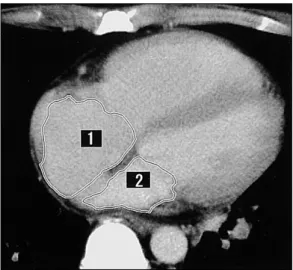
To measure the volumes of the right and left atria precisely, each atrial volume was calculated by
encircling the region of interest at the workstation (Fiji Photo Film Co., Ltd., Tokyo, Japan), multi- plying each area by its slice thickness (8 mm) and adding the results together (Fig. 4). The right atrial volume was 124 mL (Normal: 110-185 mL), but the left atrial volume markedly decreased to 53 mL (Normal: 100-130 mL).
6Based on the find- ings of the ECG, the echocardiogram, and CT, a diagnosis of massive pulmonary embolism with hemodynamic compromise was made. Magnetic resonance venography revealed a deep vein throm- bosis in the left popliteal vein. An inferior vena cava filter (TrapEase; Cordis, Miami, FL, USA) was placed via the right superficial femoral vein to prevent further thromboembolism. Intrave- nous digital subtraction angiography demon- strated intraluminal filling defects in both of the central pulmonary arteries. The pulmonary artery pressure was 58 mmHg. The mutant tissue-type plasminogen activator, monteplase (Cleactor, Eisai
Fig. 1.Electrocardiogram at the time of admission showed an SI, QIII, TIII pattern.
Fig. 2. Apical four-chamber view of the echocardiogram demonstrated marked dilatation of the right ventricle and right atrium. The interventricular septum and the atrial septum were deviated towards the left ventricle and left atrium respectively. RV, right ventricle; RA, right atrium; LV, left ventricle; LA, left atrium.
Fig. 3.A. Contrast-enhanced CT performed at the level of the right main pulmonary artery showed a large emboli in the bilateral main pulmonary arteries. B. Contrast- enhanced CT performed at the level of the descending interlobar pulmonary arteries showed low-attenuation emboli outlined by high attenuation contrast-enhanced flowing blood.
A
B
Small Left Atrium in Pulmonary Embolism
Yonsei Med J Vol. 46, No. 5, 2005
Co., Ltd., Tokyo, Japan) (1,600,000U), diluted in normal saline (20 mL), was administered directly into the pulmonary trunk using a 4-French cathe- ter. Anticoagulation therapy consisted of the ad- ministration of intravenous heparin (120,000 U/
day) followed by oral warfarin (4 mg/day). The target international normalized ratio (INR) was 2.0. Two weeks after the initiation of the anticoa- gulation therapy, the patient's dyspnea was al- leviated and the oxygen saturation increased to 98% on room air. The patient had an uneventful posttreatment course and was discharged 3 weeks after the referral.
Follow-up CT scans demonstrated that the large emboli were dissolved almost completely and the sizes of the right and left atria were normalized.
The right atrial volume became 115 cm
3and the left atrial volume became 111 cm
3. He appeared well and stable during the 10 months of follow- up.
The mortality of PE is usually due to circulatory failure from right heart failure (acute cor pul- monale).
7Although life-threatening PE traditio- nally has been equated with anatomically massive PE (defined as a> 50% obstruction of the pul-
monary vasculature or the occlusion of two or more lobar arteries), it has been considered that the outcome from PE is a function of both the size of the embolus and the underlying cardiopul- monary function.
8A pressure overload of the right ventricle secondary to pulmonary arterial hypertension initially results in right ventricular dysfunction, which may progress to right ventri- cular failure and circulatory collapse. A sudden increase in the right ventricular afterload results in elevated right ventricular wall tension, right ventricular dilatation, and eventually right ventri- cular dysfunction. Dilatation of the right ventricle causes the interventricular septum to shift to- wards the left ventricle and results in a decreased left ventricular diastolic volume. Right ventricular contractile dysfunction and tricuspid regurgitation due to the elevated pressures within the right ventricle cause a decreased output from the right ventricle, also contributing to underfilling of the left ventricle. Underfilling of the left ventricle results in decreased cardiac output, and, if severe, decreased systemic blood pressure and perfu- sion.
7-10In such a hemodynamically compromised state, the left atrial cavity is decreased in size. CT can precisely assess both the size of the embolus and the underlying cardiovascular function.
7-10Some investigators suggested that the severity of PE can be quantitatively assessed with CT. Right ventricular dilatation, and straitening of the inter- ventricular septum or the septum toward the left ventricle may be seen, indicating hemodynamic compromise.
7-10A small left atrium is a general manifestation of a decreased pulmonary venous return and not a specific finding of PE. However, to our knowledge, no previous report describes the clinical significance of a small left atrium or atrial septal bowing toward the left atrium
11.
Echocardiography is an operator/patient-de- pendent modality and certain patient charac- teristics such as obesity and respiratory distress may not permit an optimal study.
7However, MDCT can clearly depict clots within the pulmo- nary artery as well as cardiac anatomy, allowing for an evaluation of the size of the ventricular and atrial cavities as well as the position of each septum.
12The quantitative measurements of each chamber volume are easily obtained when using the picture archiving and communication system
Fig. 4.Contrast-enhanced CT at the mid-ventricular level showed that the interventricular septum was straightened and the right ventricle and right atrium were dilated, in- dicating a raised pressure on the right side of the heart.
The left atrium was markedly decreased in size, indi- cating underfilling of the left side of the heart. The region of interest was determined using contrast-enhanced CT scans. Each area (1 = right atrium; 2 = left atrium) was multiplied by its slice thickness (8 mm) and the results were added together using the workstation (Fiji Photo Film Co., Ltd., Tokyo, Japan).
Yukihiro Hama, et al.
Yonsei Med J Vol. 46, No. 5, 2005
and a computer workstation.
13At present, there is no consensus on the optimal strategy for diag- nosing acute PE with hemodynamic compromise.
This paper suggested the usefulness of MDCT for the evaluation of hemodynamically compromised PE and it emphasized the clinical importance of a small left atrium in such a state.
In conclusion, a single case cannot be gener- alized to others without additional scientific veri- fications. However, a small left atrial size in com- bination with a right ventricular pressure over- load seems to be an adjunctive sign of hemodyna- mically compromised massive PE.
REFERENCES
1. Dalen JE, Alpert JS. Natural history of pulmonary embolism. Prog Cardiovasc Dis 1975;17:257-70.
2. Barritt DW, Jordan SC. Anticoagulant drugs in the treatment of pulmonary embolism. A controlled trial.
Lancet 1960;1:1309-12.
3. De Monye W, Pattynama PMT. Contrast-enhanced spi- ral computed tomography of the pulmonary arteries;
an overview. Semin Thromb Hemost 2001;7:33-9.
4. Van Strijen MJ, de Monye W, Kieft GJ, Pattynama PM, Huisman MV, Smith SJ, et al. Diagnosis of pulmonary embolism with spiral CT as a second procedure fol- lowing scintigraphy. Eur Radiol 2003;13:1501-7.
5. Blachere H, Latrabe V, Montaudon M, Valli N, Couffinhal T, Raherisson C, et al. Pulmonary embolism revealed on
helical CT angiography: comparison with ventila- tion-perfusion radionuclide lung scanning. Am J Roent- genol 2000;174:1041-7.
6. Hirasawa K, Okamoto M. Cardiovascular anatomy. In:
Buntankaibougaku. Tokyo, Japan: Kanehara & Co., Ltd.;
1982. p.6-17 (in Japanese).
7. Contractor S, Maldjian PD, Sharma VK, Gor DM. Role of helical CT in detecting right ventricular dysfunction secondary to acute pulmonary embolism. J Comput Assist Tomogr 2002;26:587-91.
8. Wood KE. Major pulmonary embolism: review of a pathophysiologic approach to the golden hour of hemo- dynamically significant pulmonary embolism. Chest 2002;21:877-905.
9. Hiorns MP, Mayo JR. Spiral computed tomography for acute pulmonary embolism. Can Assoc Radiol J 2002;
53:258-68.
10. Mastora I, Remy-Jardin M, Masson P, Galland E, Delannoy V, Bauchart JJ, et al. Severity of acute pulmo- nary embolism: evaluation of a new spiral CT angio- graphic score in correlation with echocardiographic data. Eur Radiol 2003;13:29-35.
11. Rosenquist GC, Kelly JL, Chandra R, Ruckman RN, Galioto FM Jr, Midgley FM, et al. Small left atrium and change in contour of the ventricular septum in total anomalous pulmonary venous connection: a morpho- metric analysis of 22 infant hearts. Am J Cardiol 1985;
55:777-82.
12. Hofmann LK, Becker CR, Flohr T, Schoepf UJ. Multide- tector-row CT of the heart. Semin Roentgenol 2003;38:
135-45.
13. Hama Y, Kusano S. Picture archiving and communica- tion system: prospective study. Hong Kong Med J 2002;8:21-5.