aAssistant Professor, İnönü University, Faculty of Dentistry, Department of Orthodontics, Malatya, Turkey.
bAssociate Professor, cAssistant Professor, Cumhuriyet University, Faculty of Dentistry, Department of Orthodontics, Sivas, Turkey.
dAssistant Professor, Cumhuriyet University, Faculty of Dentistry, Department of Endodontics, Sivas, Turkey.
Corresponding author: Fırat Öztürk.
İnönü University, Dişhekimliği Fakültesi, Ortodonti AD. 44280 Malatya, Turkey.
+90 5326571572; e-mail, [email protected].
Received January 12, 2009; Last Revision July 8, 2009; Accepted July 10, 2009.
DOI:10.4041/kjod.2009.39.6.393
Effects of direct and indirect bonding techniques on bond strength and microleakage after thermocycling
Fırat Öztürk, DDS, PhD,
aHasan Babacan, DDS, MS,
bRuhi Nalçacı, DDS, PhD,
cAlper Kuştarcı, DDS, PhD
dObjective: The purpose of this study was to compare the shear bond strength (SBS) of brackets and micro-
leakage of a tooth-adhesive-bracket complex bonded with a direct and an indirect bonding technique after thermocycling. Methods: Fifty non-carious human premolars were divided into two equal groups. In the di- rect bonding group a light-cured adhesive and a primer (Transbond XT) was used. In the indirect-bonding group, a light-cured adhesive (Transbond XT) and chemical-cured primer (Sondhi Rapid Set) were used.
After polymerization, the teeth were kept in distilled water for 24 hours and thereafter subjected to thermal cycling (500 cycles). For the microleakage evaluation, 10 teeth from each group were further sealed with nail varnish, stained with 0.5% basic fuchsin for 24 hours, and examined under a stereomicroscope. Fifteen teeth from each group were used for SBS testing with the universal testing machine and adhesive remnant index (ARI) evaluation. Data were analyzed using the Mann-Whitney U test, Chi-square test, and Fisher’s exact test. Results: There were no statistical differences on SBS and microleakage between the two bond- ing techniques. The indirect bonding group had a significantly lower ARI score. Bracket failures were ob- tained between enamel-resin interfaces. Conclusions: The type of bonding technique did not significantly affect the amount of microleakage and SBS. (Korean J Orthod 2009;39(6):393-401)
Key words: Indirect bonding, Microleakage, Shear bond strength, ARI score
INTRODUCTION
The use of phosphoric acid to adhere acrylic materi- als to enamel was first introduced by Buonocore.
1In 1964, Newman
2described bonding of orthodontic at- tachments to the etched enamel surface with epoxy- derived resin. In orthodontic practice, brackets can be
bonded directly or indirectly. There are some inherent
shortcomings with the direct bonding technique, includ-
ing poor visualization of posterior teeth, greater possi-
bility of moisture contamination, and increased doctor
chair time. In 1972, Silverman and Cohen
3introduced
the indirect bonding technique to place brackets on
teeth more accurately and efficiently in the clinic. This
technique involves a two-stage process of bracket
placement in the laboratory on a plaster model and
transfer of these attachments to the patient’s mouth by
means of a tray, where they are bonded to the etched
enamel surface. Most current indirect bonding techni-
ques are based on a method described by Thomas.
4Initially, bond failure rates for indirect bonding
(13.9%) were higher when compared with direct bond-
ing (2.5%). However, with modifications and improve-
ments to the technique, the two systems now have
similar bond strengths and failure rates.
5-7Linn et al.
8compared the shear bond strength (SBS) and the sites of bond failure for brackets bonded to teeth between the direct bonding technique (Transbond XT) and in- direct bonding technique (Transbond XT/Sondhi Rapid Set). The study showed that reduction of SBS was also observed in the indirect bonding technique group; and there were no statistically significant differences be- tween the two techniques. When the Adhesive Rem- nant Index (ARI) was determined, the indirect bonding technique was found to have a significantly lower ARI score. There was no strong correlation between SBS and ARI scores. Hocevar and Vincent
7reported that 44% of direct-bonded brackets fractured at the brack- et-adhesive interface, whereas 72% of the indirect- bonded brackets failed at the adhesive-enamel inter- face. Daub et al.
9evaluated the SBS of one direct (Group 1 - Transbond XT) and two indirect bonding (Group 2 - Transbond XT/Sondhi Rapid Set, Group 3 - Enlight LV/Orthosolo) methods/adhesives after ther- mocycling. Each sample was thermocycled between 5
oC and 55
oC for 500 cycles. The mean SBS in Group 1, 2 and 3 were not statistically significantly different.
The authors also determined the ARI scores and found that Group 2 had a significantly higher percentage of bond failures at the adhesive-enamel interface.
The polymerization shrinkage of the adhesive mate- rial may cause gaps between the adhesive material and enamel surface and contribute to microleakage, permit- ting the passage of bacteria and oral fluids, which may initiate white spot lesions under the bracket surface area.
10Polymerization shrinkage also varies from com- posite to composite and depends on the percentage of filler, the diluents, and the percentage of monomer conversion in the specific composite resin.
11From an orthodontic point of view, microleakage may lead to lower clinical SBS and white spot lesions.
10Arhun et al.
10showed that metal brackets cause more microleakage compared with ceramic brac- kets. Ulker et al.
12compared the microleakage of the brackets bonded with high-intensity light curing lights and conventional halogen lights. This study showed that high-intensity curing units did not cause more mi- croleakage than conventional halogen lights.
There have been no studies that investigated the ef- fects of different bonding techniques on microleakage.
The purpose of this study was to evaluate the SBS, the mode of bond failure, and microleakage of direct and indirect bonding techniques after thermocycling.
MATERIAL AND METHODS
Fifty human maxillary premolars, extracted for or- thodontic reasons at the Cumhuriyet University, Facul- ty of Dentistry, with no decay, restorations, or surface defects, were collected. After extraction, the teeth were stored in 0.5% chloramines T solution for one week and transferred to distilled water. The teeth were stored in distilled water at room temperature until the experi- ments took place (a maximum of 4 months). They were randomly separated into two groups of 25.
Immediately before bonding, the teeth were prepared by removing soft-tissue remnants, calculus and plaque and mounted in cold-cure acrylic in groups of five with interproximal surfaces of adjacent teeth in contact.
All teeth were bonded using a Mini Master upper bi- cuspid bracket (American Orthodontics, Sheboygan, WI, USA) with a projected base surface area of 10.25 mm
2. The whole laboratory process was performed by the same author (Öztürk F) with 6 years of experience.
In the direct bonding group, teeth were bonded di- rectly according to the manufacturer’s recommenda- tions using a light-cured adhesive and primer (Trans- bond XT, 3M Unitek, Monrovia, CA, USA). In the in- direct bonding group, teeth were bonded indirectly with light-cured adhesive (Transbond XT) and a filled resin primer (Sondhi Rapid Set A/B Primer, 3M Unitek, Monrovia, CA, USA).
The surface of each tooth was polished for one mi- nute using a combination of a polishing agent and a brush at a low speed. A 37% phosphoric acid gel (3M Dental Products, St Paul, MN, USA) was used for acid etching for 30 seconds. The teeth were rinsed with wa- ter for 30 seconds and dried with an oil-free source for 20 seconds. In all etched cases, the frosty white ap- pearance of the etched enamel was apparent.
For the direct bonding group, the brackets were
bonded by the direct method, one at a time. A thin
layer of Transbond XT light-cured primer was applied
to the tooth, Transbond XT adhesive was applied to
the bracket base, and the bracket was pressed lightly
in the desired position onto the tooth. The bracket was placed in the center of the crown, with the center of the bracket over the long axis of the tooth. The excess adhesive was removed with a hand instrument, and the bracket was cured with plasma arc curing light (Apollo 95E, Denmed Technologies, Inc, Westlake Village, CA, USA) for 3 seconds from the mesial side and 3 seconds from the distal.
In the indirect bonding group, alginate impressions were taken, and hard-stone working models were ob- tained. Two coats of Al-Cote separating medium (Dentsply International Inc, York, PA, USA) were painted on the models and allowed to dry. Transbond XT adhesive was applied to the bracket base, and the bracket was pressed lightly in the desired position onto the cast. The bracket was placed in the center of the crown, with the center of the bracket over the long ax- is of the tooth. The excess adhesive was removed with a hand instrument, and the bracket was cured with a plasma arc curing light for 15 seconds from the mesial and 15 seconds from the distal. This extended curing period was chosen to achieve complete polymerization of the adhesive on the plaster model.
13,14Before form- ing the indirect bonding trays, the undercut areas, such as hooks, were blocked out with a soft transparent sili- cone (Emiluma, Ortho Kinetics, Vista, CA, USA) for indirect bonding. After the silicone hardened, transfer trays were made from 0.040-inch (1 mm) vacuum- formed essix (Raintree Essix Inc., Los Angeles, CA, USA). After the transfer tray material had set, the specimens were soaked in water for 20 minutes to dis- solve the separating medium. The transfer tray was re- moved, and the adhesive bases were gently sandblasted avoiding any disturbance in the base resin, washed, and dried as advised by Sondhi.
15A thin layer of pri- mer A (Sondhi Rapid Set, 3M Unitek, Monrovia, CA, USA) was painted on each tooth and a thin layer of resin B (Sondhi Rapid Set, 3M Unitek, Monrovia, CA, USA) was painted on each bracket’s adhesive base.
The transfer tray was placed on the experimental tooth and held with finger pressure for 30 seconds and then left on the teeth without any pressure for two minutes before removal of the tray.
Teeth were stored at 37
oC in distilled water for 24 hours. After 24 hours, the samples were thermocycled
according to the ISO 11405 recommendation.
16Each specimen underwent 500 complete cycles in distilled water between 5
oC and 55
oC, with a dwell time of 30 seconds in each bath and a transfer time of 15 seconds between baths.
After thermocycling was applied, 10 teeth of each group were used for evaluation of the microleakage and 15 teeth were used for the evaluation of the SBS and ARI score.
Evaluation of shear bond strength and ad- hesive remnant index
The teeth were removed from the acrylic block after bonding and embedded in phenolic rings using autopo- lymerizing polymethyl methacrylate. A mounting jig was used to align the facial surface of the tooth to be perpendicular to the bottom of the mold and its labial surface parallel to the force during the SBS test. A universal testing machine (LF Plus, Ametek LLOYD Instruments, West Sussex, England) was used for the shear bond test at a crosshead speed of 0.5 mm/min.
The standard knife-edge was positioned to make con- tact between the tie wing and the bracket base as close to the base as possible and directed parallel to the long axis of the crown of the tooth. The load at failure was recorded by a personal computer connected to the test machine. SBS values were calculated as the recorded failure load divided by the surface area (bracket base) and expressed in megapascals (MPa).
After debonding, the enamel surface of each tooth
and the bracket bases were examined under 10 X mag-
nification with a stereomicroscope (SMZ 800, Nikon,
Tokyo, Japan). ARI scores
17were assigned to each
specimen. An ARI score of 0 indicated that no adhe-
sive was left on the tooth in the bonded area; 1 in-
dicated that less than half of the adhesive was left on
the tooth; 2 indicated that more than half was left on
the tooth; and 3 indicated that all the adhesive re-
mained on the tooth, with a distinct impression of the
bracket mesh. Examples of assignments of ARI scores
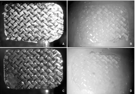
are shown in Fig 1.
Fig 1. Examples of assignments of adhesive remnant index scores (with 10 × magnification). A, B, ARI score of 3 indicated that all the adhesive remained on the tooth; C, D, ARI score of 2 indicated that more than half was left on the tooth.
Microleakage evaluation
Two consecutive layers of nail varnish were applied to the entire surface of the tooth, except for an area of approximately 1 mm away from the brackets. The teeth were immersed in 0.5% solution of basic fuch- sine for 24 hours at room temperature. After rinsing with distilled water, the samples were air-dried and each specimen was sliced longitudinally with a low- speed diamond saw (Isomet, Buehler, Lake Bluff, IL, USA) under water coolant in the buccolingual direc- tion. All sections were examined by two calibrated in- vestigators with a stereomicroscope (30 X magnifica- tions) for dye penetration. Each section was scored from both the occlusal and gingival margins of the brackets between the bracket-adhesive and the adhe- sive-enamel interfaces. Scoring was made according to the following criteria:
Score 0: No dye penetration between the brack- et-adhesive or adhesive-enamel interface.
Score 1: Dye penetration restricted to 1 mm of the bracket-adhesive or adhesive-enamel interface.
Score 2: Dye penetration into the inner half (2 mm) of the bracket-adhesive or adhesive-enamel interface.
Score 3: Dye penetration into 3 mm of the brack- et-adhesive or adhesive-enamel interface.
In cases of disagreement between scoring, consensus was obtained by using the greater score. Fig 2 demon- strates individual examples of scoring.
Statistical analyses
The data was analyzed using SPSS for Windows, version 14.0 (SPSS Inc., Chicago, IL, USA).
The assessments of SBS data were analyzed using
the Mann-Whitney U test. ARI and microleakage
scores were evaluated with the Chi-square test, and
Fisher’s exact test. Also, the relationship among SBS
Data, ARI, and microleakage scores was analyzed with
correlation analysis. The level of significance was set
at p < 0.05.
Fig 2. Microleakage evaluation between the bracket-adhesive and the adhesive-enamel interfaces. A, A specimen bonded with the direct bonding technique (30 × magnification). The adhesive-tooth interface scores are 0 for both gingival and occlusal sides. For the adhesive-bracket interface, scores are 0 for the occlusal side and 1 for the gin- gival side; B, a specimen bonded with the indirect bonding technique (30 × magnification). No microleakage found under the bracket.
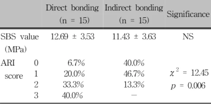
Direct bonding (n = 15)
Indirect bonding
(n = 15) Significance SBS value
(MPa)
12.69 ± 3.53 11.43 ± 3.63 NS
ARI score
0 6.7% 40.0%
χ2 = 12.45 p = 0.006
1 20.0% 46.7%
2 33.3% 13.3%
3 40.0% −
NS, Not significant. The SBS values were analyzed using the Mann-Whitney U test and ARI scores were evaluated with the Chi-square test (p < 0.05). ARI scores: 0, no adhesive left on the tooth; 1, less than half of the adhesive left on the tooth; 2, more than half of the adhesive left on the tooth; 3, all adhesive left on the tooth.
Table 1. Comparison of the shear bond strength (SBS) and the adhesive remnant index (ARI) score between di- rect and indirect bonding groups
RESULTS
The mean SBS and standard deviation values are shown in Table 1. Brackets directly bonded with Transbond XT showed mean bond strength of 12.69 ± 3.53 MPa. Brackets bonded indirectly and chemically cured had a mean SBS of 11.43 ± 3.63 MPa. There were no statistical differences between two groups (p
> 0.05).
The ARI scores are given in Table 1. Comparisons
of resin remnants between two groups showed statisti- cally significant differences (p < 0.05). The ARI scores showed that the indirect bonding group had sig- nificantly higher percentage of bond failures at the ad- hesive-enamel interface.
The comparison of microleakage scores between oc- clusal and gingival sides for enamel-adhesive and ad- hesive-bracket interfaces are shown in Table 2. Statisti- cal comparisons of the microleakage scores showed no significant differences between two groups (p > 0.05).
Microleakage scores among the groups showed that the direct bonding group had statistically higher micro- leakage scores in the enamel-adhesive interface at the gingival side (p < 0.05).
On the basis of the results of the correlation analy- sis, in the direct bonding group, there was an adverse relationship between ARI scores and SBS values (r =
−0.78). The quantity of this relationship is sig- nificantly important (p < 0.05). It is observed that as the ARI scores increase, the SBS values decrease.
Correlation analysis showed that there was no relation-
ship between microleakage scores and SBS values in
the direct bonding group. In the indirect bonding
group, an adverse relationship was found between SBS
and microleakage scores at the adhesive-bracket inter-
face, both occlusal (r = −0.64) and gingival margins
(r = −0.68). The quantity of this relationship is sig-
nificantly important (p < 0.05). When the micro-
leakage scores between the adhesive and the bracket
Bracket-adhesive interface Adhesive-enamel interface Direct bonding
(n = 10)
Indirect bonding
(n = 10) Sig Direct bonding (n = 10)
Indirect bonding
(n = 10) Sig Occlusal
scores
0 80% 60%
NS
90% 70%
1 20% 40% 10% 20% NS
2 − − − 10%
3 − − − −
Gingival scores
0 40% 40%
NS
30% 20%
1 60% 60% 60% 50% NS
2 − − 10% 30%
3 − − − −
NS, Not significant; microleakage scores: Score 0, no dye penetration between the bracket-adhesive or adhesive- enamel interface; Score 1, dye penetration restricted to 1 mm of the bracket-adhesive or adhesive-enamel interface;
Score 2, dye penetration into the inner half (2 mm) of the bracket-adhesive or adhesive-enamel interface; Score 3, dye penetration into 3 mm of the bracket-adhesive or adhesive-enamel interface.
Table 2. Comparison of microleakage scores between bracket - adhesive surfaces and adhesive - enamel surfaces from occlusal-gingival sides
interface increase (both occlusal and gingival), the SBS values decrease. According to the correlation analysis there were no relationship between ARI scores and SBS values in the indirect bonding group.
DISCUSSION
This study compared the SBS, mode of bond failure (ARI), and microleakage of direct and indirect bonding techniques after thermocycling.
The results from this study showed that there were no significant differences in shear bond strength be- tween direct and indirect bonding techniques. Reynolds and von Fraunhofer
18indicated that to overcome nor- mal orthodontic forces shear bond strengths should be in the range of 5.9 to 7.9 MPa. The mean SBS in this study was 12.69 ± 3.53 MPa for direct bonding and 11.43 ± 3.63 MPa for the indirect bonding group. The SBS values of both groups were over this clinically ac- ceptable range.
Several studies compared the SBS of direct and in- direct bonding methods. Polat et al.
19showed that there were no statistically differences between the direct bonding group (Transbond XT) and indirect bonding Group I (Therma Cure/Custom IQ), whereas both yielded significantly higher SBS values compared with the indirect bonding Group II (Transbond XT/Sondhi
Rapid Set) in vitro. In the in vivo parts of the same study, failure rates of the brackets were followed after nine months and there were no differences found be- tween the two indirect bonding groups. Klocke et al.
20showed that there were no significant differences in SBS between the indirect bonding groups (Transbond XT/Sondhi rapid set and Phase II/Maximum cure) and direct bonding group (Transbond XT). The bond strength of thermally cured indirect bonding group (ThermaCure), however, was significantly lower. Yi et al.
21found no statistically significant differences in SBS between indirectly bonded brackets (Transbond XT and sondhi rapid set) and light-cured directly bond- ed brackets (Transbond XT). In these previous studies, samples were not thermocyled. Orthodontic adhesives are routinely exposed to temperature variations in the oral cavity. Air temperature, humidity, and air velocity when breathing can also alter resting mouth tem- perature. Bishara et al.
22have suggested that thermal cycling should be a part of the testing protocol of new adhesives. Daub et al.
9compared the shear bond strengths of direct bonding (Transbond XT) and in- direct bonding (Transbond XT/Sondhi Rapid Set) tech- niques after thermocycling. Their study showed that there were no statistically significant differences be- tween the two techniques.
Previous studies in which samples were nonther-
mocycled showed that the indirect bonding method had significantly lower ARI scores than the direct bonding method.
7,8,23Daub et al.
9also showed that there was no statistically significant difference in the location of bond failure in the direct bonding (Transbond XT) and light-cured indirect bonding (Enlight LV/Orthosolo) groups after thermocycling. Based on their findings, the chemical-cured indirect bonding group (Transbond XT/Sondhi Rapid Set) was statistically different from the direct bonding and the light-cured indirect bonding groups after thermocycling. In accordance with these studies, our study showed significantly higher bond failure rates at the adhesive-enamel interface in the in- direct bonding group. This reduced remnant resin on the tooth is clinically desirable because it requires few- er cleanups on debonding and reduces the risk of en- amel damage.
23The difference of our study from similar previous studies is the evaluation of microleakage. The potential of white spot lesion formation around orthodontic brackets has become a particular clinical problem for orthodontic treatment. Microleakage is the seeping and leaking of fluids and bacteria between the tooth and restoration in restorative dentistry. James et al.
11were the first to point out the increased risk of decal- cification caused by microleakage around orthodontic brackets. Both the area around the brackets and the area under the brackets need attention to determine the risk of caries formation.
Different techniques have been introduced to assess microleakage around restorations in dentistry. In the present study, microleakage of the bonded specimens was determined by the dye penetration method, which is one of the most common microleakage assessment methods.
12,24-27We used plasma arc curing light and Transbond XT adhesive in both groups. James et al.
11showed that there were significant differences in mi- croleakage among plasma arc light, argon laser, and conventional halogen light when the adhesive pre- coated (APC) adhesive system was used. However, there was no significant difference in microleakage be- tween groups when the Transbond XT adhesive system was used. In the current study microleakage was exam- ined using specimens that were sliced longitudinally.
Although we did not find any significant differences
between microleakage scores, these specimens may not represent the whole enamel-adhesive-bracket interface.
This status was a limitation of our study and in a fu- ture project we will try to overcome this problem.
Ulker et al.
12showed that the type of light-curing unit (plasma arc curing light, light-emitting diode, and con- ventional halogen light) did not significantly affect the amount of microleakage.
The type of the bonding technique did not sig- nificantly affect the amount of microleakage at the gin- gival or occlusal margins of the adhesive-enamel and adhesive-bracket interfaces according to our results.
Studies in restorative dentistry have demonstrated that curing composites causes polymerization shrinkage and microleakage.
28,29Polymerization shrinkage also varies from composite to composite and depends on the per- centage of the filler, diluents, and monomer conversion in the specific composite resin and the photo- polymerization type.
30In restorative dentistry, compo- site resin is placed in the cavity in large amounts, and curing can create excessive shrinkage and gap for- mation. In contrast, orthodontic adhesive layers are very thin, and there is some adhesive at the edges of the bracket to absorb some shrinkage. Because the bracket is free floating, the shrinkage can pull the bracket closer to the enamel.
31Therefore, polymer- ization shrinkage and subsequent microleakage is less important in orthodontic applications than it is in re- storative dentistry.
11Ramoglu et al.
25observed higher microleakage scores at both adhesive interfaces at the gingival sides for all specimens, which were cured with resin-mo- dified glass ionomer or conventional resin. In this study, although there were higher microleakage scores found at the gingival side between both interfaces, there was a statistically significant difference in micro- leakage scores only between adhesive-tooth interfaces at the gingival side in the direct bonding group. Arhun et al.
10and Ramoglu et al.
25attributed these differences to surface curvature anatomy, which may result in rela- tively thicker adhesive at the gingival margin.
Factors such as the adhesive system, composite
composition, photopolymerization type, and exposure
time affect the bond strength of brackets. In restorative
dentistry literature, it was established that durability of
bond strength could be affected by microleakage.
32,33When we look from the orthodontic perspective; in the case of microleakage occurring on the adhesive-tooth interface, there is a risk of formation of a white spot lesion, and in the case of microleakage occurring on the adhesive-bracket interface, bracket failure proba- bility may increase due to bond degradation.
34Al- though James et al.
11could not demonstrate any corre- lation between microleakage and bond strength, Arhun et al.
10reported that metal brackets cause more leakage between the adhesive-bracket interface than ceramic brackets, which may lead to lower clinical SBS and white spot lesions. Our study showed that there is an adverse relationship between SBS and microleakage at the occlusal and gingival side between adhesive- bracket interfaces in the indirect bonding group.
CONCLUSION
This study showed that indirect bonding and direct bonding techniques produce clinically acceptable bond strengths. The type of the bonding technique did not significantly affect the amount of microleakage. In or- thodontic practice, our results indicate that the indirect bonding method - with its significant advantages - can be conveniently used. This study was an in vitro study;
thus, further clinical studies are needed to strengthen the validity of our results.
- 국문초록 -
직접 부착법과 간접 부착법이 열순환 후 부착강도와 미세누출에 미치는 영향에 대한 연구
Fırat Öztürk,a Hasan Babacan,b Ruhi Nalçacı,c Alper Kuştarcıd본 연구는 직접 또는 간접 부착법으로 부착한 교정용 브라 켓을 열순환 처리 후에 전단 결합 강도(shear bond strength, SBS)와 치아-접착제-브라켓 복합체의 미세 누출을 비교하기 위하여 시행하였다. 50개의 치아우식증이 없는 사람 소구치 를 구하여 동등한 2개의 군으로 나누었다. 직접 부착군에서 는 광중합 접착제 및 primer (Transbond XT)를 사용하였고 간접 부착군에서는 광중합 접착제와(Transbond XT) 화학중 합 primer (Sondhi Rapid Set)를 사용하였다. 중합 후에 치 아는 24시간 동안 증류수에 보관하였고 이후 500회 열순환
처리를 하였다. 미세누출의 평가를 위해 각 군에서 10개의 치아에 nail varnish로 추가 봉연을 실시하고 0.5% basic fuchsin에 24시간 동안 염색한 후 입체현미경으로 검경하였 다. 만능시험기를 이용하여 각 군의 치아 15개로 SBS를 계 측하였고 adhesive remnant index (ARI)를 평가하였다. 결 과 자료는 Mann-Whitney U test, Chi-square test, Fisher’s exact test로 통계 분석하였다. 직접 부착군과 간접 부착군 사이에 SBS 및 미세누출의 통계적으로 유의한 차이는 보이 지 않았으며 간접 부착군에서 통계적으로 유의하게 ARI 점 수가 낮았다. 브라켓의 탈락은 법랑질-레진 계면에서 발생하 였다. 이상의 결과로 보아 부착법의 차이는 미세누출의 양과 SBS에 유의한 영향을 미치지 않는다.
주요 단어:
간접 부착법, 미세누출, 전단 결합 강도, ARI 점수
REFERENCES
1. Buonocore MG. A simple method of increasing the adhesion of acrylic filling materials to enamel surfaces. J Dent Res 1955;34:849-53.
2. Newman GV. Bonding plastic orthodontic attachments to tooth enamel. JNJ Dent Soc 1964;35:346-58.
3. Silverman E, Cohen M. Current adhesives for indirect bracket bonding. Am J Orthod 1974;65:76-84.
4. Thomas RG. Indirect bonding: simplicity in action. J Clin Orthod 1979;13:93-106.
5. Miles PG. Weyant RJ. A comparison of two indirect bonding adhesives. Angle Orthod 2005;75:1019-23.
6. Aguirre MJ, King GJ, Waldron JM. Assessment of bracket placement and bond strength when comparing direct bonding to indirect bonding techniques. Am J Orthod 1982;82:269-76.
7. Hocevar RA, Vincent HF. Indirect versus direct bonding: bond strength and failure location. Am J Orthod Dentofacial Orthop 1988;94:367-71.
8. Linn BJ, Berzins DW, Dhuru VB, Bradley TG. A comparison of bond strength between direct- and indirect-bonding methods.
Angle Orthod 2006;76:289-94.
9. Daub J, Berzins DW, Linn BJ, Bradley TG. Bond strength of direct and indirect bonded brackets after thermocycling. Angle Orthod 2006;76:295-300.
10. Arhun N, Arman A, Cehreli SB¸ Arıkan S, Karabulut E, Gülşahi K. Microleakage beneath ceramic and metal brackets bonded with a conventional and an antibacterial adhesive system. Angle Orthod 2006;76:1028-34.
11. James JW, Miller BH, English JD, Tadlock LP, Buschang PH.
Effects of high-speed curing devices on shear bond strength and microleakage of orthodontic brackets. Am J Orthod Dentofacial Orthop 2003;123:555-61.
12. Ulker M, Uysal T, Ramoglu SI, Ertas H. Microleakage under orthodontic brackets using high-intensity curing lights. Angle Orthod 2009;79:144-9.
13. Klocke A, Shi J, Vaziri F, Kahl-Nieke B, Bismayer U. Effect of time on bond strength in indirect bonding. Angle Orthod
2004;74:245-50.
14. Klocke A, Tadic D, Vaziri F, Kahl-Nieke B. Custom base pre- aging in indirect bonding. Angle Orthod 2004;74:106-11.
15. Sondhi A. Efficient and effective indirect bonding. Am J Orthod Dentofacial Orthop 1999;115:352-9.
16. International Organization for Standardization Technical Speci- fication Report (ISO/TS 11405:2003).
17. Artun J, Bergland S. Clinical trials with crystal growth con- ditioning as an alternative to acid-etch enamel pretreatment.
Am J Orthod 1984;85:333-40.
18. Reynolds IR, von Fraunhofer JA. Direct bonding of ortho- dontic brackets - a comparative study of adhesives. Br J Orthod 1976;3:143-6.
19. Polat O, Karaman AI, Buyukyilmaz T. In vitro evaluation of shear bond strengths and in vivo analysis of bond survival of indirect-bonding resins. Angle Orthod 2004;74:405-9.
20. Klocke A, Shi J, Kahl-Nieke B, Bismayer U. Bond strength with custom base indirect bonding techniques. Angle Orthod 2003;73:176-80.
21. Yi GK, Dunn WJ, Taloumis LJ. Shear bond strength compar- ison between direct and indirect bonded orthodontic brackets.
Am J Orthod Dentofacial Orthop 2003;124:577-81.
22. Bishara SE, Ajlouni R, Laffoon JF. Effect of thermocycling on the shear bond strength of a cyanoacrylate orthodontic ad- hesive. Am J Orthod Dentofacial Orthop 2003;123:21-4.
23. Sinha PK, Nanda RS, Duncanson MG, Hosier MJ. Bond strengths and remnant adhesive resin on debonding for ortho- dontic bonding techniques. Am J Orthod Dentofacial Orthop 1995;108:302-7.
24. Taylor MJ, Lynch E. Microleakage. J Dent 1992;20:3-10.
25. Ramoglu SI, Uysal T, Ulker M, Ertas H. Microleakage under ceramic and metallic brackets bonded with resin-modified
glass ionomer. Angle Orthod 2009;79:138-43.
26. Gillgrass TJ, Millett DT, Creanor SL, MacKenzie D, Bagg J, Gilmour WH, et al. Fluoride release, microbial inhibition and microleakage pattern of two orthodontic band cements. J Dent 1999;27:455-61.
27. Mehl A, Hickel R, Kunzelmann KH. Physical properties and gap formation of light-cured composites with and without
‘softstart-polymerization’. J Dent 1997;25:321-30.
28. Ferracane JL, Mitchem JC. Relationship between composite contraction stress and leakage in class V cavities. Am J Dent 2003;16:239-43.
29. Calheiros FC, Sadek FT, Braga RR, Cardoso PE.
Polymerization contraction stress of low-shrinkage composites and its correlation with microleakage in class V restorations.
J Dent 2004;32:407-12.
30. Burgess JO, DeGoes M, Walker R, Ripps AH. An evaluation of four light-curing units comparing soft and hard curing. Pract Periodontics Aesthet Dent 1999;11:125-32.
31. Oesterle LJ, Newman SM, Shellhart WC. Rapid curing of bonding composite with a xenon plasma arc light. Am J Orthod Dentfacial Orthop 2001;119:610-6.
32. Celiberti P, Lussi A. Use of a self etching adhesive on pre- viously etched intact enamel and its effect on sealant micro- leakage and tag formation. J Dent 2005;33:163-71.
33. Kubo S, Yokota H, Sata Y, Hayashi Y. Microleakage of self etching primers after thermal and flexural load cycling. Am J Dent 2001;14:163-9.
34. Arıkan S, Arhun N, Arman A, Cehreli SB. Microleakage be- neath ceramic and metal brackets photopolymerized with LED or conventional light curing units. Angle Orthod 2006;76:
1035-40.